ISSN (print) 1226-8496 ISSN (online) 2288-3819
Clinical Features and Correlation With Congenital Missing Teeth of
Delayed First Permanent Molar
Myeongyeon Lee1, Hyoseol Lee2, Jeseon Song1, Jaeho Lee1, Byungjai Choi1, Seongoh Kim1, Seunghye Kim3
1Department of Pediatric Dentistry, College of Dentistry, Yonsei University 2Department of Pediatric Dentistry, School of Dentistry, Kyung Hee University
3Department of Dentistry, Ajou University School of Medicine
Delayed eruption of the first molar, without a generalized or localized cause, is usually associated with delayed development of the affected tooth. The aim of this study was to investigate the clinical features of the first per-manent molar showing delayed development and eruption, and its association with developmental anomalies of other teeth.
Panoramic radiographs of 40 healthy children showing delayed development and eruption of first permanent molars were analyzed. The clinical features of affected first molars and developmental anomalies of other teeth (except third molars) were evaluated.
Delayed first molars were more frequent in the maxilla. The incidence of bilateral delayed development of first molars was greater than that of unilateral cases in female patients. In contrast, male patients showed unilateral delayed development of the first molar more frequently.
A higher incidence of congenitally missing teeth was observed in patients with delayed first molar.
In each case, delayed development or congenital absence was observed in the second molar adjacent to the de-layed first molar.
Overall, delayed first molar seems to be associated with congenital absence of additional teeth. Understanding the developmental mechanisms of this phenomenon requires further studies.
Key words :Tooth, Unerupted; Anodontia; Molar, First
Abstract
Ⅰ. Introduction
Delayed tooth eruption is the retarded emergence of a tooth into the oral cavity. There are two methods for es-timating the timing of tooth eruption. One method,
sug-gested by Grøn1), evaluates the extend of dental root
de-velopment, and denotes the tooth eruption stage as the
time point at which 3/4 of the root is formed. The second method estimates the tooth eruption stage based on the
chronological age. According to Suri et al.2)
, it is defined that delayed eruption occurs when the chronological age is delayed more than two standard deviations from the mean eruption age.
The first permanent molar erupts at the age of 6.12
-Corresponding author : Seunghye Kim
Department of Dentistry, Ajou Univeristy School of Medicine, 164 World cup-ro, Yeongtong-gu, Suwon, 16499, Korea Tel: +82-31-219-4112 / Fax: +82-31-219-4112 / E-mail: [email protected]
6.39 years3). Grover4) reported that delayed eruption of
the first permanent molar is rare compared to other teeth, with an incidence of 0.01% in the general popula-tion. Delayed eruption of the first permanent molar in the maxilla and mandible accounted for 5.84% and 2.76%, respectively, of the total delayed eruption in
Korean children5). Most of the cases of delayed eruption
were attributable to local factors, such as early loss of a primary tooth, loss of eruption space, and presence of
supernumerary teeth5).
Delayed eruption of the first permanent molar without systemic or local causes was reported to accompany de-layed tooth development or congenital absence of the
ad-jacent second permanent molar6-9). Rasmussen7) named
the first permanent molar showing delayed eruption as
the “9-year-molar”, because the first permanent molar
often erupted at approximately 9 years of age.
In a previous case study, delayed development of the adjacent second molar was also observed in 7 out of 9 cases with delayed eruption of the first permanent
molar8). In addition, congenital missing of the third
mo-lar was observed in all 9 cases in the same study8)
.
Nakano et al.8) proposed that delayed development of
the first permanent molar could be resulted from con-genital absence of the first permanent molar and mesial migration of second permanent molar. However, the higher rate of congenitally missing teeth cannot be ex-plained in patient with delayed development of the first molar. Furthermore, there are no clinical studies proving the association of congenitally missing teeth with de-layed development of the first permanent molar.
The aim of this study was to investigate the clinical
aspects of ‘delayed eruption and development of the first
permanent molar (DFM)’without systemic or local causes and to demonstrate its association with delayed development or congenital absence of other teeth.
Ⅱ. Materials and Methods
1. Subjects
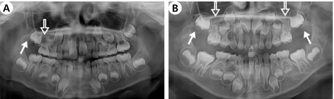
The study subjects were first selected from those who visited the Department of Pediatric Dentistry, Dental Hospital, Yonsei University between January 2008 and December 2014. The patient selection inclusion criterion was a clinical and radiographical evidence of localized delayed eruption of the first permanent molar. The ex-clusion criteria were medically compromised patients and patients with localized causes of delayed tooth eruption (such as, odontoma or ectopic impacted teeth). In all cases, clinically delayed eruption accompanied clear de-layed development of the first permanent molar, when compared to erupted first permanent molars in other quadrants (Fig. 1). A total of 40 subjects (15 males, 25 females) with DFM were selected; they were aged 6 - 10 years (mean age of 8.62 years) were selected for this study.
2. Methods
The panoramic radiographs of the study subjects were examined by two investigators. The location and laterali-ty of DFM, delayed development or congenital absence of the adjacent second permanent molars, and congenital
Fig. 1. Panoramic radiographs of patients with DFM. (A) Unilateral occurrence of DFM and congenital absence of adjacent second permanent molar
(closed arrow) in the right maxilla of a male patient (aged 8 years, 2 months). The patient showed congenital absence of the right maxillary second premolar (open arrows). (B) Bilateral occurrence of DFM and congenital absence of adjacent second permanent molar (closed arrow) in the maxilla of a female patient (aged 6 years, 8 months). The patient showed congenital absence of both maxillary second premolars (open arrows).
absence of other teeth were examined on panoramic ra-diographs of the subjects. The dental age of the subjects’ was determined by the calcification stage according to Nolla’s method10)
. Delayed development of teeth was di-agnosed when the dental age was delayed by more than 2 standard deviations of the mean calcification stage for Korean children11). The interclass correlation coefficient
between two examiners was 0.95.
Statistical analysis was performed using Excel 2010 (Microsoft, Redmond, Washington, USA) and R (x64 3.13, R Foundation for Statistical Computing, Vienna, Austria). Statistical significance was determined at a level of p < 0.05. The Fisher’s exact test was used to compare the laterality of DFM according to the gender of the subjects, and the chi-square test was used to assess the differences in the frequency of congenital missing teeth between the DFM affected quadrant and the nor-mal quadrant.
Ⅲ. Results
In this study, 69 cases of DFM were found in 40 sub-jects; 21 cases were found in 14 male subjects, while 48 were in 26 female subjects.
1. Distribution
DFM was detected in the maxilla of 32 out of 40 (80.0%) subjects, while it was found in the mandible of 5 out of 40 (12.5%) subjects. In total, 7.5% of the
pa-tients (3 out of 40) exhibited bimaxillary manifestation of DFM.
2. Laterality
Sixteen subjects (40.0%) showed unilateral DFM in the maxilla (30.0%, 12 subjects) or mandible (10.0%, 4 subjects), 21 subjects (52.5%) showed bilateral involve-ment of DFM in the maxilla (20 patients, 50.0%) or mandible (1 patient, 2.5%). In 3 subjects (7.5%), DFMs were found in 3 quadrants (Table 1).
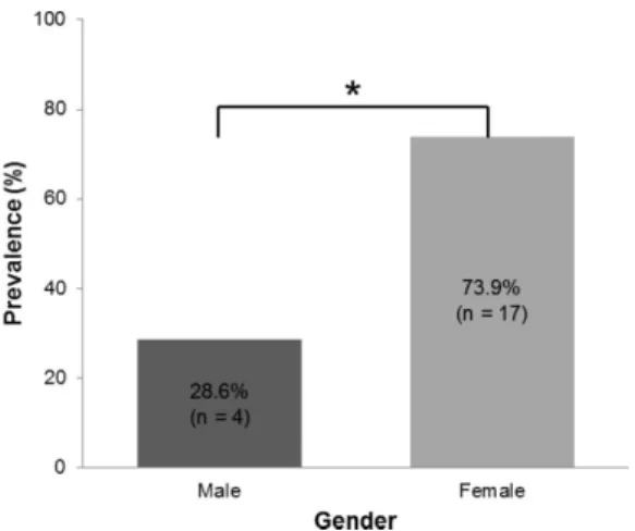
Among the 37 subjects (14 males and 23 females), DFM was unilateral in 16 subjects (43.2%, 10 males and 6 females) and bilateral in 21 (56.8%, 4 males and 17 females). Three female subjects with DFM in three quadrants were excluded from this analysis. There was a statistically significant difference in the laterality of DFM in male and female patients. Unilateral manifesta-tion of DFM was significantly frequent in male patients. In contrast, female patients exhibited significantly high-er frequency of bilathigh-eral presentation of DFM (Fig. 2, 3).
Fig. 3. Correlation of unilateral DFM with gender. The incidence of
uni-lateral DFM was significantly higher in male patients than in female patients. (Fisher's exact test, * : p < 0.05).
DFM = delayed eruption and development of the first permanent molar
Table 1. Distribution of DFM in relation to location
Laterality Location Number of patients (%)
Unilateral Maxilla 12 (30.0)
Mandible 4 (10.0)
Bilateral Maxilla 20 (50.0)
Mandible 1 (2.5)
More than 3 quadrants 3 (7.5)
Total 40 (100)
DFM = delayed eruption and development of the first permanent molar
Fig. 2. Correlation of bilateral DFM with gender. The incidence of
bilat-eral DFM was significantly higher in female patients than in male patients (Fisher's exact test, * : p < 0.05).
3. Developmental abnormalities of adjacent second permanent molars.
In 69 cases of DFM, delayed development of the sec-ond permanent molar was seen in 39 cases, whereas congenital absence of the adjacent second permanent molar was observed in 30 cases.
4. Positive correlation between DFM and prevalence of congenital missing teeth
Congenital absence of permanent teeth, except the third molar, was evaluated. If the tooth germ was miss-ing in the panoramic views of subjects (aged 6 years and older), the tooth was considered to be congenitally miss-ing.
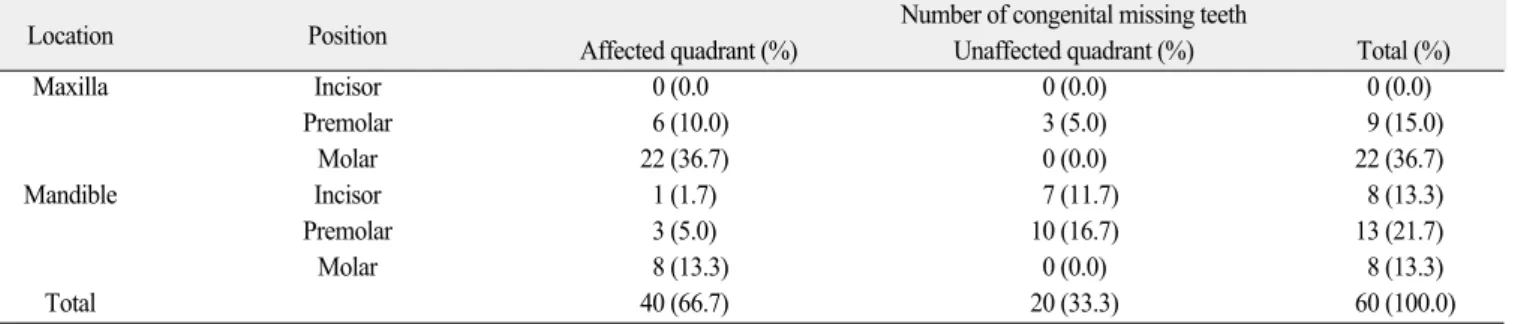
Based on this criteria, congenital absence of perma-nent teeth was seen in 23 subjects (57.5%). Among them, 9 were male (60% of all male subjects), while 14 were female (56.0% of all female subjects). The total number of congenital missing teeth was 60, including the congenital absence of the second permanent molar next to the DFM. Distribution of the missing teeth was as follows: 22 (36.7%) upper second molars, 13 (21.7%) lower premolars, 9 (15.0%) upper premolars, 8 (13.3%) lower second molars, and 8 (13.3%) lower incisors (Table 2).
Among the 60 congenitally missing teeth, 40 were lo-cated in the same quadrant as the DFM. The most fre-quently absent tooth was the second permanent molar (30 teeth, 50.0% of the total missing teeth) in the same quadrant of the DFM.
Regarding the number of congenitally missing teeth, 5 subjects (21.7%) showed only one missing tooth while 18 subjects (78.3%) showed 2 or more missing teeth.
There was a statistically significant correlation be-tween the prevalence of congenitally missing teeth and the DFM-affected quadrants (Fig 4). The quadrants in which DFM was noted showed a higher prevalence for congenitally missing teeth.
Ⅳ. Discussion
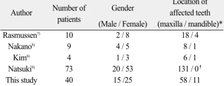
This study was conducted to investigate the clinical features of DFM on the basis of radiographic analysis of 40 children who showed no underlying general or local factors causing delayed tooth eruption. DFM occurred more frequently in the maxilla (32 subjects, 80%) com-pared to the mandible. This result is in accordance with
previous studies. For instance, Rasmussen7)
reported 22 cases of DFM in 10 subjects, with 18 of them in the
maxilla and 4 in the mandible. Nakano et al.8)reported
Table 2. Distribution of congenitally missing teeth in relation to DFM
Location Position Number of congenital missing teeth
Affected quadrant (%) Unaffected quadrant (%) Total (%)
Maxilla Incisor 0 (0.0 0 (0.0) 0 (0.0) Premolar 6 (10.0) 3 (5.0) 9 (15.0) Molar 22 (36.7) 0 (0.0) 22 (36.7) Mandible Incisor 1 (1.7) 7 (11.7) 8 (13.3) Premolar 3 (5.0) 10 (16.7) 13 (21.7) Molar 8 (13.3) 0 (0.0) 8 (13.3) Total 40 (66.7) 20 (33.3) 60 (100.0)
DFM = delayed eruption and development of the first permanent molar
Fig. 4. Correlation of congenitally missing teeth with DFM-affected
quadrants. The prevalence of congenital missing teeth was significantly higher in DFM-affected quadrants. (Chi-square test, * : p < 0.001) DFM = delayed eruption and development of the first permanent molar
9 cases, with 8 occurring in the maxilla and 1 in the
mandible. Sano et al.9)
reported 131 cases in the maxilla
of 73 subjects. In Korea, Kim et al.6)reported 7 cases in
4 subjects, with 6 found in the maxilla and only 1 in the mandible. The results from these studies support the conclusion that the incidence of DFM is higher in the maxilla compared to the mandible, and is more frequent-ly seen in female than in male patients (Table 3).
Regarding the laterality of DFM, the results of this study did not reveal a difference between the incidence
of unilateral and bilateral DFM6,9)
. This is in contrast to previous studies, in which it was reported that DFM of-ten occurs bilaterally. However, laterality of occurrence differed according to gender. In female patients, bilateral of DFM was higher than that of unilateral DFM. In con-trast, unilateral DFM was more frequent in male pa-tients.
There was significant correlation between the location and prevalence of the DFM in relation to congenitally missing teeth. According to a previous study in Korean children, the incidence of congenitally missing teeth was
reported to be 4.7 - 8.8%12-15)
. In the present study, con-genital missing teeth were observed in 62.5% of patients (25 out of 40 subjects) who had DFM, and 66.7% of the congenitally missing teeth (40 teeth) were found in the same quadrant as DFM. While the most commonly con-genitally missing teeth are the mandiblular lateral in-cisor (followed by maxilla and mandible second premo-lar), 50% (30 teeth) of the congenitally missing teeth in the present study involved the second permanent molar in the same quadrant as DFM. In addition, while the rate of absence of single tooth was 43.3 - 53.6% in Korean children12-14)
, the proportion of multiple teeth that were congenitally absent (78.3%) was higher than that of single teeth missing (21.7%) in children showing
DFM. Nakano et al.8) reported that 2 of 9 patients with
DFM had congenitally absence of the second premolar, as well as second and third permanent molars in the
af-fected quadrant. Kim et al.6)
also reported a case where bilateral DFM accompanied the bilateral congenital ab-sence of lateral incisors. These studies indicate that DFM may not be the result of a localized abnormality, but rather a developmental problem in the molar field where DFM is located.
In a previous study, DFM and the congenital absence of secondary molars were generally interpreted as con-genitally absence of the first permanent molar and early eruption of the mesially drifted second permanent molar
at the age of nine years old8). Nakano et al.8)interpreted
the results based on frequent association of decrease in the size of distolingual cusp, congenital absence of the ipsilateral third molar, and the close proximity of the time of first molar eruption to the normal eruption of the second permanent molar. The decrease in the size of dis-tolingual cusp is a characteristic phenomenon that is
ob-served frequently on the maxillary second molar16), the
incidence of which becomes higher in the absence of a
third molar17). However, there are limitations in this
con-jecture in light of previous studies regarding odontogenic development. First, the congenital absence of a tooth has a high correlation with the reduction in tooth size and number of cusps18-20). Loss of distolingual cusp
ob-served in DFM may be related to the reduction in tooth size associated with a congenitally missing tooth. Moreover, several studies have reported that congenital absence of a tooth is accompanied by generalized delay
in tooth development21,22). As congenitally missing teeth
are observed in cases of DFM, this phenomenon may oc-cur as a result of a generalized developmental delay or an anomaly in the affected molar field. Although calci-fication of the third molar varies with the ethnic or geo-graphic variables, the mean age of radiogeo-graphic appear-ance of third molar calcification has been reported as 9
years of age based in the Korean children23,24).
Radiographic examination may be inappropriate for the detection of the third molar because the age of our sub-jects ranged from 6 to 10 years. Further longitudinal study of our sample group is required to verify the asso-ciation between DFM and developmental anomaly of the third molar.
Congenitally missing teeth originate from local factors (such as infection25), trauma26), drug side effects27),
radia-tion therapy28)) and genetic factors29). Genetic mutations
Table 3. An overview of previous studies about DFM
Number of Gender Location of
Author
patients affected teeth
(Male / Female) (maxilla / mandible)*
Rasmussen7) 10 2 / 8 18 / 4
Nakano8) 9 4 / 5 8 / 1
Kim6) 4 1 / 3 6 / 1
Natsuki9) 73 20 / 53 131 / 0�
This study 40 15 /25 58 / 11
*(Number of affected teeth)
�This study only considered maxillary permanent first molar. DFM = delayed eruption and development of the first permanent molar
related to epithelial-mesenchymal interaction lead to
agenesis of teeth without a syndrome30)
. Cooperative ge-netic interaction models for odontogenesis suggest that interaction between mesenchymal cells from neural crest and oral epithelium activates the transcription of several
genes including homeobox31). Mutations of Msx1 and
Pax9, which are the causative genes for congenital ab-sence of teeth, are also involved in general developmen-tal defects of the teeth such as, eruption distur-bance29,30,32). In our data, all second molars adjacent to
DFM were either congenitally absent or showed delayed development. The prevalence of congenitally missing teeth in DFM-affected quadrants was significantly high. This indicates that DFM may be a result of defects in genetic or signal transduction processes involved in tooth development of the affected field, rather than single de-fects of the first molar. We suspect that genetic muta-tions affecting molar field development may be the basis of this developmental defect. Considered comprehensive-ly, genetic pathways can be used to explain the etiology and development of DFM.
Ⅴ. Conclusion
The study proved that DFM correlates with the con-genital absence of additional teeth, especially in DFM-affected quadrants, with every second permanent molar next to the DFM exhibiting delayed development or be-ing congenitally absent. In conclusion, a close follow-up examination should be performed to assess the temporal development of dentition in patients with DFM. However, a complete understating of the underlying de-velopmental mechanism responsible for development DFM requires further studies.
References
1. Grøn AM : Prediction of tooth emergence. J Dent
Res, 41:573-585, 1962.
2. Suri L, Gagari E, Vastardis H : Delayed tooth erup-tion: Pathogenesis, diagnosis, and treatment. A
lit-erature review. Am J Orthod Dentofacial Orthop,
126:432-445, 2004.
3. Kwon JH, Choi BJ, Lee JH, et al. : Eruption time and sequence of permanent teeth in students from
E-elementary school. J Korean Acad Pediatr Dent,
36:253-261, 2009.
4. Grover PS, Lorton L : The incidence of unerupted
permanent teeth and related clinical cases. Oral
Surg Oral Med Oral Pathol Oral Radiol, 59:420-425, 1985.
5. Lee JB, Jang CH, Kim CC, et al. : Eruption distur-bances of teeth in Korean children. J Korean Acad Pediatr Dent, 34:13-18, 2007.
6. Kim JM, WhangBo M, Kim JY, et al. : A clinical
review on the delayed eruption of 1st molars. J
Korean Acad Pediatr Dent, 21:555-560, 1994. 7. Rasmussen P : “9-year-molars”aberrantly
develop-ing and eruptdevelop-ing: report of cases. J Clin Pediatr Dent, 22:151-153, 1998.
8. Nakano K, Matsuoka T, Takahashi A, et al. :
Delayed development or congenital absence of a sin-gle first permanent molar in Japanese child patients. Int J Paediatr Dent, 9:271-276, 1999.
9. Sano N, Kameda T, Terashima Y, et al. : Formation and development of maxillary first molars with delayed eruption. Odontology, 1-9, 2014.
10. Nolla CM : The Development of the permanent teeth. J Dent Child, 27:254-266, 1960.
11. Shin M, Song J, Lee J, et al. : Evaluation of the
developmental age of permanent teeth by the Nolla method. J Korean Acad Pediatr Dent, 43:1, 2016. 12. Lee JM, Lee Sang Rae : A clinical and radiographic
study of congenitally missing teeth. Imaging Sci
Dent, 21:275, 1991.
13. Jeong HK, Yang YM, Kim JG, et al. : A clinical
study of congenital missing teeth. J Korean Acad Pediatr Dent, 36:245-252, 2009.
14. Jeon H, Yang Y, Baik B, Kim J : Prevalence and distribution of congenitally missing teeth in patients visiting the department of pediatric dentistry of
Chonbuk National University hospital. J Korean
Acad Pediatr Dent, 40:274-282, 2013.
15. Lee JH, Yang BH, Lee SM, et al. : A study on the prevalence of dental anomalies in Korean dental-patients. Korean J Orthod, 41:346-353, 2011. 16. Dahlberg AA : The dentition of the American Indian.
Papers on the physical anthropology of the American Indian. New York: Viking Fund, p138-176, 1951. 17. Keene HJ : The relationship between third molar
agenesis and the morphologic variability of the molar teeth. Angle Orthod, 35:289-298, 1965.
18. Brook AH : A unifying aetiological explanation for
anomalies of human tooth number and size. Arch
Oral Biol, 29:373-378, 1984.
non-syndromic hypodontia patients. The Angle Orthodontist, 83:16-21, 2013.
20. Ramazanzadeh BA, Ahrari F, Hajian S : Evaluation of tooth size in patients with congenitally-missing teeth. J Dent Res Dent Clin Dent Prospects, 7:36, 2013.
21. Uslenghi S, Liversidge H, Wong F : A radiographic study of tooth development in hypodontia. Arch Oral Biol, 51:129-133, 2006.
22. TunçE , Bayrak , Koyutürk AE : Dental
develop-ment in children with mild-to-moderate hypodontia. Am J Orthod Dentofacial Orthop, 139:334-338, 2011.
23. Cho SM, Hwang CJ : Skeletal maturation evalua-tion using mandibular third molar development in adolescents. Korean J Orthod, 39:120-129, 2009. 24. Kang KY, Yang KH, Choi NK, Kim SM : Skeletal
maturity and mandibular third molar development in Class III malocclusion. J Korean Acad Pediatr Dent, 35:235-242, 2008.
25. Gullikson J : Tooth morphology in rubella syndrome children. ASDC J Dent Child, 42:479-482, 1974. 26. Schalk-Van der Weide Y, Steen W, Bosman F :
Distribution of missing teeth and tooth morphology
in patients with oligodontia. ASDC J Dent Child,
59:133-140, 1991.
27. Axrup K, d’Avignon M, Hellgren K, et al. : Children
with thalidomide emrryopathy: Odontological obser-vations and aspects. Acta Odontol Scand, 24:3-21, 1966.
28. Maguire A, Craft AW, Evans RG, et al. : The long-term effects of treatment on the dental condition of
children surviving malignant disease. Cancer, 60:
2570-2575, 1987.
29. De Coster PJ, Marks LA, Martens LC, Huysseune A : Dental agenesis: genetic and clinical perspectives. J Oral Pathol Med, 38:1-17, 2009.
30. Ramos Boeira Junior B, Echeverrigaray S : Dentistry and molecular biology: a promising field for tooth agenesis management. Tohoku J Exp Med, 226:243-249, 2012.
31. Mitsiadis TA, Smith MM : How do genes make teeth to order through development? J Exp Zool B Mol Dev Evol, 306:177-182, 2006.
32. Pani SC : The genetic basis of tooth agenesis: basic
concepts and genes involved. J Indian Soc Pedod
주요어: 맹출 지연, 선천성 결손치, 제1대구치