Taenia solium metacestode
fasciclin-like proteins are reactive
with inactive neurocysticercosis
Joon Sup Yeom
Department of Medicine
Taenia solium metacestode
fasciclin-like proteins are reactive
with inactive neurocysticercosis
Directed by Professor June Myung Kim
Doctoral Dissertation
submitted to the Department of Medicine
the Graduate School of Yonsei University
in partial fulfillment of the requirements for the
degree of Doctor of Philosophy
Joon Sup Yeom
This certifies that the Doctoral
Dissertation of Joon Sup Yeom is
approved.
---
Thesis Supervisor : June Myung Kim
---
Thesis Committee Member#1 : Yoon Kong
---
Thesis Committee Member#2 : Tai Soon Yong
---
Thesis Committee Member#3 : Bong Ki Lee
---
Thesis Committee Member#4 : Won Taek Lee
The Graduate School
Yonsei University
ACKNOWLEDGEMENTS
In 2006, with Professor Kong, I had a chance to visit
Dibrugarh, India where many parasitic infections exist.
Everything was exciting and new to me and isolating cyst of
Taenia solium from infected pig was one of the unforgettable
experiences. This experience had leaded me to this work. A
lot of work was done by people around me and I want to give
my appreciation to all of them. My mentor, Professor June
Myung Kim, always taught me and supported me all the way
to where I am now. Most of this work was done under
Professor Kong’s advice and I would like express my special
gratitude to him. I also would like to thank Professor Sun
Hee Kim and Young-An Bae and all the other members of
the Molecular Parasitology Lab in Sungkyunkwan University
School of Medicine for their help. Without them, this work
couldn’t become possible. Professor Tai Soon Yong, Bong
Ki Lee and Won Taek Lee have served as committee
member and given me important advice to make this better
article.
Finally, I would like to express my gratitude to my wife Sa
Yun, my daughter Suji, Jiwon and my parents for their
encouragement, love and support.
<TABLE OF CONTENTS>
I. INTRODUCTION ... 3
II. MATERIALS AND METHODS ⋅⋅⋅⋅⋅⋅⋅⋅⋅⋅⋅⋅⋅⋅⋅⋅⋅⋅⋅⋅⋅⋅⋅⋅⋅⋅⋅⋅⋅⋅⋅⋅⋅⋅⋅⋅⋅⋅⋅⋅⋅⋅⋅⋅⋅⋅⋅⋅⋅⋅⋅⋅⋅ 6
1. Chemicals and reagents ... 6
2. Parasite ...6
3. Serum samples... 6
4. Partial purification of TsM protein molecules that react with sera
from inactive patients ... 7
5. Sodium dodecyl sulfate (SDS)-polyacrylamide gel electrophoresis
(PAGE) and immunoblotting ... 7
6. Two-dimensional electrophoresis (2-DE), image analysis and
MALDI-TOF MS ... 8
7. N-terminal amino acid sequencing ... 9
8. Cloning and sequence analysis ... 9
9. Expression and purification of the recombinant proteins ... 10
10. Immunohistochemistry ... 10
III. RESULTS... 12
1. Identification and partial purification of proteins that react with
sera from inactive NC patients ... 12
2. Identification of protein spots reactive with sera from inactive
NC patients by 2-dimensional electrophoresis/immunoblot ... 13
3. Cloning of the genes encoding for the TsM fasciclin-like protein
... 16
4. Expression of the recombinant TsMFc (rTsMFc) and antibody
reactivity of the rTsMFc ... 17
5. Immunohistochemical localization of the TsMFc ... 21
IV. DISCUSSION ... 23
VI. REFERENCES... 27
ABSTRACT (IN KOREAN)... 32
LIST OF FIGURES
Figure 1. Immunoblot analyses of sera from inactive
neurocysticercosis (NC) patients with crude CF (A) and
partially purified fraction (B) ...13
Figure 2. Identification of protein spots reactive with sera
from inactive NC patients by 2-dimensional electrophoresis
and immunoblot ...15
Figure 3. Deduced amino acid sequence of the fasciclin-like
protein of Taenia solium metacestode ...16
Figure 4. Physical map and expression strategy of T. solium
metacestode fasciclin...17
Figure 5. Amplification of fasciclin-like gene for cloning into
a expression plasmid, pET28a(+) ...17
Figure 6. Agarose gel analysis of colony PCR...18
Figure 7. Expression and purification of T. solium metacestode
recombinant fasciclin (rTsMFc) ...18
Figure 8. Expression and purification of truncated rTsMFc
...19
Figure 9. Assessment of diagnostic applicability of rTsMFc
...20
Figure 10. Generation of polyclonal antibody against rTsMFc
and localization of fasciclin-like protein in the TsM ...22
LIST OF TABLES
Table 1.
Identification of pots by peptide mass finger printing
...14
Table 2.
Result of N-terminal amino acid sequencing...15
Table 3. Reactivity of the rTsMFc with sera from various
parasitic disease and normal control ...21
1
<ABSTRACT>
Taenia solium metacestode fasciclin-like proteins are reactive with
inactive neurocysticercosis
Joon Sup Yeom
Department of Medicine
The Graduate School, Yonsei University
(Directed by Professor June Myung Kim)
Neurocysticercosis (NC), an infection to the central nervous system
with larval pork tapeworm Taenia solium metacestodes (TsM), invokes a
significant neurological disorder. It is a leading cause of adult onset
seizures in endemic countries. The disease has been classified in active
and inactive stages, according to the viability of the invasive worms.
Differential diagnosis of active NC from inactive cases is important for
the proper management of the patients. Active cases are treated with
anti-parasitic agents while chronic stage should be treated with
anti-convulsant and symptomatic treatment to prevent and reduce the
intractable epileptic seizures. Although several antigens have been used
for specific serologic assay, most of them failed to detect antibodies in
sera of inactive NC. In this study, 65 and 83 kDa proteins which reacted
with patient sera from inactive NC were purified by fast performance
liquid chromatography from the TsM cyst fluid. Proteomic analysis of
the 65 and 83 kDa proteins revealed that these two proteins shared a high
identity with mammalian fasciclin. A cDNA encoding this TsM
fasciclin-like protein (TsMFc) was isolated from a cDNA library. The
cloned cDNA contained open-reading frame of 766-amino acid
polypeptides. The recombinant protein was expressed using pET-28a
vector system and western blot analysis was performed. Eighty percent
(32/40) of sera from inactive stage neurocysticercosis cases showed
strong reactivity. It also showed some false-positive reactions against
sera
from
sparganosis
(30%)
and
paragonimiasis
(32.5%).
Immunohistochemical staining using polyclonal antibodies against
TsMFc showed that TsMFc was localized in scolex and bladder wall. In
addition, the secreted TsMFc were clustered around host immune cells in
the granuloma. TsMFc identified and characterized in this study showed
that it could give additional potential value for serologic differentiation
of inactive NC from active NC if it is used with other previously
developed TsM proteins.
---
Key words : Taenia solium, neurocysticercosis, diagnosis, serologic test
3
Taenia solium metacestode fasciclin-like proteins are reactive with
inactive neurocysticercosis
Joon Sup Yeom
Department of Medicine
The Graduate School, Yonsei University
(Directed by Professor June Myung Kim)
I. INTRODUCTIONThe life cycle of Taenia solium (pork tapeworm) involves two kinds of hosts. Humans (definitive host) harbor the adult tapeworm in their intestine. The worms produce numerous eggs, which pass out with gravid segments. When swine (intermediate host) ingest the eggs, they develop into T. solium metacestode (TsM). Humans, however, also serve as an intermediate host, and can be infected with T. solium eggs, which then develops into TsM. TsM preferentially infects subcutaneous tissues, but may invade the central nervous system and cause neurocysticercosis (NC)1. NC constitutes one of the common and major causes of global public health problems1. Clinical manifestation of the disease is often nonspecific; however, major symptoms included headache, seizure, and other focal neurologic deficits. Approximately 25–50 million people are infected worldwide2-6. In some countries of Asia, Latin America, and Africa where the disease is endemic, NC accounts for 10–12% of all hospital admissions to the neurological or psychiatric wards. Socioeconomic status and cultural background of the patients are important risk factors for the disease1,5. A significant proportion of adult-onset seizures are attributable to NC where the disease is prevalent. It is a leading cause of epileptic disorder of parasitic origin7-9. When NC is coupled with chronic intractable seizures, the burden of the disease is exacerbated substantially by the associated social stigma. Therefore, the early identification of affected patients may have a great impact not only on the treatment of infected individuals, but also on the reduction of disease burdens in certain communities where the disease is prevalent. A recent epidemiological study also demonstrated that NC is being increasingly
recognized especially in childhood, thereby significantly worsened its morbidity10. The disease is endemic in several regions of developing countries. However, NC has recently gained recognition in industrialized countries as an important communicable disease due to an immigration of many people from endemic areas11.
The current clinical diagnosis of NC depends largely on combined studies of serological tests and neuroimaging scans3,12. However, neuroimaging scans often fail to differentiate cerebral infections from other space-occupying lesions, due mainly to the ambiguity of the interpretation. In addition, neuroimaging findings of NC itself are highly protean, depending on the locations and status of the affected parasite. One of the characteristic signs of cysticercosis is subcutaneous nodule(s), which represent the presence of TsM in underlying tissues. These nodules are, however, nonspecific and could be easily caused by several other etiologies. Moreover, many NC cases may not be associated with subcutaneous lesions. In these cases, the etiological differentiation of the lesions is a prerequisite for the proper management of patients. A group of low-molecular weight proteins (LMWPs) of the TsM ranged between 7-50 kDa have previously been identified and/or purified, using several biochemical approaches13-15 , and have been extensively analyzed to establish the standard serodiagnosis of NC, through which several candidate molecules have been well characterized and, in part, their recombinant forms were shown to be useful for serodiagnosis12.
Previous studies determined that TsM cyst fluid (CF) harbored two major macromolecular proteins, of which sizes are 120-kDa and 150-kDa. The 120-kDa protein was found to be comprised of six subunits, ranging from 14-38 kDa in size, which originated from 14- and 18-kDa precursor molecules16. The 150-kDa protein was shown to be composed of three subunits of 7-, 10- and 15-kDa by SDS-PAGE analysis, but our recent experiment involving 2-DE demonstrated that the 150-kDa protein was composed of at least 16 subunits, which originated from two multi-gene families encoded by 7- and 10-kDa molecules17. These glycoproteins were shown to be highly effective for
5
detection of active stage NC, while these proteins did not show positive reactions, when the parasites undergo degeneration by calcification or during which the parasites are completely calcified16,17.
Many cases with early active NCC can be treated with chemotherapeutic agents. Specific treatment can shorten and diminish the symptoms caused by inflammation associated with active stage NCC, and reduces the risk of headache and late-recurred refractory seizure18-20. In contrast, chronic inactive stage or acephalic budding cysticercosis in the ventricles do not respond to anti-helmintics currently available and often require surgical or symptomatic treatment1,21. Therefore, identification and characterization of the protein molecule that can detect the specific antibodies circulating in inactive NC cases are highly required.
II. MATERIALS AND METHODS 1. Chemicals and reagents
Urea, CHAPS, DTT, IPG strips (Immobiline DryStrip, pH 3-10, 13 cm), IPG-buffer (pH 3-10) and ECL detection reagent were all purchased from Amersham Biosciences (Uppsala, Sweden). Acetone, acetonitrile, 2-propanol, TFA and CBB G-250 were obtained from Merck (Darmstadt, Germany). All other chemicals and reagents were purchased from Sigma (St. Louis, MO, USA), unless otherwise specified.
2. Parasite
Taenia solium metacestodes (TsM) were obtained from the muscles of naturally infected pigs in endemic area, Dibrugarh, Assam, India. After removing the surrounding host tissue and granuloma wall, intact worms were washed with physiological saline more than 10 times. CFs were collected by puncturing the intact individual cysts in the presence of protease inhibitor cocktail (1 tablet/10 ml CF, Complete, Roche, Germany), followed by centrifugation at 20,000 g for 1 hr. All the procedures were done at 4oC, and the samples were stored at -80oC or in liquid nitrogen until use.
3. Serum samples
A total of 40 serum samples from inactive NC patients were used for evaluation. Diagnosis of inactive NC was done on the basis of their clinical manifestations, neuroimaging findings, and concomitant positive antibody
reactions in serum/CSF by ELISA/immunoblot done with crude TsM CF
antigen7. Inactive cases were defined when multiple calcifications were recognized in their neuroimages. To observe cross-reactions, sera from patients infected with adult T. solium were included. The patients with adult worm infections were diagnosed on the basis of morphological characteristics of the scolices and the gravid proglottids discharged, and the presence of
specific bands by random amplified polymorphic DNA (RAPD) analysis13. In
7
echinococcosis (CE), paragonimiasis, clonorchiasis, and normal controls who denied any possible exposure to parasitic infections were tested. Sparganosis patients were diagnosed by surgery. Other patients were diagnosed by either ELISA together with typical radiological findings (AE, CE, and paragonimiasis) or stool examination (clonorchiasis).
4. Partial purification of TsM protein molecules that react with sera from inactive patients
In order to partially purify the proteins that react with sera from chronic inactive cases, crude CF proteins were fractionated using an AKTA fast-performance liquid chromatograpy (Amersham Biosciences, Piscataway,
NJ, USA) and a HiLoad column (16 × 60 ㎠) packed with Superdex 200 Prep
grade. A total of 3 ml (10 mg) of crude CF was eluted (1 ml/1 min) with sodium phosphate buffer (20 mM, pH 7.2) containing 150 mM NaCl. A total of 85 fractions which contained 1.5 ml allocations were collected, after which each fraction was analyzed via SDS-PAGE and immunoblotting as described in the following section.
5. Sodium dodecyl sulfate (SDS)-polyacrylamide gel electrophoresis (PAGE) and immunoblotting
SDS-PAGE was conducted with various gel concentrations under reducing conditions. The gels were stained with Coomassie blue G-250 (CBB) or silver nitrate, and were further processed by immunoblot analysis. For immunoblotting, the proteins resolved by SDS-PAGEs were blotted to nitrocellulose membranes. The membranes were blocked in PBS/T (0.05% Tween 20 in PBS) containing 3% skim milk for 1 hr at room temperature, and then incubated with 1:200 diluted pooled or individual patient sera for 4-5 hr at room temperature. The membranes were subsequently incubated overnight with 1:1,000 or 1:5,000 diluted peroxidase conjugated host-specific IgG. The immunoreactive proteins were visualized using either SuperSignal Chemiluminescence kit (Pierce, Rockford, IL, USA) or 4-chloro-1-naphthol
chromogen (Sigma, MO, USA).
6. Two-dimensional electrophoresis (2-DE), image analysis and MALDI-TOF MS
The partially purified proteins were precipitated with an equal volume of ice-chilled 20% (w/v) TCA and then washed twice in cold acetone prior to subject air-dry in a speedvac. The dried sample was mixed with lysis buffer consisting of 7 M urea, 2 M thiourea, 4% CHAPS, 1% DTT and 0.5% IPG buffer (pH 3-10). The sample (30 µg) was loaded onto an IPG strip (pH 3-10) with a cup-loading instrument on an IPGphor system (Amersham Biosciences) and focused via the application of 32 kVh. After equilibration, the IPG strip was processed by 15% SDS-PAGE (160 × 160 × 1 mm). The proteins, separated on 2-DE gels, were visualized with colloidal CBB G-250. The CBB-stained and/or immunoblotted spots were digitalized with a UMAX image scanner (Umax Technologies, Dallas, TX, USA). The obtained images were analyzed using Progenesis software (Nonlinear Dynamics, Newcastle, UK). The gel images were calibrated for Mr and pI using 2-D SDS-PAGE
standards (Bio-Rad, Hercules, CA, USA). The CBB stained spots of interest were excised from the 2-DE gels and the proteins were subjected to in-gel trypsin digestion. The tryptic peptide mixtures were analyzed with a Voyager-DE STR MALDI-TOF (PerSeptive Biosystems, Framingham, USA) and Ultraflex MALDI-TOF/TOF MS systems (Bruker Daltonics GmbH, Bremen, Germany). For the MALDI-TOF MS analysis, I applied the solution-phase nitrocellulose method to the target samples. Mass spectra were obtained in the reflectron/delayed extraction mode, with a 20 kV accelerating voltage and 120 ns delay time. PMFs acquired by the summation of 150 laser shots were then subjected to 2-point internal calibration with tryptic autodigestion peaks (m/z 842.5099 and 2211.1046), using the Data Explorer software (PerSeptive Biosystems). Monoisotopic peptide masses were selected in a range between 600-3,500 Da and the proteins were identified by PMF, using the Matrix Science Mascot (http://www.matrixscience.com) and
9
National Center for Biotechnology Information protein sequences database (http://www.ncbi.nlm.nih.gov). Mass tolerance was ±30 ppm and one missed cleavage site was allowed. Both the carbamidomethylation of cysteine and methionine oxidation were considered in this search.
7. N-terminal amino acid sequencing
The protein spots separated by 2-DE gel were then transferred onto polyvinylidene difluoride (PVDF) membranes (Millipore, Billerica, MA, USA) and stained with CBB. The spots were excised and used for protein sequencing on an ABI model 477A protein sequencer and an ABI model 120A PTH analyzer (Perkin Elmer, Foster City, CA, USA) at the Korea Basic Science Institute (Daejeon, Korea).
8. Cloning and sequence analysis
On the basis of MALDI-TOF analysis, TsM fasciclin-like protein was found to reactive with sera from chronic patients. In order to clone this protein, two primer sets were designed to match the 5′ regions. The sense primers, that
contained a BamH I site were 5′-CCGGGATCCTTTGAACAGAATCCCAG
-3′ and 5′-CGGGATCCGATCGTCCAGAATGCGC-3′; the antisense primer
with the Not I sites were 5′-GCGGCCGCACAAATCTTTCAGCG-3′ and
5′-GCGGCCGCACTAGTGTTCGTGACCC-3′. These primers were
combined with a T7 promoter primer to amplify each target gene from the TsM cDNA library, which had been constructed as previously described7, by polymerase chain reaction (PCR). Amplification reactions were performed with a thermal cycling profile of 94oC for 4 min, 35 cycles at 94oC for 50 sec, 58oC for 50 sec, and 72oC for 1 min, followed by a final extension at 72oC for 10 min. The amplified PCR products were separated on a 1% agarose gel and visualized by ethidium bromide staining. The amplicons were gel purified using a QIAquick Gel Extraction Kit (Qiagen), and then ligated into pET-28a vector (Promega, Madison, WI, USA). The plasmid DNAs were transformed into competent Escherichia coli DH5α cell (Takara, Shiga, Japan). The
nucleotide sequences of the cloned genes were automatically determined from both strands by using the BigDye Terminator cycle Sequencing Ready Reaction kit (Perkin-Elmer Corporation, Foster City, CA, USA), and an ABI PRISM 377 DNA sequencer (Applied Biosystems, Foster City, CA, USA). The obtained nucleotide sequences were deduced into amino acid sequences and used in sequence alignments. The homology patterns were determined by non-redundant database of GenBank using BLAST algorithms. A search for recognition site for cleavable N-terminal signal peptidase was conducted using SignalP program (http://www.cbs.dtu.dk/service/SignalP).
9. Expression and purification of the recombinant proteins
The putative mature domain of each gene was PCR amplified from the corresponding T vector. The specific primer pairs contained restriction sites for BamH I and Not I at the 5′- and 3′-end, respectively. The PCR conditions were identical as described above. The purified PCR products were digested with corresponding restriction enzymes, and ligated into the pET-28a(+) vector with the clone. The orientation of the inserted DNA was confirmed by DNA sequencing. These plasmids were transformed into competent E. coli BL21 cells. Expression of these recombinant proteins was induced by addition of 0.1 mM IPTG. The expressed recombinant proteins were purified by
Glutathione Sepharose 4B or nickel-nitrilotriacetic acid affinity
chromatography.
10. Immunohistochemistry
Fresh TsM was fixed overnight in PBS containing 4% paraformaldehyde at 4 oC. The worms were dehydrated with a graded ethanol series, embedded in paraffin blocks and stored in desiccators until use. Sections (4㎛-thickness) were mounted on slide glasses, deparaffinized, rehydrated and rinsed with PBS. The sections were treated for 5 min with 3% hydrogen peroxide, after which they were blocked for 1h with 1% BSA. The sections were incubated overnight with anti-mouse TsM-fasciclin 1 antibody diluted to 1:200 in PBS
11
supplemented with 1% BSA at 4oC. The reactions were visualized with a streptavidin-biotin system (DAKO LSAB+, DAKO Cytomation, Glostrup, Denmark) with a 3-amino-9-ethylcarbazole (AEC) substrate. Normal mouse/rabbit serum diluted to the same ratio was employed as a control. The slides were covered and observed under light microscope.
III. RESULTS
1. Identification and partial purification of proteins that react with sera from inactive NC patients
I first examined immunoreactivity of the TsM CF in detecting specific antibodies circulated in the patients’ sera with inactive NC by immunoblot analyses. Figure 1 showed an immunoblot outcome of the TsM CF against individual sera from patients with chronic inactive NC cases, in which sera from inactive NC patients reacted positively against the crude CF, especially against high molecular weight proteins. Moreover, the patients' sera showed strong and constant antibody reactions against 65 and 83 kDa bands. For further purification, the crude TsM CF was fractionated by molecular sieve fast-performance liquid chromatography and 85 fractions were obtained. The elution profile displayed 4 main peaks, which were designated groups I to IV, between fractions 25 and 61. The two highest peaks, groups III and IV, were located between fractions 40 and 42 and fractions 48 and 50, respectively (data not shown).
Comparison of the elution profiles with the molecular weights of the standard proteins indicated that groups III and IV eluted proteins corresponding to sizes of 150 kDa and 120 kDa, respectively. Characteristics of 150 kDa and 120 kDa proteins were previously described12,16. However, 83 kDa and 65 kDa proteins were eluted in between group III and IV. We further evaluated whether these proteins invoked strong antibody reactions against sera from inactive NC patients. The partially purified CF proteins were separated by 10% SDS-PAGE in reducing conditions, after which processed by immunoblot probed with individual serum samples from inactive NC patients. Indeed, the patients' sera showed strong antibody reactions against 65 and 83 kDa bands (Fig. 1B).
13
Fig. 1. Immunoblot analyses of sera from inactive neurocysticercosis patients with crude CF (A) and partially purified fraction (B). The crude CF and partially purified CF proteins using gel filtration chromatography were separated by 15% SDS-PAGE in reducing conditions, after which further processed by immunoblot probed with individual serum samples from chronic NC patients. The patients' sera showed strong antibody reactions against 65 and 83 kDa bands.
2. Identification of protein spots reactive with sera from inactive NC patients by 2-dimensional electrophoresis/immunoblot
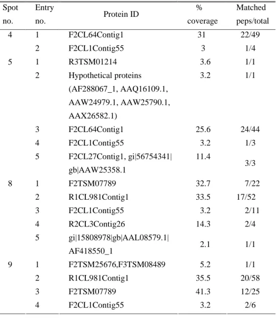
In order to further characterize the structural components of the TsM 83 and 65 kDa protein, I performed 2-DE, MALDI-TOF MS and N-terminal sequencing. I separated the proteins on IPG strips with pH values of 4-7, followed by 10% SDS-PAGE (Fig. 2A). These proteins were found to be identical proteins seen in 1-dimensional SDS-PAGE, comparing their immunoreactivity (Fig. 2B) A total of 12 protein spots were excised from the 2-D gel and analyzed by MALDI-TOF MS (Lower small panel of Fig. 2B). The peptides generated by the trypsin-digestion of the spots were separated by liquid chromatography and analyzed via tandem mass spectrophotometry. Results of peptide mass finger printing revealed that most of proteins in spot 4
and 5 were identified as fasciclin-like protein (Table 1).
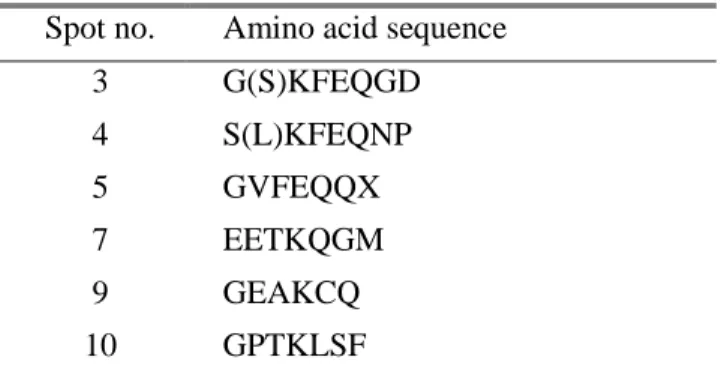
I further analyzed the protein spots by internal N-terminal amino acid sequencing. As shown in Table 2, Spot 4, 5, 8 and 9 showed highest sequence coverage with TsM cDNA library. The N-terminal amino acid sequences of the spots 3 and 4 were determined to be KFEQNP, which was consistent with that of fasciclin.
Table 1. Identification of protein spots by peptide mass finger printing Spot no. Entry no. Protein ID % coverage Matched peps/total 1 F2CL64Contig1 31 22/49 4 2 F2CL1Contig55 3 1/4 1 R3TSM01214 3.6 1/1 2 Hypothetical proteins (AF288067_1, AAQ16109.1, AAW24979.1, AAW25790.1, AAX26582.1) 3.2 1/1 3 F2CL64Contig1 25.6 24/44 4 F2CL1Contig55 3.2 1/3 5 5 F2CL27Contig1, gi|56754341| gb|AAW25358.1 11.4 3/3 1 F2TSM07789 32.7 7/22 2 R1CL981Contig1 33.5 17/52 3 F2CL1Contig55 3.2 2/11 4 R2CL3Contig26 14.3 2/4 8 5 gi|15808978|gb|AAL08579.1| AF418550_1 2.1 1/1 1 F2TSM25676,F3TSM08489 5.2 1/1 2 R1CL981Contig1 35.5 20/58 3 F2TSM07789 41.3 12/25 9 4 F2CL1Contig55 3.2 2/6
15
A B
Fig. 2. Identification of protein spots reactive with sera from inactive NC patients by 2-dimensional electrophoresis and immunoblot. The partially purified proteins (30 µg) were isoelectrically focused using IPGphore (pH 4-7) followed by 10% SDS-PAGE (A). The proteins were transblotted to PVDF membrane and probed with pooled serum of 10 inactive NC patients. The patients' sera showed strong antibody reactions against 65 and 83 kDa bands. The protein spots (numbered from 1-12 as shown in inset) were excised and proceeded for MS-target preparation.
Table 2. Result of N-terminal amino acid sequencing
Spot no. Amino acid sequence
3 G(S)KFEQGD 4 S(L)KFEQNP 5 GVFEQQX 7 EETKQGM 9 GEAKCQ 10 GPTKLSF
3. Cloning of the genes encoding for the TsM fasciclin-like protein
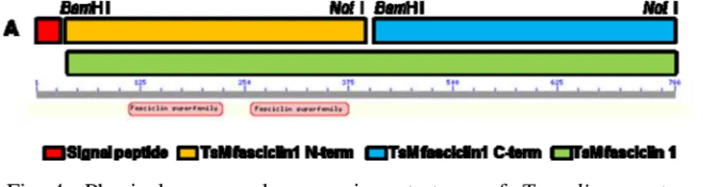
Based on both the N-terminal amino acid sequences of the 83 kDa proteins I designed a set of primers to clone the genes encoding the 83 kDa protein. The PCR amplification of the TsM cDNA library using these primers allowed us to isolate the cDNA sequences encoding the mature portion of the 83 kDa TsM fasciclin-like proteins. Figure 3 demonstrated a deduced amino acid sequence of the cloned gene. The full-length open reading frame was composed of 766 amino acid residues. Multiple sequence alignment revealed that this protein shared a high sequence homology (63-82%) with fasciclin from other organisms. I designated this protein as Taenia solium metacestode fasciclin-like protein (TsMFc). Brief physical map and expression strategy are show in figure 4.
Fig. 3. Deduced amino acid sequence of the fasciclin-like protein of Taenia
solium metacestode. The full-length open reading frame was composed of 766
17
Fig 4. Physical map and expression strategy of T. solium metacestode fasciclin.
4. Expression of the recombinant TsMFc (rTsMFc) and antibody reactivity of the rTsMFc
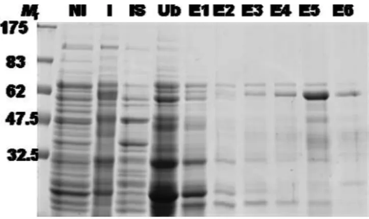
In order to produce a recombinant TsMFc (rTsMFc), I introduced the expression plasmid into pET-28a expression vector (Fig. 5), after which the recombinant plasmids were transformed into competent E. coli and the positive clones were selected by colony PCR (fig. 6). The cells were grown at 37oC, and induced with 0.1 mM β-isopropyl thiogalactoside (IPTG). Considerable amount of the recombinant proteins were expressed as a soluble form as shown in figure 7. I purified these recombinant proteins by using nickel-nitrilotriacetic acid affinity chromatography. Figure 8 showed an expression, purification and immunoblot analysis of the truncated rTsMFc protein.
Fig 5. Amplification of fasciclin-like gene for cloning into a expression plasmid, pET28a(+). The amplicons were digested with the corresponding enzymes and resolved in a 1% agarose gel.
Fig. 6. Agarose gel analysis of colony PCR. (A) Identification of N-terminal region of TsMFc1 in pET 28a (+) plamid. (B) A gene encoded C-terminal domain of TsMFc1 in pET 28a(+) plamid. (C) A gene putatively coding for full-length TsMFac1 is identified by colony PCR. The DNAs were analyzed on 1% agarose gel
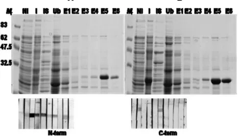
Fig. 7. Expression and purification of T. solium metacestode recombinant fasciclin (rTsMFc). The expression plasmid was introduced into pET-28a,
grown at 37oC, and then the recombinant protein was induced with 0.1 mM
β-IPTG. The recombinant proteins were purified by nickel-nitrilotriacetic acid affinity chromatography, after which analyzed by 10% SDS-PAGE in reducing conditions. Mr; molecular marker NI:not induced I; induced IS:insoluble, Ub; unbind, E; elution
19
Fig. 8. Expression and purification of truncated rTsMFc. (A) N-terminal domain of TsMFc was introduced into pET-28a, grown at 37oC, after which
the recombinant proteins were induced with 0.1 mM β-IPTG. The
recombinant proteins were purified by nickel-nitrilotriacetic acid affinity chromatography and analyzed by 10% SDS-PAGE. Lower panel shows an immunoblot analysis with sera from inactive NC cases. (B) A plasmid harboring a C-terminal domain of TsMFc was introduced into pET-28a, and the protein was expressed and analyzed as in A. Lower panel shows an immunoblot analysis with sera from inactive NC cases. Mr; molecular marker NI:not induced I; induced IS:insoluble, Ub; unbind, E; elution
Immunoblot analysis of N-terminal domain for the rTsMFc with sera from inactive NC cases showed strong reactivity with the patients’ sera. In contrast, the C-terminal domain of rTsMFc was not reactive with sera from inactive NC cases. This result indicated that epitope specificity of the rTsMFc was present in the N-terminal domain. Figure 9 assessed a diagnostic applicability of rTsMFc employing various helminthic infection sera. When the blots containing rTsMFc were incubated with individual serum samples from
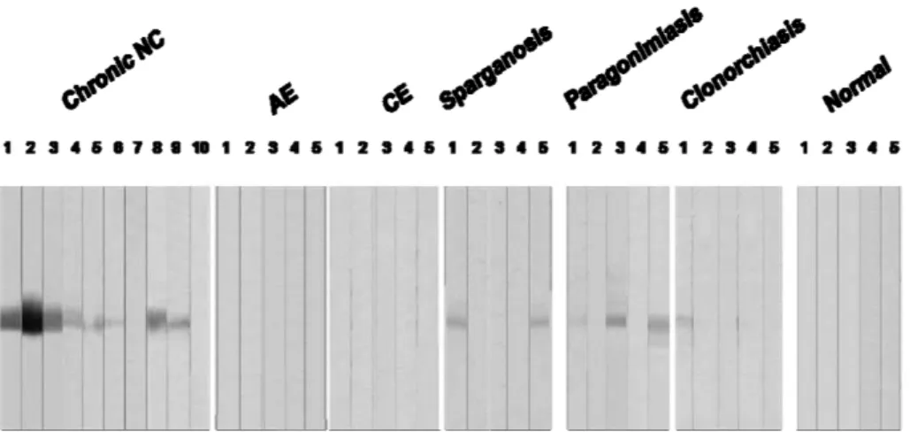
inactive NC cases together with sera from other helminthic infection and those from normal controls, approximately 80% (32/40 cases examined) of sera from inactive NC showed positive reactions (table 3). rTsMFc did not cross react with those from alveolar echinococcosis nor cystic echinococcosis, which usually show cross reaction with cysticercosis sera. However, granuloma-forming tissue invasive parasitic infections exhibited some degree of positive reactions (table 3).
Fig. 9. Assessment of diagnostic applicability of rTsMFc. The rTsMFc separated by 10% SDS-PAGE in reducing condition was transferblotted onto PVDF membrane. The membrane was cut into strips and incubated with individual serum samples (1:200 dilutions) overnight. The strips were
subsequently incubated with 1:1,000 diluted horseradish
peroxidase-conjugated anti-human IgG for 4 hr. The blots were developed with 4-chloro-1-naphthol chromogen. 1-10 indicated individual serum samples. NC, neurocysticercosis; AE, alveolar echinococcosis; CE, cystic echinococcosis.
21
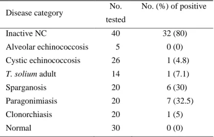
Table 3. Reactivity of the rTsMFc with sera from various parasitic infections and normal control
Disease category No.
tested No. (%) of positive Inactive NC 40 32 (80) Alveolar echinococcosis 5 0 (0) Cystic echinococcosis 26 1 (4.8) T. solium adult 14 1 (7.1) Sparganosis 20 6 (30) Paragonimiasis 20 7 (32.5) Clonorchiasis 20 1 (5) Normal 30 0 (0)
5. Immunohistochemical localization of the TsMFc
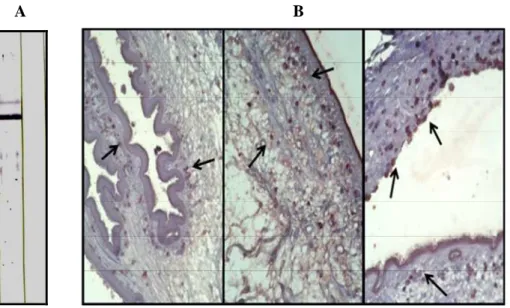
In order to observe the specific locality of the TsMFc in infected tissue and parasite tissue, we generated specific antibodies by immunizing BALB/c mice with rTsMFc. Figure 10A showed an immunoblot analysis of the generated antibodies. When the partially purified CF proteins were separated by 10% SDS-PAGE and further processed by immunoblot probed with the anti-sera, specific antibody reaction against 83 kDa TsMFc was clearly observed. I subsequently performed immunohistochemical staining using the whole worm sections of TsM with surrounding granuloma wall. As shown in figure 10B, the proteins were localized in scolex and bladder walls as enclosed in vesicle-like organelles, while the proteins secreted from the parenchyma are clustered around host immune cells in the granuloma wall.
A B
Fig. 10. Generation of polyclonal antibody against rTsMFc and localization of fasciclin-like protein in the TsM. (A) The partially purified CF proteins using gel filtration chromatography were separated by 10% SDS-PAGE in reducing conditions, after which further processed by immunoblot probed with 1:1,000 diluted anti-mouse antiserum against rTsMFc. The blot was developed with 4-choloro-1-naththol chromogen. (B) Immunohistochemical staining of TsMFc. The whole worm sections of TsM with surrounding granuloma wall were reacted with the polyclonal antibody and positive signals were visualized streptavidin-biotin system with a 3- amino-9-ethylcarbazole substrate. The proteins are localized in scolex and bladder walls as enclosed in vesicle-like organelles (left and middle panels, X200), while the proteins secreted from the parenchyma are clustered around host immune cells in the granuloma wall (right panel, X200).
23 IV. DISCUSSION
NC has been classified in active and inactive stages, according to the associated immune response and the radiologic findings of the central nervous system14. In active forms, degenerating parasites induce inflammatory response in the involved tissue but in inactive forms, parasites are calcified and immune response is minimal23-25. Due to different immunologic reaction occurring during these stages, most of the currently used serologic test can detect only active stage of NC patients and development of serologic test that can detect inactive stage has been a challenge. Distinction between active and inactive NC is important for the treatment of the patients1. Active cases are treated with anti-parasitic agents while chronic inactive stage should be treated with anti-convulsant and symptomatic treatment to prevent and reduce the intractable seizures. But access to neuroimaging is very limited due its cost and time in many endemic countries, making diagnosis of NC based on serological test26. So, identification of protein molecule that can detect the specific antibodies circulating in inactive NC cases is highly required.
Several TsM proteins have been used for the diagnosis of human
NC12,13,15,27-31. However, most of the results showed that proteins found in TsM
were reactive in active stage sera while not with inactive stage. And molecular masses of these proteins were in rather lower range. Recently, several studies trying to differentiate active and inactive stages had been published. Ferrer et al. used Taenia saginata oncosphere adhesion protein expressed in baculovirus and evaluated its diagnostic applicability24. Sensitivity of this protein was against inactive NC sera was 64.5% which is a much better result than previously reported TsM proteins. Using excreted/secreted antigens of TsM for ELISA tests, Molinari et al. showed that the CSF samples of active NC cases had significantly higher positive indexes than inactive cases30. If the viable TsM secrete proteins into its surrounding tissue and microenvironments, specific antibodies against these proteins might be produced in the host. However, when parasites undergo degeneration, they may not secrete these
proteins and the host response to these proteins may not be occurred, which underlies the implication that this might be the reason for excreted/secreted antigens of TsM showing reactivity only with the sera from active stage NC. However, many previous studies have shown that when crude extract of TsM were used, most sera from chronic inactive NC cases showed marginal levels of specific antibody titers, which suggested that this discrepancy might be attributed to the prolonged release of somatic antigens in the process of parasite destruction34. Barcelos et al. used IgG antibodies of crude TsM extracts to differentiate between active and inactive NC sera and cerebrospinal fluid35. They demonstrated that patient sera from active and inactive sera showed some reactivity to 80, 86, 95 kDa bands but they also reacted with control sera. TsMFc purified in this study is a secretory protein contained in cystic fluid and shows strong reaction against inactive NC sera. Molecular masses of these proteins were in similar range with the TsMFc, but TsMFc did not reacted against normal controls sera. TsMFc also has some limitation as to be developed as a diagnostic marker. Although it showed 80% positive reaction against inactive sera, it also showed strong reaction against active NC sera (data not shown). Therefore, using TsMFc alone as a target protein cannot differentiate between active and inactive cases. But when TsMFc is used in combination with the low molecular proteins that react only with active sera as a serologic test, it can contribute in differentiating active and inactive NC cases only by serologic test. Recently, new approach such as DNA-based diagnostic test using patients CSF is under investigation to overcome the problem of low sensitivity of conventional serologic assays in inactive or solitary and some of the results are reported.Almeida et al. reported that PCR amplification of the parasite DNA in the CSF showed identification of 29 case out of 30 cases examined (96.7%)38. Harrington et al reported using global DNA screening platform dot diagnosis of NC patients with solitary lesion in brain biopsy tissue39. Although these new approach using PCR technique may
25
have better sensitivity and specificity than conventional serologic assay, but they are not applicable in resource limited settings and need more invasive procedure to obtain specimen. In this context, TsMFc has many advantages if it can be used as a diagnostic marker for NC cases together with low molecular weight proteins recently characterized40.
False-positive reaction had been a problem for TsM proteins especially when crude extract of TsM were used18,36,37. It occurs frequently with other helminthic infections especially in those involved with metacestodes of the
Echinococcus species. But western blot analysis of the TsMFc showed some
cross reaction mostly with granuloma-forming tissue invasive parasitic infections such as paragonimiasis and sparganosis. Although this finding is different from other low molecular weight TsM proteins, cross reaction between sera of sparganosis, paragonimiasis and NC has been reported41,42. It was presumed to be a result of the common antigenic components shared by larval cestodes. Problems related with cross reactions should be resolved prior to application of the TsMFc as a serologic antigen.
Fasciclin-like protein has never been reported in Taenia solium. Fasciclin is reported as ortholog of vertebrate neural cell adhesion molecule in
Drosophilia. It belongs to immunoglobulin superfamily and is expressed as
several protein isoforms. Although function of TsMFc is not investigated in this work, I think it might have some immunologic functions in the pathogenesis of NC. Immunohistochemical localization might support this idea since the TsMFc secreted from the TsM parenchyma were clustered around host immune cells in the granuloma wall. Further investigations concerning these issues will properly clarify its functional role.
V. CONCLUSION
In conclusion, a protein molecule from the TsM CF that showed strong antibody responses against sera of inactive NC cases was indentified. Molecular size of the protein was 83 kDa. Proteomic analysis and N-terminal sequencing revealed that it has a high sequence homology with protein called fasciclin. The TsMFc composed of 766-amino acid residues. Immunoblot analysis of the TsMFc showed that it could give additional potential value for serologic differentiation of inactive NC from active NC if it is used with other previously developed TsM proteins.
27 VI. REFERENCES
1. White Jr AC. Neurocysticercosis: updates on epidemiology, pathogenesis, diagnosis, and management. Ann Rev Med 2000; 51:187-206.
2. Bern C, Garcia HH, Evans C, Gonzalez AE, Verastegui M, Tsang VC et al. Magnitude of the disease burden from neurocysticercosis in a developing country. Clin Infect Dis 1999; 29:1203-09.
3. Del Brutto OH, Sotelo J, Roman GC eds. Neurocysticercosis: a clinical handbook. Lisse: Swets & Zeitlinger Publishers, 1998.
4. Gemmell MA, Johnston PD. Efficacy of praziquantel against ovine cysticercosis caused by Taenia hydatigena. Res Vet Sci 1983; 34:199-204.
5. Tsang VCW, Wilson M. Taenia solium cysticercosis: An
under-recognized but serious public health problem. Parasitol Today 1995; 11:124-6.
6. White AC Jr., Robinson P, Kuhn R. Taenia solium cysticercosis: host-parasite interactions and immune response. Chem Immunol 1997; 66:209-30.
7. Carpio A. Neurocysticercosis: an update. Lancet Infect Dis 2002; 2:751-62.
8. Phiri IK, Ngowi H, Afonso S, Matenga E, Boa M, Mukaratiwa S, Githigia S, Saimo M, Sikasunge C, Maingi N, Lubega GW, Kassuku A, Michael L, Siziya S, Krecek RC, Noormahomed E, Vilhena M, Dorny P, Willingham Al 3rd. The emergence of Taenia solium cysticercosis in eastern and southern Africa as a serious agricultural problem and public health risk. Acta Trop 2003; 87:13-23.
9. Garcia HH, Pretell EJ, Gilman RH, Martinez SM, Moulton LH, Del Brutto OH et al. A trial of antiparasitic treatment to reduce the rate of seizures due to cerebral cysticercosis. N Eng J Med 2004; 350:249-58. 10. Mandal J, Singhi PD, Khandelwal N, Malla N. Evaluation of ELISA and
dot blots for the serodiagnosis of neurocysticercosis, in children found to have single or multiple enhancing lesions in computerized tomographic scans of the brain. Ann Trop Med Parasitol 2006; 100:39-48.
11. Roman G, Sotelo J, Del Brutto OH, Flisser A, Dumas M, Wadia N et al. A proposal to declare neurocysticercosis an international reportable disease. Bull World Health Organ 2000; 78:399-406.
12. Chung JY, Bahk YY, Huh S, Kang SY, Kong Y, Cho SY. A recombinant 10-kDa protein of Taenia solium metacestodes specific to active neurocysticercosis. J Infect Dis 1999; 180:1307-15.
13. Yang HJ, Chung JY, Yun D, Kong Y, Ito A, Ma L et al. Immunoblot analysis of a 10 kDa antigen in cyst fluid of Taenia solium metacestodes. Parasite Immunol 1998; 20:483-8.
14. Restrepo BI, Obregon-Henao A, Mesa M, Gil DL, Ortiz BL, Mejia JS et al. Characterisation of the carbohydrate components of Taenia solium metacestode glycoprotein antigens. Int J Parasitol 2000; 30:689-96. 15. Haslam SM, Restrepo BI, Obregon-Henao A, Teale JM, Morris HR, Dell
A. Structural characterization of the N-linked glycans from Taenia solium metacestodes. Mol Biochem Parasitol 2003;126:103-7.
16. Lee EG, Bae YA, Jeong YT, Chung JY, Je EY, Kim SH et al. Proteomic analysis of a 120 kDa protein complex in cyst fluid of Taenia solium metacestode and preliminary evaluation of its value for the serodiagnosis of neurocysticercosis. Parasitology 2005; 131:867-79.
17. Lee EG, Kim SH, Bae YA, Chung JY, Suh M, Na BK et al. A hydrophobic ligand-binding protein of the Taenia solium metacestode mediates uptake of the host lipid: implication for the maintenance of parasitic cellular homeostasis. Proteomics 2007; 7:4016-30.
18. Sotelo J, del Brutto OH, Penagos P, Escobedo F, Torres B, Rodriguez-Carbajal J et al. Comparison of therapeutic regimen of anticysticercal drugs for parenchymal brain cysticercosis. J Neurol 1990; 237:69-72.
19. Vazquez V, Sotelo J. The course of seizures after treatment for cerebral cysticercosis. N Engl J Med 1992; 327:696-701.
20. Baranwal AK, Singhi PD, Khandelwal N, Singhi SC. Albendazole therapy in children with focal seizures and single small enhancing computerized
29
tomographic lesions: a randomized, placebo-controlled, double blind trial. Pediatr Infect Dis J 1998; 17:696-700.
21. Carpio A, Escobar A, Hauser W. Cysticercosis and epilepsy: a critical review. Epilepsia 1998; 39:1025-40.
22. Eom KS, Jeon HK, Kong Y, Hwang UW, Yang I, Li X, et al. Identification of Taenia asiatica in China: molecular, morphological, and epidemiological analysis of a Luzhai isolate. J Parasitol 2002; 88:758-64. 23. Sotelo J, Guerrero V, Rubio F. Neurocysticercosis: a new classification based on active and inactive forms. A study of 753 cases. Arch Intern Med 1985: 145;442-5.
24. Salgado P, Rojas R, Sotelo J. Cysticercosis. Clinical classification based on imaging studies. Arch Intern Med 1997: 157;1991-7.
25. Castilho M. Imaging of neurocysticercosis. Seminars Roentgenology 2004: 39;205-13.
26. Flisser A, Sarti E, Lightowlers M, Schantz P. Neurocysticercosis: regional status, epidemiology, impact and control measures in the Americas. Acta Trop 2003; 87:43-51.
27. Ko RC, Ng TF. Specificity of isoelectric focusing-purified antigens in the dianosis of human cysticercosis. Parasitol Res 1988: 84;565-69.
28. Tsang VC, Brand JA, Boyer AE. An enzyme-linked
immunoelectrotransfer blot assay andglycoprotein antigens for diagnosing human cysticercosis (Taenia solium). J Infect Dis 1989: 159;50-9.
29. Sako Y, Nakao M, Ikejima T, Piao XZ, Nakaya K, Ito A. Molecular
characterization and diagnostic value of Taenia solium
low-molecular-weight antigen genes. J Clin Microbiol 2000: 38;4439-44. 30. Hancock K, Khan A, Williams FB, Yushak ML, Pattabhi S, Noh J et al.
Characterization of the 8-kilodalton antigens of Taenia solium metacestodes and evaluation of their use in an enzyme-linked immunosorbent assay for serodiagnosis. J Clin Microbiol 2003: 41;2577-86.
neurocysticercosis using synthetic 8-kD proteins: Comparison of assay formats. Am J Trop Med Hyg 2005: 73;771-6.
32. Hancock K, Pattabhi S, Whitfield FW, Yushak ML, Lane WS, Garcia HH et al. Characterization and cloning of T24, a Taenia solium antigen diagnostic for cysticercosis. Mol Biochem Parasitol 2006: 147;109-17. 33. Ferrer E, Gonzalez LM, Martinez-Escribano JA, Gonzalez-Barderas ME,
Cortez MM, Davila I et al. Evaluation of recombinant HP6-Tsag, an 18 kDa Taenia saginata oncoshperal adhesion protein for the diagnosis of cysticercosis. Parasitol Res 2007: 101;517-25.
34. Molinari JL, Garcia-Mendoza E, de la garza Y, Ramirez JA, Sotelo J, Tato P. Discrimination between active and inactive neurocysticercosis by metacestode excretory/secretory antigens of Taenia solium in an enzyme-linked immunosorbent assay. Am J Trop Med Hyg 2002; 66:777-81.
35. Barcelos ISC, Moura LP, Costa VP, Ferreira MS, Costa-Cruz JM. Taenia
solium metacestode immunodominant peptides recognized by IgG
antibodies in cerebrospinal fluid and serum paired samples from patients with active and inactive neurocysticercosis. Mem Inst Oswaldo Cruz 2007: 102;713-17.
36. Gekeler F, Eichenlaub S, Mendoza EG, Sotelo J, Hoelscher M, Loscher T. Sensitivity and specificity of ELISA ans immunoblot for diagnosing neurocysticercosis. Eur J Clin Microbiol Infect Dis 2002: 21;227-9. 37. Gottenstein B, Tsang VC, Sanchez PM. Demonstration of specific and
cross-reactive components of Taenia solium metacestode antigens. Am J Trop Med Hyg 1986: 35;308-13.
38. Almedia CR, Ojopi EP, Nunes CM, Machado LR, Takayanagui OS, Livramento JA et al. Taenia solium DNA is present in the cerebrospinal fluid of neurocysticercosis patients and can be used for diagnosis. Eur Arch Psychiatry Clin Neurosci 2006: 256;307-10.
39. Harrington AT, Creutzfeldt CJ, SenGupta DJ, Hoogestraat DR, Zunt JR, Cookson BT. Diagnosis of neurocysticercosis by detection of Taenia
31
solium DNA using a global DNA screening platform. Clin Infect Dis 2009: 48;86-90.
40. Bae YA, Jeong YT, Kim SH, Mahanta J, Feng Z, Chong CK, Kim TS, Kong Y. A recombinant chimeric antigen toward a standardized serodiagnosis of Taenia solium neurocysticercosis. Proteomics Clin Appl 2008;2:1596-610.
41. Yeo IS, Yong TS, Im KI. Serodignosis of human sparganosis by a monoclonal antibody-based competition ELISA. Yonsei Med J 1994: 35;43-8.
42. Yong TS, Seo JH, Yeo IS. Serodignosis of human paragonimiasis by ELISA-inhibition test using monoclonal antibodies. Korean J Parasitol 1993: 31;141-7.
< ABSTRACT(IN KOREAN)>
비활동성 신경유구낭미충증 환자 혈청과 반응하는
유구낭미충 fasciclin 유사 단백질
<지도교수 김 준 명>
연세대학교 대학원 의학과
염 준 섭
신경유구낭미충증은 유구조충 유충 (유구낭미충)이 중추신경
계를 침범한 질병으로 심각한 신경계 질환을 유발한다. 이
질환이 토착화된 국가에서는 성인에서 발병하는 간질의 가장
흔한 원인이다. 신경유구낭미충증은 침입한 유구조충의 생존
여부에 따라 활동성과 비활동성으로 구분된다. 활동성과 비활동
성 신경유구낭미충증을 구분하는 것은 치료 측면에서 매우
중요한데 그 이유는 활동성 신경유구낭미충증은 항기생충약물로
치료가 가능하지만 비활동성 상태는 난치성 간질경련의 예방 및
치료를 위해 항경련제와 대증적 치료를 하기 때문이다. 그 동안
혈청학적 진단을 위해 여러 가지 특이 항원을 이용한 검사 방법
이 개발되었으나 지금까지 개발된 항원 대부분은 비활동성
신경유구낭미충증 환자의 혈청과는 반응하지 않는 문제를 가지
고 있다. 본 연구에서는 유구낭미충 낭액으로 fast performance
liquid chromatography를 시행하여 비활동성 신경유구낭미충증
환자의 혈청과 반응하는 65 kDa과 83 kDa의 단백질을 정제
하였다. 정제된 65 kDa과 83 kDa 단백질을 프로테옴 분석과
N-말단 염기서열분석을 시행하였고 분석 결과 83 kDa 단백질은
fasciclin으로 알려진 단백질과 매우 상동성이 높은 단백질인
것을 알 수 있었다. 유구낭미충 cDNA 라이브러리에서 유구낭미
충 fasciclin 유사 단백질 (TsMFc)을 암호화하는 유전자를 클로닝
한 결과 이 단백질은 766개 아미노산으로 이루어진 단백질이
었다. pET-28a vector 시스템을 이용하여 재조합 단백질을 만들고
33