Effect of purinergic receptor agonists
on mucin secretion in human middle
ear epithelial cell
Jae Young Choi
Department of Medicine
Effect of purinergic receptor agonists
on mucin secretion in human middle
ear epithelial cell
Directed by Professor Joo-Heon Yoon
The Doctoral Dissertation
submitted to the Department of Medical Science,
the Graduate School of Yonsei University
in partial fulfillment of the requirements for the
degree of Doctor of Philosophy
Jae Young Choi
This certifies that the
Doctoral Dissertation of Jae Young
Choi is approved.
Thesis Supervisor : Joo-Heon Yoon
Won Sang Lee: Thesis Committee Member#1
Dong Goo Kim: Thesis Committee Member#2
Eun Chang Choi:Thesis Committee Member#3
Min Goo Lee: Thesis Committee Member#4
The Graduate School
Yonsei University
감사의 글
만학이라는 말이 본인에게는 해당되지 않을 줄 알았는데 박사 학위를 하면서 만학도의 어려움을 가끔 느낍니다.초등학교 때 부터 20여년을 책을 보아왔지만 학문이 무엇인지 최근에 깨달 은 것 같습니다.능력 없는 사람이지만 한 가지 다행스럽게 생 각되는 점이 있습니다.항상 좋은 스승이 제 곁에 계신다는 것 입니다.제가 교직에 뜻을 같게 동기를 주셨던 최 은창 선생 님,수술을 처음부터 가르쳐 주셔서 의사로서의 자신감을 심어 주셨던 이 원상 선생님,학문하는 방법을 알려주시고 의학이라 는 목표에 뛰어 들게 지도해주신 윤 주헌 선생님,그리고 가장 근래에 체계적인 연구방법을 배울 기회를 주신 이 민구 선생 님,그리고 주 남수 선생님을 통해 격려의 말씀 전해주시던 김 동구선생님 이러한 분들이 모두 제 학위를 지도해 주신 것이 제게는 크나큰 행운이었습니다.그리고 돈 많이 못 버는 의사 남편을 자랑스럽게 여겨주는 처와 내게 항상 기쁨을 주는 딸 수아에게 감사합니다.또한 물심양면으로 학위를 지원해 주신 부모님께도 감사의 말씀드리고자 합니다. 저자씀TA B LE OF CON TEN TS
Abstract --- ⅲ
I. INTRODUCTION --- 3
II. MATERIALS AND METHODS--- 6
1. Cell culture --- 6
2. Chemicals and Solutions --- 6
3. Measurement of [Ca2+]i --- 7
4. Fluorescent immunohistochemistry --- 8
5. Quantitation of mucin secretion --- 8
6. Statistical Analysis --- 9
III. RESULTS --- 10
1. Regulation of [Ca2+]i by purinergic agonists --- 10
2. Localization of the P2Y2 receptor --- 11
3. Contribution of intra- and extra-cellular stores to UTP-induced calcium mobilization --- 12
4. Ca2+ dependency of mucin secretion--- 13
5. Involvement of PLC and IP3 in the signaling pathway of the UTP-induced mucin secretion. --- 14
6. Effect of caffeine, an another IP3 inhibitor, on UTP-induced [Ca2+]i and mucin secretion--- 16
IV. DISCUSSION --- 18
V. CONCLUSION --- 21
LIST OF FIGURES
Figure 1. Mobilization of the [Ca2+]i by the purinergic agonists
in cultured normal human middle ear epithelial cells.--- 10
Figure 2. Immunohistochemical analysis of the normal human middle ear epithelial (NHMEE) cells using antibody against the P2Y2 receptor. --- 11
Figure 3. Effect of the inhibition of the Ca2+ uptake from external and internal stores on the UPT-induced Ca2+influx
in NHMEE cells. --- 12
Figure 4. Effect of intracellular calcium on mucin secretion
in NHMEE cells. --- 14
Figure 5. Effect of the inhibitors on the UTP-induced mucin secretion in human middle ear epithelial cells --- 15
Figure 6. The Effect of caffeine on the UTP-induced Ca2+
mobilization in NHMEE cells. --- 16
ABSTRACT
Effect of purinergic receptor agonists on mucin secretion in
human middle ear epithelial cell
JJJaaaeeeYYYooouuunnngggCCChhhoooiii DepartmentofMedicine
TheGraduateSchool,YonseiUniversity
(DirectedbyProfessorJoo-HeonYoon)
Puringeric agonists regulate mucin secretion in the airway epithelial cells. This study examined the effects of the purinergic agonists on Ca2+ influx ([Ca2+]i), and mucin secretion along with their underlying
signaling pathway in normal human middle ear epithelial (NHMEE) cells. The effects of caffeine, an inositol 1,4,5-triphosphate (IP3)
receptor inhibitor, on the UTP induced [Ca2+]i and mucin secretion in
with various purinergic agonists, including UTP, and the [Ca2+]i was measured using a miniature Ussing double perfusion chamber. P2Y2
receptor in NHMEE cells was also localized by immunohistochemistry. UTP-induced mucin secretion was quantified by an immunoblotting assay. The order of the purinergic agonist potency with respect to
[Ca2+]i determined in this study was ATP=UTP>2-MeSATP> ADP>>adenosine which is consistent with that obtained from P2Y2
receptor activation. The P2Y2 receptor is expressed in the apical and
basal cell layers of cultured NHMEE cells. UTP-induced [Ca2+]i was inhibited by 2-aminoethoxydiphenyl borate(2-APB 100µM/ml) but not by ryanodine(10µM). UTP-induced mucin secretion was inhibited by a Ca2+ chelating agent, BAPTA-AM, and was stimulated by inomycin. UTP-induced mucin secretion was also suppressed by U73122 and 2-APB while Calphostin C suppressed it to a small extent and PD98059 was ineffective. Caffeine also inhibited the UTP-induced [Ca2+]i and mucin secretion. These results suggest that the P2Y2 receptor is
expressed in NHMEE cells, and plays a major role in modulating the
[Ca2+]i. from the IP3-sensitive intracellular Ca
2+
store. UTP-induced mucin secretion in NHMEE cells is strongly dependent on Ca2+- and IP3.
mucin, caffeine, signaling transduction, IP3, Ca 2+
Effect of purinergic receptor agonists on mucin secretion
in human middle ear epithelial cell
JaeYoungChoi DepartmentofMedicine
TheGraduateSchool,YonseiUniversity (DirectedbyProfessorJoo-HeonYoon)
I. IN TROD U CTION
Otitis media with effusion is the most common cause of hearing impairment in children. Moreover, the results of conservative treatment are disappointing, when the effusion changes into mucoid fluid and surgical intervention is needed. The hallmark of mucoid otitis media is mucus hypersecretion from middle ear mucosa1. However, the underlying mechanisms of mucus hypersecretion are poorly understood.
Nucleotides such as uridine-5-triphosphate (UTP) and adenosine triphosphate (ATP) regulate mucin secretion by activating the
purinergic receptors in the airway epithelial cells2,3. Recently, we showed that purinergic receptors are expressed in the cultured normal human middle ear epithelial (NHMEE) cells and that the agonist, UTP, stimulates mucin secretion4. However, it is unclear what purinergic receptor subtypes are functionally active in terms of the intracellular calcium level in NHMEE cells.
In contrast to the apparently universal activation of the exocytotic mechanism by Ca2+, the role of Ca2+and the underlying signaling pathways in nucleotide-induced mucin secretion are unclear. UTP up-regulate mucin gene expression and stimulate mucin secretion via Ca2+-independent, diacylglycerol (DAG)-Protein Kinase C (PKC) and extracellular signal regulated kinase (ERK) Mitogen-activated protein kinase (MAPK) pathway in hamster tracheal epithelial cells5,6. However, depletion of intracellular Ca2+ strongly suppressed the UTP-induced mucin secretion in our preliminary study. This result is consistent with the reports on conjunctival epithelium7, cholangial epithelium8 where nucleotide-induced mucin secretion was reported to be Ca2+-dependent. These results suggest that the Ca2+-dependency of mucin secretion by nucleotides might depend on the cell type or species. In addition, Ca2+ release from the endoplasmic reticulum (ER) is endowed with two types of Ca2+ release channels, the inositol 1,4,5-triphosphate (IP3) sensitive channel and the ryanodine-sensitive,
Ca2+ induced Ca2+ release (CICR) 9.
This study first examined the effects of the purinergic agonists on Ca2+ influx ([Ca2+]i) and the underlying release mechanism from the ER
in NHMEE cells. Secondly, Ca2+-dependency of UTP-induced mucin secretion and the involvement of signal molecules such as IP3 and PKC
in UTP-induced mucin secretion were investigated. Lastly, this study aimed to confirm the effect of IP3 inhibition using caffeine, an another
inhibitor of IP3 on UTP induced [Ca 2+
]i and mucin secretion in NHMEE cells.
II. MA TERIA LS A N D METHOD S
1. Cell culture
The isolation, expansion, cryopreservation, and the serial passaged culture of NHMEE cells were performed as described previously (5, 22). Briefly, the passage-2 NHMEE cells (105 cells/well) were seeded in Transwell clear culture inserts (Costar, Cambridge, MA), and cultured in a 1:1 mixture of the bronchial epithelial cell basal medium and Dulbecco's modified Eagle's medium containing all the supplements 10. Cells were maintained in an incubator in a humidified 95% air/5% CO2
atmosphere for 7 days until confluent. An air-liquid interface was then created by removing the apical medium and feeding cells basolaterally.
2. Chemicals and Solutions
Fura-2-AM was purchased from Molecular Probes (Eugene, OR). Calphostin C, and heparin were obtained from Calbiochem (La Jolla,
CA). All other chemicals including UTP, ATP, UDP,
2-methylthioadenosine 5-triphosphate (2MeS-ATP), adenosine, 2-bis (2-aminophenoxy) ethane-N,N,N',N'-tetraacetic acid-acetoxymethyl ester (BAPTA-AM) and caffeine were acquired from Sigma chemical (St. Louis, MO). The standard perfusion solution contained (in mM) 140 NaCl, 5 KCl, 1 MgCl2, 1 CaCl2, 10 D-Glucose, and 10 HEPES (pH
3. Measurement of [Ca2+]i
The [Ca2+]i measurements in monolayers of the NHMEE cells were performed using previously reported protocols with slight modification
11
. Briefly, cells were loaded with Fura-2 by incubating them for 30 min in a medium containing 3 M Fura-2-AM. A membrane bearing Fura-2-loaded NHMEE cells was then mounted in a miniature Ussing chamber (AKI Institute, Uni. of Copenhagen, Denmark) attached to the stage of an inverted microscope. A transparent coverslip was placed at the bottom of the perfusion chamber, which allowed fluorescence measurements from the dye-loaded monolayer using objective lenses having a long working distance (more than 2 mm). Both luminal and basolateral perfusates were heated to 37C and delivered to the chamber by gravity flow (rate = 35 ml/min). Fura-2 fluorescence was recorded (PTI Delta Ram, Photon Technology International, NJ)at excitation wavelengths of 350 nm and 380 nm, and the 350/380 fluorescence ratioswere calibrated by exposing the luminal surface of the cells to solutions containing various purinergic agonists and blockers. When we examined the effect of caffeine on [Ca2+]i , the cells were pretreated with caffeine for 1 min, and then stimulated with UTP.
4. Fluorescent immunohistochemistry
Fluorescent immunohistochemistry was performed in the cultured NHMEE cells by using an anti-P2Y2 receptor antibody. Briefly, cells
were fixed with 4% paraformaldehyde for 24 hours, cryoprotected with sucrose and stored in a deep freezer until required. Frozen samples were
then sectioned at 10m and stained by dropping 20µL (10ng/mL)of
primary rabbit antibody directed against P2Y2 (1:500, Alomone Labs,
Jerusalem, Israel)onto a histologic section. After incubating for 1 hour, NHMEE cells were washed 3 times for 10 minutes with PBS. The section were then incubated with Fluorescein isothiocyanate (FITC)-conjugated goat anti-rabbit immunoglobulin G secondary antibody (1:200, Jackson
Immunoresearch, PA) for 30 mim in a dark room,washed with
PBS,andmountedwith 10µL glycerol.Sections so treated were
then examined under a fluorescent microscope (high-performance cooled charge-coupled device (CCD) imaging systems Apogee Instruments Inc). Negative controls were performed routinely an antisense peptide.
5. Quantitation of mucin secretion
Before collecting "nucleotide-treated" samples and their corresponding "time-controls", accumulated secretion was removed from the apical surface of the cells by thoroughly washing cells with PBS. To collect "time-control" samples, NHMEE cells were incubated in medium alone for 30 min, and cellular secretionsin the apical medium
were collected and saved. These cells were then exposed to medium containing 100µM UTP and cellular secretions in medium werecollected as "treated" samples. The incubation times of these treated samples were the same as those of the "time-control" samples. In experiments designed to examine the effects of BAPTA-AM(50µM), caffeine and other inhibitors, they were added 10 min prior to UTP treatment thus extending the total treatment period to 40 min. To remove cellular debris, collected samples were centrifuged at 2,500 r.p.m. for 3 min and supernatants were assayed for mucin by dot-blotting method using 17Q2 mucin monoclonal antibody(Covance, CA)10.
6. Statistical Analysis
The results are expressed as a mean ± standard deviation based on the triplicate experiments and at least two independent experiments from two different primary tissues. The data was analyzed using a Student's t-test for the paired and unpaired values. P value <0.05 was considered significant.
III. RESU LTS
1. Regulation of [Ca2+]i by purinergic agonists
The cultured NHMEE cells were stimulated with various purinergic agonists. Both ATP (100 µM) and UTP (100 µM) induced relatively large [Ca2+]i responses. 2MeATP (100µM) also induced [Ca2+]i but adenosine (100µM) did not (Fig. 1). The agonist profile obtained for the increase in [Ca2+]i ,i.e., ATP>UTP>2MeATP>>adenosine is consistent
with that obtained from P2Y2 receptor activation 12
. In contrast, UDP (100µM), a P2Y6 agonist, barely induced [Ca
2+
]i (Fig. 1).
Fig. 1. Mobilization of the [Ca2+]i by the purinergic
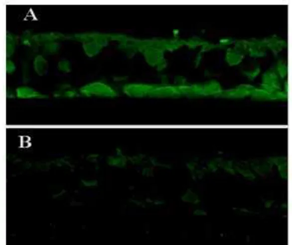
2. Localization of the P2Y2 receptor
The P2Y2 receptor was immunolocalized using confocal fluorescence
microscopy, and was observed in the apical and basal cell layers of the cultured NHMEE cells, but not in the intermediate cell layers. No signals were observed in the samples treated with the anti-sense peptide (Fig 2).
Fig. 2 Immunohistochemical analysis of the normal human middle ear epithelial (NHMEE) cells using antibody against the P2Y2 receptor. The
NHMEE cells showed positive immunofluorescent reactions in the apical and basal cell layers (A), whereas, the negative controls, which were treated with the anti-sense peptides, showed no immunoreactivity (B)
3. Contribution of intra- and extra-cellular stores to UTP-induced calcium mobilization
This study further investigated the pattern of UTP-induced [Ca2+]i in NHMEE cells. The increase in [Ca2+]i evoked by UTP persisted in the Ca2+-free external solutions, suggesting that the increase in [Ca2+]i was due to its release from the intracellular Ca2+store (Fig. 3A). The application of 10 µM ryanodine, a CICC antagonist, did not inhibit UTP-induced [Ca2+]i . In contrast, 2-aminoethoxydiphenyl borate (100µM), an IP3 receptor antagonist significantly inhibited the UTP
induced [Ca2+]i . (Fig. 3B and C) This suggests that UTP stimulates
[Ca2+]i from the internal stores, presumably via the IP3-sensitive Ca 2+
channels but not through the ryanodine-sensitive CICC.
1 1.5 2 0 100 200 300 400 500 1 1.5 2 0 100 200 300 400 500 1 1.5 2 0 100 200 300 400 500 0 0 0 0 CaCaCaCa2+2+2+2+ 100µM UTP 100µM UTP 2 22
2---APB(100 -APB(100 APB(100 APB(100 µµµµM))))
100µM UTP 100µM UTP Ryanodine RyanodineRyanodine Ryanodine (10(10(10(10µµµµM) 100µM UTP 100µM UTP A B C R a ti o3 5 2 /3 8 0 sec 1 1.5 2 0 100 200 300 400 500 1 1.5 2 0 100 200 300 400 500 1 1.5 2 0 100 200 300 400 500 0 0 0 0 CaCaCaCa2+2+2+2+ 100µM UTP 100µM UTP 2 22
2---APB(100 -APB(100 APB(100 APB(100 µµµµM))))
100µM UTP 100µM UTP Ryanodine RyanodineRyanodine Ryanodine (10(10(10(10µµµµM) 100µM UTP 100µM UTP A B C R a ti o3 5 2 /3 8 0 sec
Fig. 3. Effect of the inhibition of the Ca2+ uptake from external and internal stores on the UPT-induced Ca2+influx [Ca2+]i in NHMEE cells.
The increase in [Ca2+]i evoked by UTP persists in a Ca2+ free solution (A). 2ABP (100µM/ml) inhibits the UTP induced [Ca2+]i but ryanodine
fail to inhibit. (B & C)
4. Ca2+ dependency of mucin secretion
The role of intracellular Ca2+ on mucin release in NHMEE cells was investigated. The UTP-induced mucin secretion (408± 16 % of control) was suppressed by the depletion of intracellular Ca2+ with 50µM BAPTA-AM (176± 19 % of the control level.) and increasing the intracellular Ca2+ level with 1µM inomycin, a Ca2+ ionophore, enhanced the UTP-induced mucin secretion (528± 21% of the control). Furthermore, the BAPTA-AM suppressed the constitutional mucin secretion (73± 9% of the control) and inomycin increased constitutional secretion (163± 12 %) (Fig. 4). Therefore, it appears likely that both UTP-induced and constitutional mucin secretion are Ca2+-dependent in NHMEE cells.
Fig. 4 Effect of intracellular calcium on mucin secretion in NHMEE cells. The depletion of intracellular Ca2+ by adding 50 µM 2-bis (2-aminophenoxy) ethane-N,N,N',N'-tetraacetic acid-acetoxymethyl ester (BAPTA-AM) suppresses both the UTP-induced mucin secretion and constitutional mucin secretion and inomycin (1 µM) enhanced the
constitutional and UTP-induced mucin secretion. BAPTA,
BAPTA-AM; Ino, inomycin. * indicates significant difference from the control and ** indicates significant difference from the UTP treated-control.
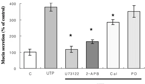
5. Involvement of PLC and IP3 in the signaling pathway of the UTP-induced mucin secretion.
UTP-induced mucin secretion (378± 19% of the control) was strongly suppressed by blocking the PLC with 10 µM of U73122 (118± 25% of the control). 2-ABP(100µM), an IP3 inhibitor, also suppressed the
UTP-induced mucin secretion (165± 19% of the control ). Calphostin C (0.1µM), a PKC inhibitor, partially inhibited the UTP-induced mucin
secretion (285± 12% of the control ), but was less potent than U73122 and heparin. However, PD98059, an ERK MAPK inhibitor, did not suppress the UTP-induced mucin secretion (352± 15% of the control). (Fig. 5) This suggests that the PLC and IP3 pathways are essential for
UTP-induced mucin secretion in NHMEE cells.
Fig. 5 Effect of the inhibitors on the UTP-induced mucin secretion in human middle ear epithelial cells. C, control; Cal, Calphostin C, PD, PD98059;2-APB, 2-aminoethoxydiphenyl borate. * indicates significant difference from UTP treated-sample.
0 100 200 300 400 C C U 73122 2-A P B C a l P D M u ci n s ec re ti o n ( % o f co n tr o l) UTP
*
**
*
*
**
*
*
**
*
UTP 0 100 200 300 400 C C U 73122 2-A P B C a l P D M u ci n s ec re ti o n ( % o f co n tr o l) UTP*
**
*
*
**
*
*
**
*
UTP6. Effect of caffeine, an another IP3 inhibitor, on UTP-induced [Ca2+]i and mucin secretion
Caffeine is widely used as a blocker of the IP3-dependent response 13
. Because IP3 is essential in UTP-induced mucin secretion, we next wanted
to confirm the effect of caffeine on UTP induced [Ca2+]i in NHMEE cells. UTP-induced [Ca2+]i is partially suppressed by 5mM caffeine. Fig 6 shows that the inhibitory effect of caffeine gradually increased in a dose-dependent manner.
Fig.6. The Effect of caffeine on the UTP-induced Ca2+ mobilization in NHMEE cells. Caffeine suppressed UTP-induced mucin Ca2+ influx in a dose-dependant manner.
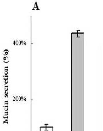
As a next, the effect of caffeine on UTP-induced mucin secretion was also evaluated. 100M of UTP promptly induced mucin secretion in NHMEE cells and caffeine suppressed this UTP-induced mucin secretion in a dose-dependent manner.(Fig 7A). More than 10mM of caffeine also suppressed constitutional mucin secretion in a dose-dependent manner (Fig 7B).
Fig. 7 Effect of caffeine on mucin secretion in NHMEE cells. Caffeine suppressed the UTP-induced mucin secretion in NHMEE cells in a dose- dependent manner.(A) More than 10mM of caffeine also suppressed the constitutional mucin secretion. (B)
IV . D ISCU SSION
Extracellular purinergic agonists play significant regulatory roles in various biological responses. They also regulate the mucociliary clearance of the airway epithelium via uracil-sensitive receptors such as P2Y2, P2Y4 and P2Y6.
2,14,15
We previously reported that P2Y2 and P2Y6
mRNA transcripts are present in middle ear epithelial cells both in vivo and in vitro 4. However, in this study, P2Y6 could not modulate [Ca
2+
]i
in NHMEE cells because its potent agonist, UDP, did not activate
[Ca2+]i. P2Y6 inactivation in NHMEE cell is a unique finding. P2Y6 was
found to be as potent as P2Y2 in the nasal and tracheobronchial
epithelium2,15. The order of potency (ATP>UTP>2MeATP>>adenosine) described earlier suggests that P2Y2 acts on [Ca
2+
]i 12. The activation of the P2Y2 receptors in the middle ear epithelium by purinergic
stimulation resembles findings in the tracheobronchial3 and nasal2 epithelium, and is also consistent with a result in the middle ear cell line
16
. It was also confirmed that the P2Y2 receptor is localized in the basal
and apical layers of cultured NHMEE cells, but not in the intermediate layers. It is unclear why the P2Y2 receptors are expressed only in these
layers.
Ca2+ from the ER in mammalian cells via the P2Y2 receptor. 17
The ERis endowed with two different types of Ca2+ release channels, i.e. IP3 and
ryanodine receptors.9 Our data suggest that the intracellular Ca2+ level is increased through the IP3 receptors in NHMEE cells, but not through the
ryanodine receptors. These results are consistent with the report by Yang et al.18 where UTP directly stimulate PLC-mediated IP3
accumulation and Ca2+ mobilization in canine tracheal epithelial cells.
There are conflicting reports about the role of Ca2+ in nucleotide-induced mucin secretion in airway epithelial cells. Mucin release by ATP does not require an increase in the intracellular Ca2+ level in primary hamster tracheal epithelial cells 6. On the other hand, nucleotide-induced mucin secretion is regulated independently with Ca2+ and the PKC pathways in human nasal epithelial cells19, the spontaneously transformedrat goblet cell line, SPOC1 cells20. These
discrepancies suggest that the Ca2+-dependency of the
nucleotide-induced mucin secretion differs according to the cell type. In this study, the removal of the intracellular Ca2+ with BAPTA-AM strongly blocked the UTP-induced mucin secretion, and inomycin enhanced the UTP-induced mucin secretion in NHMEE cells. These results suggest that UTP-induced mucin secretion is more Ca2+-dependent in NHMEE cells than in the other airway epithelial cells.
There is some controversy regarding the signaling downstream of nucleotide-induced mucin secretion. The activation of the P2Y2 receptor
by nucleotide has previously been shown to trigger the activation of PLC, and subsequently PKC. Both of these enzymes have been linked to mucin secretion in hamster6, human14 treacheonchial cultures or SPOC1 cells20. However, the stimulatory effect of PKC varies according to the cell type.2 This study found that U73122 and 2-ABP almost completely inhibited the UTP-induced mucin secretion, while Calphostin C suppressed it slightly and PD98059 was ineffective, suggesting that PLC and IP3 are important signaling molecules in the
UTP-induced mucin secretion in NHMEE cells. The crucial role of IP3
in nucleotide-induced mucin secretion has not yet been reported in other airway epithelial cells, and a more detailed role of IP3 needs to be
elucidated.
An additional aim of this study was to confirm the effect of caffeine on the UTP induced [Ca2+]i and mucin secretion in NHMEE cells.
Caffeine, although best known as an activator of the ryanodine receptor, also inhibits the opening of the IP3 receptor.
12
In this study, caffeine partially inhibited the UTP-induced [Ca2+]i and mucin secretion in NHMEE cells, which confirming that NHMEE cells have a caffeine-sensitive IP3 receptor, and IP3 plays an important role in the
UTP-induced mucin secretion in NHMEE cells.
V . CON CLU SION
The P2Y2 receptor was found to be expressed in NHMEE cells, and
play a major role in modulating the [Ca2+]i. from the ER. UTP-induced mucin secretion in NHMEE cells is strongly Ca2+- and IP3-dependent.
Reference
1. Alessandra R, Alfio F. The pathology and clinical features of glue ear: a review. Eur Arch Otorhinolaryngol 2000;257:300-303.
2. Knight DE, H von Grafenstein, CM Athayde. Calcium-dependent and calcium-independent exocytosis Trends Neurosci 1989;12:451-458.
3. Kim KC, Park HR. Shin CY, Akiyama T, Ko KH. Nucleotide-induced mucin release from primary hamster tracheal surface epithelial cells involves the P2u purinoreceptor. Eur Respir J 1996 9:542-548.
4. Choi JY, Cho KN, Shin JH, Yoon.JH.. Effect of Uridine 5'-Triphosphate on Mucin and Lysozyme Expression in Human Middle Ear Epithelial Cells. Acta otolaryngologica(Stockh) 2003;123: 362-366.
5. Chen Y, Zhao YH, Wu R. Differential regulation of airwaymucin gene expression and mucin secretion by extracellular nucleotide triphosphates. Am J Respir Cell Mol Biol 2001;25:409-417.
6. Ko KH, Jo M, McCracken K, Kim KC. ATP-induced mucin release from cultured airway goblet cells involves, in part, activation of protein
kinase C. Am J Respir Cell Mol Biol 1997;16:194-198.
7. Dartt DA, Rios JD, Kanno H, Rawe IM, Zieske JD, Ralda N,
Hodges RR, Zoukhri D, Rios JR. Regulation of conjunctival goblet cell
secretion by Ca(2+)and protein kinase C.Exp Eye Res 2000;71:619-628.
8. Dray-Charier N, Paul A, Combettes L, Bouin M, Mergey M,
Balladur P et al. Regulation of mucin secretion in human gallbladder
epithelial cells: predominant role of calcium and protein kinase C.
Gastroenterology 1997;112:978-990.
9. Ehrlich BE, Kaftan E, Bezprozvannaya S, Bezprozvanny I. The pharmacology of intracellular Ca(2+)-release channels. Trends
Pharmacol Sci 1994;15(5):145-149.
10. Yoon JH, Kim KS, Kim SS, Lee JG, Park IY. Secretory differentiation of serially passaged normal human nasal epithelial cells by retinoic acid: expression of mucin and lysozyme. Ann Otol Rhinol
Laryngol 2000;109:594-601.
11. Ko WH, Law VWY, Wong HY, Wilson SM. The simultaneous measurement of epithelial ion transport and intracellular free Ca2+ in cultured equine sweat gland secretory epithelium. J Membr Biol
1999;170: 205-211.
12. King BF, Townsend-Nicholson A, Burnstock G Metabotropic receptors for ATP and UTP: exploring the correspondence between native and recombinant nucleotide receptors. Trends Pharmacol Sci 1998;19(12):506-514.
13. Osipchuk YV, Wakui M, Yule DI,Gallacher DV; Petersen OH. Cytoplasmic Ca2+oscillations evoked by receptor stimulation, G-protein activation, internal application of inositol trisphosphate or Ca2+: simultaneous microfluorimetry and Ca2+ dependent Cl- current recording in single pancreatic acinar cells. EMBO 1990;9:697-704.
14. Kemp PA, Sugar RA, Jackson AD. Nucleotide-mediated mucin secretion from differentiated human bronchial epithelial cells. Am J
Respir Cell Mol Biol.2004; 31(4):446-455.
15. Lazarowski EA, Paradiso AM, Watt WC, Harden TK, Boucher
RC.UDP activates a mucosal-restricted receptor on human nasal
epithelial cells that is distinct from the P2Y2 receptor. Proc Natl Acad
Sci USA 1997;94:2599-2603.
modulates ion transport via P2Y purinoceptors in a middle-ear epithelial cell line. ORL J Otorhinolaryngol Relat Spec 1997;59(3):170-175.
17. Ralevic V, Burnstock G. Receptors for purines and pyrimidines.
Pharmacol Rev 1998;50(3):413-492.
18. Yang CM, Wu WB, Pan SL, Tsai YJ, Chiu CT, Wang CC. P2Y(2) receptor-stimulated phosphoinositide hydrolysis and Ca(2+) mobilization in tracheal epithelial cells. Am J Physiol Lung Cell Mol
Physiol 2000;279(2):L235-241.
19 Abdullah LH, Conway JD, Cohn JA, Davis CW. Protein kinase C and Ca2+ activation of mucin secretion in airway goblet cells. Am J Physiol 1997;273:L201-210.
20. Scott CE, Abdullah LH, Davis CW. Ca2+and protein kinase C activation of mucin granule exocytosis in permeabilized SPOC1 cells.
국문 요약
인
인
인체
체
체중
중
중이
이
이점
점
점막
막
막 상
상
상피
피
피세
세
세표
표
표에
에
에서
서
서 퓨
퓨
퓨린
린
린수
수
수용
용
용체
체
체 길
길
길항
항
항제
제
제가
가
가
점
점
점액
액
액분
분
분비
비
비에
에
에 미
미
미치
치
치는
는
는 영
영
영향
향
향
<지도교수 윤 주헌> 연세대학교 대학원 의학과 최 재영 퓨린수용체는 기도상피세포에서 점액분비를 조절하는 것으로 알려져 있다 . 본 연구에서는 인체중이점막상피세포에서 퓨린수용 체의 자극제가 Ca2+의 세포내 유입 ([Ca2+ ]i) 과 점액 분비에 미치는 영향을 그 작용기전과 함께 알아보고자 한다 . 또한 연구자는inositol 1,4,5-triphosphate (IP3)의 길항제인 caffeine이 UTP에 의한
[Ca2+]i 와 점액분비에 미치는 영향을 알아보았다. 인체중이점막 상
피세포를 UTP를 포함한 여러 퓨린수용체 자극제로 자극한수
miniature Ussing double perfusion chamber를 이용하여 [Ca2+]i를 측
정하였다 . 또한 P2Y2수용체 항체를 이용하여 인체중이점막상피세
포에서 이 수용체의 분포양상을 확인하였다 . UTP에 의한 점액분비 는 immunoblotting assay를 통하여 정량하였다 . 여러 수용체 자극제
의 자극 강도는 ATP=UTP>2-MeSATP>ADP>>adenosine순이었으
며 , 이는 P2Y2 수용체의 자극과 일치한다 . P2Y2수용체는 중이점막
상피세포의 첨부와 기저부에서 발현되었다 . UTP 에 의한 [Ca2+
]i 는
2-APB(100µM)에 의해 억제되었으나 , ryanodine(10µM)에 의해서는
억제되지 않았다 .UTP에 의한 점액분비는 Ca2+chelating agent인
BAPTA-AM에 의해 억제되고 , inomycin에 의해 촉진되었다 . UTP에
의한 점액분비는 U73122 와 2-APB에 의해서는 억제되었으나 ,
Calphostin C와 PD98059에 의해서는 억제되지 않았다 . Caffeine은
UTP에 의한 [Ca2+]i 와 점액분비를 억제하였다 . 이상과 같은 결과는
P2Y2 수용체가 인체중이점막상피세포에 발현하며 , IP3-sensitive
intracellular Ca2+ store를 자극하여 세포내 [Ca2+]i.을 유도함을 알 수
있었다 .
핵심 되는 말 점액 , 카페인 , 신호전달 , IP3, Ca 2+
![Fig. 1. Mobilization of the [Ca 2+ ]i by the purinergic](https://thumb-ap.123doks.com/thumbv2/123dokinfo/5073241.72162/16.892.215.586.573.852/fig-mobilization-ca-i-purinergic.webp)