The effect of
The effect of
The effect of
The effect of bacterial
bacterial
bacterial
bacterial
γ
γ
γ
γ-
-
-
-glutamyltranspeptidase
glutamyltranspeptidase
glutamyltranspeptidase
glutamyltranspeptidase
on osteoclastogenesis
on osteoclastogenesis
on osteoclastogenesis
on osteoclastogenesis
Bong
Bong
Bong
Bong-
-
-Ju Kim
-
Ju Kim
Ju Kim
Ju Kim
The Graduate School
The Graduate School
The Graduate School
The Graduate School,
,
, Yonsei University
,
Yonsei University
Yonsei University
Yonsei University
Department of Dentistry
Department of Dentistry
Department of Dentistry
Department of Dentistry
The effect of bacterial
The effect of bacterial
The effect of bacterial
The effect of bacterial
γ-
-
-
-glutamyltranspeptidase
glutamyltranspeptidase
glutamyltranspeptidase
glutamyltranspeptidase
on osteoclastogenesis
on osteoclastogenesis
on osteoclastogenesis
on osteoclastogenesis
Bong
Bong
Bong
Bong-
-
-
-Ju Kim
Ju Kim
Ju Kim
Ju Kim
The Gradua
The Gradua
The Gradua
The Graduate
te
te
te School
School
School
School,
,
,
, Yonse
Yonse
Yonse
Yonse University
University
University
University
Department of Dentistry
Department of Dentistry
Department of Dentistry
Department of Dentistry
The effect of bacterial
The effect of bacterial
The effect of bacterial
The effect of bacterial
γ
γ
γ
γ-
-
-
-glutamyltranspeptidase
glutamyltranspeptidase
glutamyltranspeptidase
glutamyltranspeptidase
on osteoclastogenesis
on osteoclastogenesis
on osteoclastogenesis
on osteoclastogenesis
A Master Thesis Submitted to the
A Master Thesis Submitted to the
A Master Thesis Submitted to the
A Master Thesis Submitted to the
Department of Dental Science and the
Department of Dental Science and the
Department of Dental Science and the
Department of Dental Science and the
Graduate School of Yonsei University
Graduate School of Yonsei University
Graduate School of Yonsei University
Graduate School of Yonsei University
In partial fulfillme
In partial fulfillme
In partial fulfillme
In partial fulfillment of the
nt of the
nt of the
nt of the
Requirement for the degree of
Requirement for the degree of
Requirement for the degree of
Requirement for the degree of
Master of Philosophy of Dental Science
Master of Philosophy of Dental Science
Master of Philosophy of Dental Science
Master of Philosophy of Dental Science
Bong
Bong
Bong
Bong-
-
-Ju Kim
-
Ju Kim
Ju Kim
Ju Kim
January 2006
January 2006
January 2006
January 2006
감사의
감사의
감사의
감사의
글
글
글
글
연구 시작단계부터 본 논문의 완성까지 세심한 지도와 많은 배려를 해 주시고 마지막까지 좋은 논문이 되도록 힘써주신 차정헌 교수님께 진심으 로 감사를 드립니다. 논문 실험 시작부터 과정 내내 깊은 관심과 조언을 해주신 유윤정 교수님과 많은 충고와 조언으로 논문을 다듬어 주신 서정 택 교수님께 감사 드립니다. 실험에 많은 도움을 주시고 항상 친절히 질 문에 답해주신 이양신 선생님, 최호길 선생님께도 감사의 마음을 전합니 다. 아울러 변함없는 사랑과 기도를 보내주신 양가 부모님, 든든한 후원자 인 아내 유수정과 사랑스런 두 아들 영준, 호준에게 감사하며 하나님께 감사 드립니다.
Table of Contents
Table of Contents
Table of Contents
Table of Contents
LIST OF FIGURES LIST OF FIGURES LIST OF FIGURES LIST OF FIGURES ⅱ LIST OF TABLE LIST OF TABLE LIST OF TABLE LIST OF TABLE ⅲ Abstract Abstract Abstract
Abstract (English) (English) (English) (English) ⅳ
Ⅰ Ⅰ Ⅰ
Ⅰ. Introduction. Introduction. Introduction. Introduction 1
Ⅱ Ⅱ Ⅱ
Ⅱ. Materials and Methods. Materials and Methods. Materials and Methods. Materials and Methods 4
1. Materials 4 2. Preparation of primary calvaria and bone marrow cells 4 3. Bacteria culture 4 4. Plasmid construction 5 5. Expression and purification of recombinant
B. subtilis GGT protein 5 6. Osteoclast formation assay 6 7. Reverse-transcription polymerase chain-reaction
(RT-PCR) method 6
Ⅲ Ⅲ Ⅲ
Ⅲ. Results. Results. Results. Results 10 1. Osteoclast-forming activity of B. subtilis
GGT in conditioned medium 10 2. Purification of B. subtilis GGT 11 3. Effect of purified GGT on expression of RANKL,
OPG, HB-EGF, AR, and COX-2 12
Ⅳ Ⅳ Ⅳ
Ⅳ. Discussion. Discussion. Discussion. Discussion 16 References References References References 19 Abstract (Korean Abstract (Korean Abstract (Korean Abstract (Korean)))) 24
L
LL
List of Figures
ist of Figures
ist of Figures
ist of Figures
Figure 1. The effect of B. subtilis GGT in conditioned medium
on osteoclast-forming activity 10 Figure 2. Overexpression of B. subtilis GGT in E. coli 11 Figure 3. The purified B. subtilis GGT using His-Tag
purification was incubated at
37oC for the autocatalytic process 12 Figure 4. The effect of the purified GGT on mRNA expression
List of Tables
List of Tables
List of Tables
List of Tables
Table 1. Bacterial strains and plasmids in this study 8 Table 2. Sequence of primers for B.subtilis GGT, RANKL,
OPG, HB-EGF, AR, and β-actin 9 Table 3. The mRNA ratios of RANKL, OPG, HB-EGF, AR and COX-2
to β-actin in the calvaria-derived osteoblasts after 3 hr incubation with the purified GGT at the
Abstract
Abstract
Abstract
Abstract
The effec
The effec
The effec
The effect of
t of
t of
t of
bacterial
bacterial
bacterial
bacterial
γ
γ
γ----glutamyltranspeptidase
γ
glutamyltranspeptidase
glutamyltranspeptidase
glutamyltranspeptidase
on osteoclastogenesis
on osteoclastogenesis
on osteoclastogenesis
on osteoclastogenesis
A novel bone-resorbing factor, γ-glutamyltranpeptidase (GGT), was recently cloned from mouse T-lymphoma cell cDNA library. Since the GGT is widely distributed in living organisms and Bacillus subtilis
GGT exhibits high similarity in its primary structure and enzymatic characteristics with the mammalian GGTs, the B. subtilis GGT was examined for the osteoclast formation. The osteoclast formation was performed in a co-culture system of mouse calvaria-derived osteoblasts and bone marrow cells, and determined by tartrate resistant acid phosphatase staining. Conditioned medium from GGT-overproducing B. subtilis culture showed significantly higher activity of osteoclast formation when compared to those from wild-type and ggt-deleted B. subtilis culture. To understand the mechanism of how GGT induce osteoclastogenesis, B. subtilis GGT was cloned, expressed in Escherichia coli and purified using His-tag purification. The purified GGT stimulated mRNA expression of the receptor activator of nuclear factor-κB ligand, cyclooxygenase-2, heparin-binding epidermal growth factor-like growth factor, and amphiregulin in mouse calvaria-derived osteoblasts.
This report is the first demonstration that bacterial GGT is able to serve as the bone-resorbing factor and the osteoclastogenesis by the GGT is mediated by a RANKL-dependent pathway. Based on this study, it can be hypothesized that GGT in periodontopathic bacteria can be an important virulence factor for bone destruction in
periodontitis.
Keywords : γ-glutamyltranspeptidase, Bacillus subtilis, osteoclastogenesis, RANKL, COX-2, HB-EGF, amphiregulin
The effect of
The effect of
The effect of
The effect of
bacterial
bacterial
bacterial
bacterial
γ
γ
γ
γ----glutamyltranspeptidase
glutamyltranspeptidase
glutamyltranspeptidase
glutamyltranspeptidase
on osteoclastogenesis
on osteoclastogenesis
on osteoclastogenesis
on osteoclastogenesis
Directed by Professor Jeong
Directed by Professor Jeong
Directed by Professor Jeong
Directed by Professor Jeong-
--
-Heon Cha
Heon Cha
Heon Cha
Heon Cha
Department of Dentistry
Department of Dentistry
Department of Dentistry
Department of Dentistry
The Graduate School, Yonsei University
The Graduate School, Yonsei University
The Graduate School, Yonsei University
The Graduate School, Yonsei University
Bong
Bong
Bong
Bong-
--
-Ju Kim
Ju Kim
Ju Kim
Ju Kim
Ⅰ
Ⅰ
Ⅰ
Ⅰ.
.
.
. Introduction
Introduction
Introduction
Introduction
Osteoclasts are multinucleated cells with a bone resorbing activity and play a crucial role in bone resorption. They are derived from hemotopoietic cells through multiple steps including proliferation, expression of tartrate resistant acid phosphatase (TRAP), and fusion of cells (Suda et al., 1997; Hofbauer et al., 2000).
Osteoclast formation requires the presence of osteoblast or stromal cells (Takahashi et al., 1999). These cells express a receptor of the nuclear factor-κB(RANK) ligand (RANKL, also known as the osteoclast differentiation factor) that promotes osteoclastogenesis. The osteoclast precursors express RANK which interacts with RANKL, through cell-cell contact, the cells then differentiate into osteoclasts (Hsu et al., 1999). Thus, Osteoblast regulates osteoclast differentiation through RANKL-RANK interaction
(Lacey et al., 1998). Osteoprotegerin (OPG), which is also secreted by osteoblast lineage cells, is a soluble detoy receptor that neutralizes the biological activity of RANKL (Simonet et al., 1997; Yasuda et al., 1998; Udagawa et al., 2000). The regulation of RANKL by OPG suggests that bone resorption may be mediated through control of RANKL and /or OPG production and that ostesclast formation is determined principally by the ratio of RANKL to OPG (Hofbauer et al., 1998; Vidal et al., 1998; Nagai et al., 1999; Brändström et al., 2001).
RANKL expression in osteobalsts is upregulated by pro-resorptive hormones and cytokines, such as 1,25-dihydroxyvitamin D3 (1 α,25(OH)2D3), parathyroid hormone, prostaglandin E₂(PGE2), and interlenkin-1 (Tsukii et al., 1998; Hofbauer et al., 1999; Walsh et al., 2003).
Recently, a novel bone-resorbing factor was isolated and cloned using an expression cloning technique from a BW5147 mouse T-lymphoma cell cDNA library. Sequencing analysis identified the factor as γ-glutamyltranpeptidase (GGT) (Niida et al., 2004). GGT, which is widely distributed in living organism (Tate et al., 1981), catalyzes the hydrolysis of γ-glutamyl compounds such as glutathione (Lieberman et al., 1995), as well as the hydrolysis of the amide bonds of glutamine to yield glutamic acid and ammonia, simillar to glutaminase. GGT can also catalyze the transfer of γ-glutamyl moieties to amino acids and peptides. ywrD might encode a second GGT and exhibit 31 and 27% identity with amino acid B. subtilis GGT.
ywrD has no signal sequence.
The addition of purified GGT protein to mouse bone marrow culture effectively induced formation of osteoclasts. An antibody against GGT inhibited Osteoclast formation but not the enzymatic activity. They also demonstrated that an inactive form of GGT, the enzymatic
activity of which had been blocked by chemical modification with a specific inhibitor, acivicin, supported osteoclast formation. These results indicate that GGT acts on osteoclast formation independent of its own enzymatic activity. Furthermore, both native GGT and inactive GGT stimulated the expression of the RANKL mRNA and protein from bone marrow stromal cells. The osteoclastogenesis biological activity of GGT protein is in a manner independent of its enzymatic activity.
Since GGT is widely distributed in living organism, bacteria also have GGT. The GGT of gram-positive bacterea such as Bacillus species are extracellular enzymes (Ogawa et al., 1991; ogawa et al., 1997; Kunst et al., 1997), so they can be purified from the culture broth and the GGT of gram-negative bacteria such as Escherichia coli is present in the periplasmic space (Suzuki et al., 1986). It was reported that Helicobacter pylori GGT upregulates cyclooxygenase (COX)-2, heparin-binding epidermal growth factor-like growth factor (HB-EGF) and amphiregulin (AR) expression in human gastric cells (Busiello et al., 2004). The COX-2 is known as a factor of PGE₂ induction. The expression of RANKL is stimulated by systemic bone resorbing factors such as PGE2. Above mechanisms suggest that the bacterial GGT may also stimulate RANKL expression and serve as a bone resorbing factor.
Thus, we examined whether bacterial GGT also has bone-resorbing activity and investigated the mechanism of how the GGT induces osteoclastogenesis.
Ⅱ
Ⅱ
Ⅱ
Ⅱ.
.
. Materials and Method
.
Materials and Method
Materials and Method
Materials and Method
1. Materials 1. Materials 1. Materials 1. Materials
The ICR mice were obtained from Bio Korea Co. (Seoul, Korea). The α-minimum essential medium (α-MEM), bovine serum albumin (BSA), and heat-inactivated fetal bovine serum (FBS) were purchased from GIBCO BRL (Grand Island, NY). LPS of Escherichia coli, and tartrate-resistant acid phosphatase (TRAP: a marker of osteoclast) staining kit were obtained from Sigma (St. Louis, MO).
2. 2. 2.
2. Preparation of primary calvaria and bone marrow cells.Preparation of primary calvaria and bone marrow cells.Preparation of primary calvaria and bone marrow cells.Preparation of primary calvaria and bone marrow cells.
The osteobblastic cells were isolated from the calvariae of 1- to 2-day old ICR mice. The calvariae were digested in 10 ml of α -MEM containing 0.2% collagenase (Wako Pure Chemicals, Osaka, Japan) and 0.1% dispase (GIBCO BRL, Grand Island, NY, USA) for 20 min at 37℃ with vigorous shaking and then centrifuged at 1,500 x g for 5 min. The first supernatant was discarded, another 10 ml of the collagenase-dispase enzyme solution was added, and the preparation was incubated for 20 min. The digestion procedure was repeated four times, and the cells isolated by the last three digestions were combined as an osteoblastic cell population. They were cultured in α-MEM containing 10 % FBS. The bone marrow cells were collected from 5– to 8-week old mice. The ends of the tibiae and femurs were removed, and each marrow cavity was flushed by slowly injecting medium at one end with a 25-gauge needle. The marrow cells were washed and used for the co-culture.
3. 3. 3.
3.Bacteria cultureBacteria cultureBacteria cultureBacteria culture....
-deleted (MH2353) and GGT-overproducing (MH2308) B. subtilis were obtained from Hideyuki Suzuki, Kyoto Univeristy and listed in Table 1. B. subtilis strains were inoculated on BHI agar plate and cultured at 37oC for 18 hr. A single colony was inoculated and cultured at 37oC for 96 hr in 10 ml Brain Heart Infusion (BHI) media. The bacteria were harvested by centrifugation and discarded. The supernatant was filtrated by 0.22 μm filter (Millipore, Bedford, Ireland). The filtrates were stored at -70oC until use.
4. Plasmid contruction.Plasmid contruction.Plasmid contruction. Plasmid contruction.
The pET21b plasmid was used for B. subtilis GGT protein expression in E. coli. The coding regions (residues 1 to 587) was amplified by PCR with B. subtilis chromosomal DNA template and primers (BsGGT-F and BsGGT-B), and cloned into pET21b using the EcoRI and XhoI sites, resulting in pET21b/BsGGT plasmid. The both strands of the insert fragment were sequenced by the Cosmogene Tec. (Cosmo, Korea) to confirm the fidelity of PCR.
5. 5. 5.
5. Expression and purification of recomExpression and purification of recomExpression and purification of recomExpression and purification of recombinant binant binant binant B. subtilisB. subtilisB. subtilisB. subtilis GGT protein GGT protein GGT protein GGT protein The recombinant B. subtilis GGT protein was expressed from pET21b/BsGGT in E. coli BL21(DE3). The recombinant B. subtilis GGT protein contained a T7 tag at its amino terminus and a His tag at its carboxyl terminus to facilitate the purification step. The overexpressed B. subtilis GGT protein formed inclusion body, which was isolated and dissolved in 8 M urea. The dissolved solution was purified by His tag affinity column as described in the manufacturer’s instructions. The purified recombinant B. subtilis
GGT protein was refolded by sequential dialytic removal of urea in 25 mM Tris buffer with 4, 2, 1, and 0 M urea steps. The amount of purified and refolded recombinant B. subtilis GGT protein was
determined by the Bradford protein assay (Bio-rad). 6.
6. 6.
6. Osteoclast formation assay.Osteoclast formation assay.Osteoclast formation assay.Osteoclast formation assay.
The isolated calvaria cells were seeded at a concentration of 106 cells per 10-cm culture dish and grown to confluence. The cells were then detached from the culture dishes with trypsin-EDTA (GIBCO BRL). Subsequently, the cells (1 x 10⁴ cells/well) were cocultured with the bone marrow cells (1 x 10⁵ cells/well) in α -MEM containing 10 % FBS in 48-welll plates (Corning Inc., Corning, N.Y.). The culture volume was adjusted to 400 μl per well with α-MEM containing 10% FBS. The conditioned media (50 or 100 μl) from wild-type, ggt-deleted, ggt and ywrD-deleted, and GGT-overporducing B. subtilis cultures or the purified GGT at the indicated concentration were added to each co-culture after the medium was exchanged on day 3. Also, BHI media (50 or 100 μl) or E. coli LPS (1 μg/ml) were added as negative and positive controls, respectively. The co-culture was then maintained for an additional 4 days. Osteoclast differentiation was monitored by using a TRAP staining kit according to the manufacturer’s instructions. TRAP-positive multinucleated cells having more than three nuclei were defined as osteoclasts. The osteoclast cells per well were counted for osteoclast formation activity.
7. 7. 7.
7. ReverseReverseReverseReverse----transcription polymeratranscription polymeratranscription polymeratranscription polymerase chainse chain-se chainse chain---reaction (RTreaction (RTreaction (RT-reaction (RT---PCR) method.PCR) method.PCR) method. PCR) method. The mRNA Expression was determined by RT-PCR. Total RNA (1 μg) isolated from nontreated or treated mouse calvaria-derived osteoblast cells for 3 hr was used as a template for cDNA synthesis in a 20-μl reaction mixture performed with an RT kit (CLONTECH, Palo Alto, Calif.) according to the manufacturer’s instructions. The RNA (1 ㎍) and oligo(dT)18 primers (1 mM) were denatured at 70℃
for 5 min and incubated for 1 to 2 min on ice. The denatured RNA and oligo(dT)18 primers were added to the reaction mixture (1 U of Moloney murine leukemia virus reverse transcriptase perμl, 1X reaction buffer, 500 μM of each dATP, dCTP, dGTP, and dTTP, 20 U of recombinant RNase inhibitor) and incubated at 42℃ for 60 min, followed by 94℃ for 5 min.
The cDNA (4 μg) was amplified by PCR in a 50-μl reaction mixture containing 1 X PCR reaction buffer, each deoxynucleoside triphosphate at a concentration of 200 µM, 200 pM forward primer, 200 pM reverse primer, and 0.5 U of Taq DNA polymerase (Amersham Pharmacia Biotech, Little Chalfont, Buckinghamshire, United Kingdom) in a DNA thermal cycler (Biometra, Gottinge, Germany). The amplification reaction was performed for 35 cycles, and the primer sequences and annealing temperatures used are shown in Table 2. The PCR products were separated by electrophoresis on a 1.5% agarose gel, stained by ethidium bromide, and detected with Multilineage Light Cabinet (Alpha Innotech Corp. San Leandro, CA). The relative intensities of the gel bands were measured by using an image-analyzing program (frog2000 version3.1.2). In order to exclude contaminating DNA from the isolated RNA, the RNA was subjected to PCR without cDNA synthesis. In all preparations, no band was detected after PCR.
Table 1. Bacterial strains and plasmids in this studyTable 1. Bacterial strains and plasmids in this studyTable 1. Bacterial strains and plasmids in this studyTable 1. Bacterial strains and plasmids in this study
Strain or plasmid Characteristic(s) B. subtilis
MH2275 B. subtilis 168 trpC2
MH2342 B. subtilis 168, but ggt- trpC2 △ggt::cat+ MH2308 plasmid with B. subtilis ggt gene (pMH2312)
in sw153; GGT over-producer
MH2350 B. subtilis 168, but ywrD- trpC2 △ywrD::cat+
MH2353 B. subtilis 168, but ggt- ywrD- trpC2 △ggt::cat+ △ywrD::tet+
Plasmid
pMH2312 the HaeⅢ-BglⅡ fragment of pPL623 containing the cam+ gene was ligated with pMH2285 cleaved with HpaⅠ
Table 2. Sequence of primers for Table 2. Sequence of primers for Table 2. Sequence of primers for
Table 2. Sequence of primers for B.subtilisB.subtilisB.subtilisB.subtilis GGT, RANKL, OPG, HB GGT, RANKL, OPG, HB GGT, RANKL, OPG, HB GGT, RANKL, OPG, HB----EGF, AR, and EGF, AR, and βEGF, AR, and EGF, AR, and ββ-β---actinactinactin actin
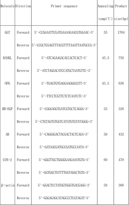
Molecule Direction Primer sequence Annealing
temp(℃) Product size(bp) GGT RANKL OPG HB-EGF AR COX-2 β-actin Forward Reverse Forward Reverse Forward Reverse Forward Reverse Forward Reverse Forward Reverse Forward Reverse 5'-CCGGAATTCGATGAAAAGAACGTGGAAC-3' 5'-CCGCTCGAGTTTACGTTTTAAATTAATGCCG-3' 5'-ATCAGAAGACACCACTCACT-3' 5'-ATCTAGGACATCCATGCTAATGTTC-3' 5'-TGAGTGTGAGGAAGGGCGTT-3' 5'-TTCCTCGTTCTCTCAATCTC-3' 5'-CGGGAGGTGATCGTGCTCAGGG-3' 5'-CTGTAGTGTGGTCATATGTGTATAGGG-3' 5'-CAGGGGACTACGACTACTCAGA-3' 5'-GATAACGATGCCGATGCCAATA-3' 5'-GGGTTGCTGGGGGAAGAAATGTG-3' 5'-GGTGGCTGTTTTGGTAGGCTGTG-3' 5'-GGACTCCTATGGTGGGTGACGAGG-3' 5'-GGGAGAGCATAGCCCTCGTAGAT-3' 55 45.3 45.5 55 59 60 58 1764 750 636 326 432 479 366
Ⅲ
Ⅲ
Ⅲ
Ⅲ.
.
.
. Results
Results
Results
Results
1. 1. 1.
1. OsteoclastOsteoclastOsteoclast-Osteoclast---forming Activity of forming Activity of forming Activity of forming Activity of B. subtilisB. subtilisB. subtilisB. subtilis GGT in conditioned GGT in conditioned GGT in conditioned GGT in conditioned medium
medium medium medium
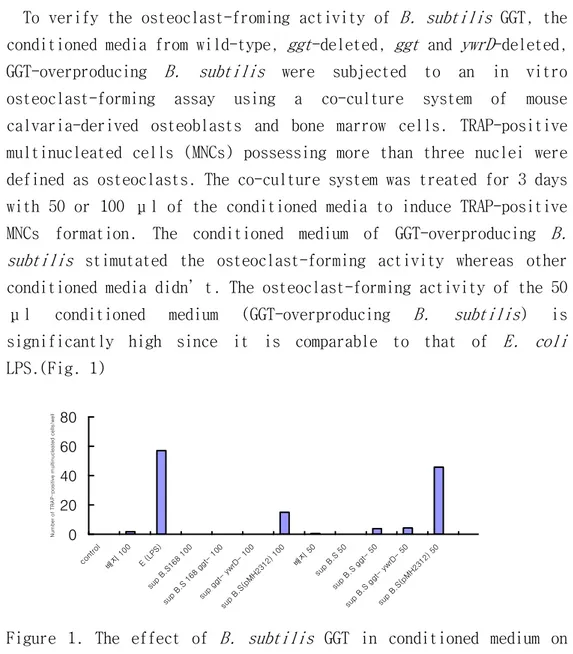
To verify the osteoclast-froming activity of B. subtilis GGT, the conditioned media from wild-type, ggt-deleted, ggt and ywrD-deleted, GGT-overproducing B. subtilis were subjected to an in vitro osteoclast-forming assay using a co-culture system of mouse calvaria-derived osteoblasts and bone marrow cells. TRAP-positive multinucleated cells (MNCs) possessing more than three nuclei were defined as osteoclasts. The co-culture system was treated for 3 days with 50 or 100 μl of the conditioned media to induce TRAP-positive MNCs formation. The conditioned medium of GGT-overproducing B. subtilis stimutated the osteoclast-forming activity whereas other conditioned media didn’t. The osteoclast-forming activity of the 50 μl conditioned medium (GGT-overproducing B. subtilis) is significantly high since it is comparable to that of E. coli
LPS.(Fig. 1)
Figure 1. The effect of B. subtilis GGT in conditioned medium on osteoclast-forming activity. 0 20 40 60 80 cont rol 배지 100 E (L PS) sup B.S1 68 1 00 sup B.S 168 gg 100 sup gg yw rD- 100 sup B.S( pMH2 312) 100 배지 50 sup B.S 50 sup B.S gg 50 sup B.S gg ywr D- 50 sup B.S( pMH2 312) 50 N u m b e r o f T R A P -p o s it iv e m u lt in u c le a te d c e lls /w e ll
Treatment with the 50 μl conditioned medium resulted in better osteoclast-forming activity than treatment with the 100 μl, suggesting that high dose of conditioned medium might be cytotoxic. 2.
2. 2.
2. Purification of Purification of Purification of Purification of B. subtilisB. subtilisB. subtilisB. subtilis GGT GGT GGT GGT
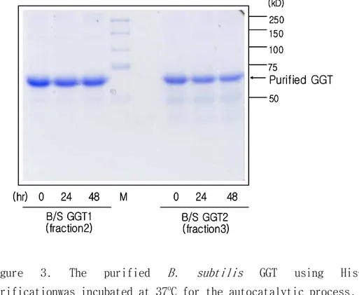
B. subtilis ggt gene was PCR-amplifed from B. subtilis chromosomal DNA and cloned in pET21-b expression vector, generating pET21b/BsGGT. The plasmid, pET21b/BsGGT containing B. subtilis ggt was screened with restriction enzymes, EcoRI and XhoI, and was sequenced to confirm the fidelity of PCR (data not shown). The plasmid was transformed, and expressed in BL21 strain. The over-expressed B. subtilis GGT was found to be in inclusion body (Fig. 2). The inclusion body was collected, denatured in 8 M urea, and purified using His-tag purification. The purified GGT was dialyzed against Tris buffer and the purified GGT was incubated at 37oC for the autocatalytic process.
Figure 2. Overexpression of B. subtilis GGT in E. coli
The autocatalytic process was not observed until 2 days. A single major band of the GGT appeared after the purification (Fig. 3).
Figure 3. The purified B. subtilis GGT using His-Tag purificationwas incubated at 37oC for the autocatalytic process.
3. 3. 3.
3. Effect of Purified GGT on mRNA Expression of RANKL, OPG, HBEffect of Purified GGT on mRNA Expression of RANKL, OPG, HBEffect of Purified GGT on mRNA Expression of RANKL, OPG, HBEffect of Purified GGT on mRNA Expression of RANKL, OPG, HB----EGF, EGF, EGF, EGF, AR, and
AR, and AR, and
AR, and COXCOXCOX-COX---2.2.2.2.
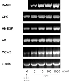
To observe the effect of the bacterial GGT on expression of RANKL, OPG, HB-EGF, AR and COX-2, the mRNA expression was measured using RT-PCR in mouse calvaria-derived osteoblasts treated for 3 hr with the purified GGT at the concentration of 0, 10, 100 and 1000 ng/ml (Figure 4 and Table 3). The RANKL mRNA expression was induced significantly by 2.1, 2.2, and 2.5 fold at the GGT concentration of 10, 100, 1000 ng/ml, respectively. In addition, since COX-2 is known as a factor to induce PGE2, pro-resorptive factor, the COX-2 mRNA
250 150 100 75 50 (kD) B/S GGT1 (hr) 0 24 48 M 0 24 48 B/S GGT2 (fraction3) (fraction2) Purified GGT 250 150 100 75 50 (kD) B/S GGT1 (hr) 0 24 48 M 0 24 48 B/S GGT2 (fraction3) (fraction2) 250 150 100 75 50 (kD) B/S GGT1 (hr) 0 24 48 M 0 24 48 B/S GGT2 (fraction3) (fraction2) Purified GGT
expression was examined and resulted in induction by approximately 1.6 fold. Interestingly, the HB-EGF mRNA expression was induced by 1.7 fold at 10 and 100 ng/ml, and 2.0 fold at 1000 ng/ml as well as AR was also induced slightly by approximately 1.4 fold.
RANKL OPG HB-EGF AR COX-2 β-actin 0 0 10 100 1000 Start point GGT ng/ml
Figure 4. The effect of the purified GGT on m RNA expression of RANKL. OPG, HB-EGF, AR and COX-2 in osteoblasts. The calvaria-derived osteoblasts were incubated with the purified GGT at the concentration of 0, 10, 100, and 1000 ng/ml for 3 hr. Total RNA was extracted and subjected to RT-PCR analysis.
However, in the case of OPG, it was not induced at the concentration of 1000 ng/ml or slightly induced by 1.2 fold at 10 and 100 ng/ml.
Therefore, the mRNA expression of RANKL, HB-EGF, AR and COX-2 were induced by the GGT concentrations of 10, 100, and 1000 ng/ml while
Table 3. The mRNA ratios of RANKL, OPG, HBTable 3. The mRNA ratios of RANKL, OPG, HB-Table 3. The mRNA ratios of RANKL, OPG, HBTable 3. The mRNA ratios of RANKL, OPG, HB---EGF, AR and COXEGF, AR and COXEGF, AR and COX-EGF, AR and COX---2 to 2 to β2 to 2 to ββ-β---actin in actin in actin in actin in the calvaria
the calvaria the calvaria
the calvaria----derived osteoblasts after 3 hr incubation with the purified GGT derived osteoblasts after 3 hr incubation with the purified GGT derived osteoblasts after 3 hr incubation with the purified GGT derived osteoblasts after 3 hr incubation with the purified GGT at the concentration of 10, 100, and 1000 ng/ml.
at the concentration of 10, 100, and 1000 ng/ml. at the concentration of 10, 100, and 1000 ng/ml. at the concentration of 10, 100, and 1000 ng/ml.
GGT concentration RANKL/ β-actin OPG/ β-actin HB-EGF/ β-actin AR/β-actin Cox2/ β-actin 0 ng/ml 1 1 1 1 1 10 ng/ml 2.072 1.232 1.667 1.307 1.535 100 ng/ml 2.211 1.234 1.730 1.390 1.646 1000 ng/ml 2.472 1.057 1.975 1.405 1.576
Ⅳ
Ⅳ
Ⅳ
Ⅳ.
.
.
. Discussion
Discussion
Discussion
Discussion
The present study showed that B. subtilis GGT is able to serve as a bone-resorbing factor through a RANKL-dependent pathway as mammalian GGT does. It is the novel biological activity demonstrated with the prokaryotic GGT. Many previous studies of bacterial GGT have focused on the enzymatic functions. These studies have revealed that GGT catalyzes the first step in the degradation of glutathione and plays an important role in glutathione metabolism (Taniguchi et ai., 1998). In addition, the expression of mammalian GGT is elevated under certain conditions, such as carcinogenesis (Hanigan et al., 1994; Haniganet al., 1999; Taniguchi et al., 1985; Taniguchi et al., 1985) and it is used as a marker enzyme for many diseases. However, other biological activities of significance of GGT have not yet been demonstrated.
This study showed that the conditioned medium from GGT-overproducing B. subtilis induced osteoclast formation in the co-culture system. Fifty μl conditioned medium showed better activity for osteoclast formation than 100 μl broth did, indicating the bacterial broth may have some cytotoxic effect.
To verify the effect of GGT in osteoclast formation activity and to investigate the mechanism how GGT induces osteoclastogenesis, B. subtilis GGT was cloned, expressed in E. coli, and purified using His-tag purification. The purified GGT produced only one protein band, indicating no autocatalytic process occurred. It is possible that T7 tag at the amino terminus and His tag at the carboxyl terminus of GGT may interfere the autocatalytic process. Without the autocatalytic process, enzyme activity of GGT is not active. Because in mammalian GGT the osteocalst formation activity was independent
of its enzymatic activity (Niida et al., 2004), we continue to investigate further with the single polypeptide GGT.
The purified GGT significantly induced by up to 2.5 fold the mRNA expression of the receptor activator of nuclear factor-κB ligand (RANKL). This result suggested that the osteoclastogenesis by the bacterial GGT is mediated by a RANKL-dependent pathway and the bacterial GGT enzymatic activity is not required for the induction of RANKL expression like the mammalian GGT. Unlike RANKL, the expression of OPG, which blocks osteoclastogenesis induced by RANKL, was not affected by the GGT treatment in the osteobalsts, suggesting that the ratio of RANKL to OPG was increased to promote the direction of bone resorption.
It was previously reported that Helicobacter pylori GGT stimulated mRNA expression of COX-2, heparin-binding epidermal growth factor-like growth factor (HB-EGF) and amphiregulin (AR) in human gastric cells (AGS) (Busiello et al., 2004). Thus, it was examined whether B. subtilis GGT induce mRNA expression of COX-2, HB-EGF and AR in mouse calvaria-derived osteoblasts. The GGT stimulated mRNA expression of COX-2 by 1.6 fold, HB-EGF by 2.0 fold and AR by 1.4 fold in the osteoblasts. It has been reported that COX-2 is a transcription factor to induce PGE2, pro-resorptive factor (Fujita et al., 2003). The RANKL-dependent pathway is essential to induce osteoclastogenesis by PGE2 and other pro-resorptive factors such as 1,25(OH3) vitamin D3, TNF-α, IL-1β and IL-6 . Therefore, it can be postulated that PGE2 is a main factor in the induction of RANKL expression in the osteoblasts by the GGT even though the induced PGE2 secretion needs to be examined in the future. It is first report that GGT induced the expression of HB-EGF and AR in the osteoblasts. They could be new player as pro-resorptive factor in osteoclastogenesis. Therefore, it is interesting to investigate new
roles of those growth factors in osteoclastogenesis.
References
References
References
References
Brändström H, T Bjorkman, and ö Ljunggren: Regulation of osteoprotegerin secretion from primary cultures of human bone marrow stromal cells. . . . Biochem Biophys Res Commun 280: 831-835, 2001. Busiello I, Acquaviva R, Popolo AD, Blanchard TG, Ricci V, Romano M, Zarrilli R. Helicobacter pylori γ-glutamyltranspeptidase upregulates COX-2 and EGF-related peptide expression in human gastric cells. Cellular Microbiology 6. 255-267, 2004.
Fujita D, Yamashita N, Amano H, Yamada S, Sakamoto K, Prostaglandin E2 induced the differentiation of osteoclasts in mouse osteoblast- depleted bone marrow cells. Prostaglandins, Leukotriens and Essential Fatty acids. 68: 351-358, 2003
Hanigan MH, Frierson HF, Brown JE, Lovell MA, and Taylor PT. Cancer Res. 54: 286-290, 1994
Hanigan MH, Frierson HF, Jr, Swanson PE, and De-Young BR. Hum. Pathol. 30: 300-305,1999
Hofbauer LC, CR Dunstan, TC Spelsberg, BL Riggs, and S. Khosla: Osteoprotegerin production by human osteoblast lineage cells is stimulated by vitamin D, bone morphogenetic protein-2, and cytokines. Biochem Biophys Res Commun 250: 776-781, 1998.
Hofbauer LC, Lacey DL, Dunstan CR, Spelsberg TC, Riggs BL, Khosla S. Interleukin-1β and Tumor necrosis factor-α , but not interleudin-6,
stimulate osteoprotegerin ligand gene expression in human osteoblastic cells. Bone 25:255-259, 1999.
Hofbauer LC, Khosla S, Dunstan CR, Lacey DL, Boyle WJ, Riggs BL. The roles of osteoprotegerin and osteoprotegerin ligand in the paracrine regulation of bone resorption. J. Bone Miner. Res. 15:2-12. 2000.
Hsu H, Lacey DL, Dunstan CR, Solovyev I, Colombero A, Timms E, Tan HS, Elliott G, Kelley MJ, Sarosi I, Wang L, Xia XZ, Elliott R, Chiu L, Black T, Scully S, Capparelli C, Morony S, Shimamoto G, Bass MB, Boyle WJ. Tumor necrosis factor receptor family member RANK mediates osteoclast differentiation and activation induced by osteoprotegerin ligand. Proc. Natl. Acad. Sci. USA. 96:3540-3545, 1999.
Kunst F, et al. The complete genome sequence of the gram-positive bacterium Bacillus subtillis. Nature 390:249-56,1997.
Lacey, DL, E Timms, H-L Tan, MJ Kelley, CR Dunstan, T Burgess, R Elliott, A Colombero, G Elliott, S Scully, H Hsu, J Sullivan, N Hawkins, E Davy, C Capparelli, A Eli, Y-X QIAN, s Kaufman, I Sarosi, V Shalhoub, G Senaldi, J Guo, J Kelaney, and WJ Boyle: Osteoprotegerin (OPG) ligand is a cytokine that regulates osteoclast differentiation and activation. Cell 93: 165-176, 1998.
Lieberman, M.W., Barrios, R., Carter, B.Z., Habib, G. M., Lebovitz, R. M., Rafagopalan, S., Sepulveda, A. R., Shi, Z.Z., and Wan, D. F. Am. J. Pathol. 147, 1175-1185,1995.
Nagai M, and N Sato: Reciprocal gene expression of osteoclastogenesis inhibitory factor and osteoclast differentiation factor regulates osteoclast formation. Biochem Biophys Res Commun 257: 719-723, 1999.
Niida S, Kawahara M, Ishizuka Y, Ikeda Y, Kondo T, Hibi T, Suzuki Y, Ikeda K, and Taniguchi N. γ-Glutamyltranspeptidase Stimulates Receptor Activator of Nuclear Factor-κB Ligand Expresssion Independent of Its Enzymatic Activity and Serves as a Pathological Bone-Resorbing Factor. The Journal of Biological Chemistry. 5752-5756, 2004.
Ogawa Y, Hosoyama H, Hamano M, Motai H. Purification and properties of γ-glutamyltranspeptidase from Bacillus subtilis (natto), Agric Biol Chem 55:2971-7,1991.
Ogawa Y, Sugiura D, Motai H, Yuasa K, Tahara Y. DNA sequence of
Bacillus subtilis (natto) NR-1 γ-glutamyltranspeptidase gene, ggt. Biosci Biotech Biochem 61:1621-5, 1997.
Simonet WS, DL Lacey, CR Dunstan, M Kelley, M-S Chang, R Lthy, HQ Nguyen, S Wooden, L Bennett, T Boone, G Shimamoto, M DeRose, R Elliott, A Colombero, H-L Tan, G Trail, J Sullivan, E Davy, NS Sander, G Van, J Tarpley, P Derby, R Lee, EST Program Amgen, and WJ Boyle: Osteoprotegerin: a novel secreted protein involved in the regulation of bone density. Cell 89: 309-319, 1997.
Suda T, Jimi E, Nakamura I, Takahashi N. Role of 1α, 25-dihydroxyvitamjin D3 in osteoclast differentiation and function. Methods Enzymol. 282:223-235, 1997.
Suzuki H, Kumagai H, Tochidura T. γ–Glutamyltranspeptidase from
Escherichia coli K-12: formation and localization. J Bacteriol 168:1332-5, 2002.
Takahashi N, Udagawa N, Suda T. A new member of tumor necrosis factor ligand family, ODF/OPGL/TRANCE/RANKL, regulates osteoclast differentiation and function. Biochem. Biophys. Res. Commun. 256: 449-455, 1999.
Taniguchi N, House S, Kuzumaki N, Yokosawa N, Yamagiwa S, Iizuka S, Makita A, and Sekiya C. J. Natl. Cancer Inst. 75: 841-847, 1985 Taniguchi N, Iizuka S, Zhe ZN, House S, Yokosawa N, Ono M, Kinoshita K, Makita A, and Sekiya C. Cancer Res. 45: 5835-5839
Taniguchi N, and Ikeda Y. Adv.Enzymol. Relat. Areas Mol. Biol. 72:239-278, 1998
Tate, S. S., and A. Meister. γ–Glutamyltranspeptidase: catalytic, structural and functional aspects. Mol. Cell. Biochem. 39:357-368,1981.
Tsukii K, Shima N, Mochizuki S, Yamaguchi K, Kinosaki M, Yano K, Shibata O, Udagawa N, Yasuda H, Suda T, Higashio K. Osteoclast differentiation factor mediated an essential signal for bone resorption induced by 1α, 25-dihydroxyvitamin D3, prostaglandin E2, of parathyroid hormone in the microenviroment of bone. Biochem. Biophys. Res. Commun. 246:337-341, 1998
Udagawa N, N Tadahashi, H Yasuda, A Mizuno, K Itoh, Y Ueno, TShinki, MT Gillespie, TJ Martin, K Higashio, and T Suda: Osteoprotegerin produced by osteoblasts is an important regulator in osteoclast development and function. Endocrinology 141: 3478-3484, 2000. Vidal, NO, K Sfogren, BI Eriksson, O Ljunggren, and C Ohlsson: Osteoprotegerin mRNA is increased by interleukin 1-a in the human osteosarcoma cell line MG-63 and in the human osteoblast-like cells. Biochem Biophys Res Com 248:696-700, 1998.
Walsh MC, Choi Y. Biology of the TRANCE axis. Cytokine Growth Factor Rev. 14: 251-263, 2003.
Yasuda H, N Shima, N Nakagawa, S-I Mochizuki, K Yano, N Fujise, Y Sato, M GOto, K Yamaguchi, M Kuriyama, T Kanno, A Murakami, E Tsuda, T Morinaga, and K Higashio: Identity of osteoclastogenesis inhibitory factor (OCIF) and osteoprotegerin (OPG): a mechanism by which OPG/OCIF inhibits osteoclastogenesis in vitro. Endocrinology 139: 1329-1337, 1998.
Yasuda H, Shima N, Nakagawa N. Yamaguchi K, Kinosaki M, Mochizuki S, Tomoyasu A, Yano K, Goto M, Murakami A, Tsuda E, Morinaga T, Higashio K, Udagawa n, Takahashi N, Suda T. Osteoclast
differentiation factor is a ligand for
osteoprotegerin/osteoclastogenesis-inhibitory factor and is identy to TRANCE/RANKL. Proc. Natl. Acad. Sci. USA. 95: 3597-3602, 1998.
국문국문국문국문 요약요약요약요약
파골세포
파골세포
파골세포
파골세포
형성에
형성에
형성에
형성에
미치는
미치는
미치는
미치는
γ
γ
γ
γ–––– glutamyltranspeptidase
glutamyltranspeptidase
glutamyltranspeptidase의
glutamyltranspeptidase
의
의
의
효과
효과
효과
효과
김 봉 주
연세대학교 대학원 치의학과
지도교수 차 정 헌
γ-glutamyltranspeptidase (GGT)는 새로운 골흡수 인자로서 최근에 쥐 T-lymphoma 세포의 cDNA library에서 클로닝되었다. GGT는 모든 생명체에
광범위하게 존재하며 특히 Bacillus subtilis GGT는 구조나 효소적 특성
이 포유동물의 GGT와 매우 흡사해 B. subtilis GGT를 가지고 파골세포형
성능을 실험하였다. 파골세포형성능은 두개골과 골수세포를 사용한 혼합 배양을 이용하였고 tartrate-resistant acid phosphatase 염색으로 평가 하였다. GGT 를 과발현하는 B. subtilis 배양액에서는 wild type이나 ggt-deleted B. subtilis 배양액보다 현저한 파골세포형성능이 나타났다.
GGT가 어떤 기전에 의해 파골세포 분화에 관여하는지 알아보기 위해 B.
subtilis GGT를 Escherichia coli에 클로닝하여 발현시키고 His-tag 정제 방법으로 다량의 정제된 GGT를 취하였다. 정제된 GGT를 쥐 두개골의 조골 세포에 처리한 결과 receptor activator of nuclear factor-κB ligand, cyclooxygenase-2, heparin-binding epidermal growth factor-like growth factor, amphiregulin의 mRNA 발현이 증가된 반면, OPG의 발현에 는 거의 영향을 미치지 않았다. 이상의 결과는 세균의 GGT도 RANKL과 관 련된 경로를 통해 골흡수와 파골세포 분화에 관여함을 나타낸다.
핵심단어 : γ-glutamyltranspeptidase, 파골세포형성능, Bacillus subtilis, RANKL, COX-2, HB-EGF, amphiregulin