저작자표시-비영리-변경금지 2.0 대한민국 이용자는 아래의 조건을 따르는 경우에 한하여 자유롭게 l 이 저작물을 복제, 배포, 전송, 전시, 공연 및 방송할 수 있습니다. 다음과 같은 조건을 따라야 합니다: l 귀하는, 이 저작물의 재이용이나 배포의 경우, 이 저작물에 적용된 이용허락조건 을 명확하게 나타내어야 합니다. l 저작권자로부터 별도의 허가를 받으면 이러한 조건들은 적용되지 않습니다. 저작권법에 따른 이용자의 권리는 위의 내용에 의하여 영향을 받지 않습니다. 이것은 이용허락규약(Legal Code)을 이해하기 쉽게 요약한 것입니다. Disclaimer 저작자표시. 귀하는 원저작자를 표시하여야 합니다. 비영리. 귀하는 이 저작물을 영리 목적으로 이용할 수 없습니다. 변경금지. 귀하는 이 저작물을 개작, 변형 또는 가공할 수 없습니다.
THESIS
FOR THE DEGREE OF MASTER OF SCIENCE
Protective Effect of Eckol against
Oxidative Stress Damaged
Mitochondria
Areum daseul Kim
Marine Life Science
GRADUATE SCHOOL
JEJU NATIONAL UNIVERSITY
1
CONTENTS
ABSTRACT... 4 1. INTRODUCTION... 6 2. MATERIALS AND METHODS... 8
2.1. Reagents 2.2. Cell culture
2.3. Mitochondrial ROS Measurement 2.4. Mitochondrial Ca2+ Measurements 2.5. Quantification of Cellular ATP Levels
2.6. Mitochondrial Succinate dehydrogenase activity 2.7. Western blot
2.8. Reverse transcriptase polymerase chain reaction 2.9. Measurement of Mn SOD Activity
2.10. Immunocytochemistry
2.11. Transient Transfection of Small RNA Interference (siRNA) 2.12. Cell viability assay
2.13. Statistical analysis
3. RESULTS... 15 3.1. Reduction of mitochondrial ROS generated by oxidative stress in eckol treated cells 3.2. The effect of eckol on intracellular Ca2+
3.3. Effect of eckol on intracellular ATP levels and succinate dehydrogenase activity 3.4. Effect of eckol on Mn SOD Enzyme
3.5. Eckol mediated on AMPK/FOXO3a signaling pathway 3.6. Eckol activated Mn SOD via of AMPK/FOXO3a
2
3.7. Involvement of Mn SOD in cell damage induced by oxidative stress
4. DISCUSSION... 34 5. REFERENCES... 38 6. ABSTRACT IN KOREAN ...46
3
LIST OF FIGURES
Fig. 1. Chemical structure of eckol...16
Fig. 2. The effect of eckol on H2O2-induced mitochondrial ROS generation...17
Fig. 3. The effect of eckol on mitochondrial Ca2+ levels………... 20
Fig. 4. The effects of eckol on intracellular ATP levels and succinate dehydrogenase activity... 22
Fig. 5. Effect of eckol on Mn SOD mRNA expression, protein expression, and enzyme activity... 24
Fig. 6. Eckol mediated on AMPK/Foxo3a signaling pathway and Foxo3a translocalization into nucleus...27
Fig. 7. Induction of Mn SOD by eckol via AMPK/FoxO3a………..…... 31
Fig. 8. The cytoprotective effect of eckol against H2O2-induced cell death…... 33
Fig.9. A proposed pathway for eckol-induced Mn SOD via up-regulation of AMPK/FoxO3a, explaining the cytoprotective effect against oxidative stress in Chang liver cells……….,………...………… 37
4
ABSTRACT
We have investigated the cytoprotective effect of eckol, which was isolated from Ecklonia
cava, a phlorotannin found in E. cava on hydrogen peroxide (H2O2)-induced mitochondrial dysfunction. When H2O2 was used to induce an increase in mitochondrial reactive oxygen species (ROS) in Chang liver cells, eckol treatment decreased high level of ROS. Eckol also attenuated intracellular Ca2+ levels that have been induced by H
2O2. Furthermore, eckol recovered ATP levels and succinate dehydrogenase activity that had been decreased by H2O2 treatment. We conclude these results suggest eckol decreased mitochondrial ROS accumulation, balanced intracellular Ca2+ levels, and improved mitochondrial energy production, thus recovering mitochondrial function. Manganese superoxide dismutase (Mn SOD) is an important antioxidant enzyme that plays a role in cytoprotection against oxidative stress induced mitochondria cell damage. Eckol recovered Mn SOD expression both at the level of mRNA and protein in Chang liver cells, resulting in increased Mn SOD activity. Eckol is able upregulate the AMP-activated protein kinase (AMPK)-mediated phosphorylation of Forkhead box O3a (FOXO3a). Eckol enhanced the level of nuclear translocation and activity of Mn SOD which was decreased by H2O2. Specific FOXO3a siRNA and AMPK siRNA attenuated Mn SOD expression, while eckol recovered it. Eckol treatment enhanced the Mn SOD protein expression which was knockdown by Mn SOD siRNA DEDTC (a Mn SOD inhibitor) and Mn SOD siRNA markedly abolished the cytoprotective effect of eckol against H2O2-induced cell damage. These studies demonstrate that eckol attenuates mitochondrial oxidative stress by activating AMPK/FOXO3a-mediated Mn SOD induction.
5
Keywords eckol · Manganese superoxide dismutase · Forkhead box O3a, AMP-activated
6
1. INTRODUCTION
Oxidative stress resulting from the increase of reactive oxygen species (ROS) such as superoxide, hydrogen peroxide (H2O2), and hydroxyl radical are produced as by-products of normal cellular metabolism. ROS can cause irreversible cellular injury and dysfunction by directly oxidizing and damaging DNA, protein, and lipids that has been associated with the underlying pathogenesis of a wide variety of diseases and also with degenerative processes associated with aging (1, 2). High concentrations of ROS can alter the balance of endogenous protective systems and attack mitochondria. Calcium ions and other apoptosis-related factors may be released from damaged mitochondria into the cytosol following dysfunction of the mitochondrial membrane that regulates cell apoptosis (3). Mitochondrial dysfunction has been correlated with diabetes mellitus (4), nonalcoholic fatty liver disease (5), and neurodegenerative diseases like Alzheimer’s disease (6, 7). Therefore, it is essential the cell have protective mechanisms that defend against damage caused by ROS. Manganese superoxide dismutase (Mn SOD), as the primary antioxidant enzyme for scavenging superoxide radicals in mitochondria, is essential for the survival of all aerobic organisms (8). Dysregulation of Mn SOD in the form of under-expression is being investigated relative to various disease including myriad neurological disorders (9) and cancer (10). Overexpression of Mn SOD has been shown to protect against oxidative stress induced cell death and tissue injury (11). The protein products of these genes that are selectively regulated through the hibernation cycle. The FOXO (forkhead box O) family of Forkhead transcription factors (FOXO1, FOXO3, F FOXO4, and FOXO6a) plays an important role in the regulation of crucial cellular processes. Recent studies have identified a number of FOXO-regulated genes involved in a variety of cellular stress responses, ranging from defence against oxidative and caloric stresses to DNA repair (12-15). In particular,
7
FOXO3a responds to cellular stress (including to oxidative stress) by inducing cell cycle arrest, repair of damaged DNA, and apoptosis via upregulation of genes that control these processes (16-20). The activation of FOXO3a is mediated by up-regulation of the transcription of the ROS scavenging enzymes superoxide dismutase 2 (SOD2, also known as Mn SOD) and catalase (21, 22).
FOXO3a transcription factors are good candidates to be regulated by AMPK, which is an important signaling pathway involved in ROS regulation. AMPK (AMP-activated protein kinase) plays a critical role in cellular responses to low energy levels by switching off pathways that consume energy and switching on those that produce it (23). AMPK is known to be activated under conditions that deplete cellular ATP and elevate AMP levels, such as glucose deprivation, hypoxia, ischaemia and heat shock (24–26). As a further mechanism of regulation, it has been recently shown that the activation of the energy sensor pathway, which is triggered by an increased AMP/ATP ratio, leads to the AMPK-mediated phosphorylation of FOXO3a (27). Besides, AMPK is also required for FOXO3a-dependent transcription of thioredoxin during oxidative stress response (28).
Therefore, we hypothesis eckol alternative pathway of AMPK activation, triggered by mitochondrial ROS and involving FOXO3a and its target genes (SOD2, catalase, PGC1α) (29). Eckol is a trimeric phloroglucinol with a dibenzeno-1, 4-dioxin skeleton, and is one of the major phlorotannins isolated from Ecklonia cava. E. cava is a brown alga (Laminariaceae) that is abundant in the subtidal regions of Jeju island, Korea. It has been reported that the Ecklonia species exhibits radical scavenging activity (30-32), cytoprotective properties against oxidative stress (33-37), and heme oxygenase-1 inducing activity (38). However, the mechanisms of eckol involved are not completely understood. Therefore, the present study was undertaken to investigate whether is upregulation of Mn SOD in Chang liver cell mediated by AMPK/FOXO3a dependent pathway.
8
2. MATERIALS AND METHODS
2.1. Reagents
Eckol (Fig. 1A) was provided by professor Nam Ho Lee of Jeju National University (Jeju, Korea). The purity of eckol was assessed by HPLC and was > 90%. Eckol was freshly dissolved in dimethyl sulfoxide (DMSO), The 2',7'-dichlorodihydrofluorescein diacetate (DCF-DA) and [3-(4,5-dimethylthiazol-2-yl)-2,5-diphenyltetrazolium] bromide (MTT) were purchased from Sigma Chemical Company (St. Louis, MO, USA). Dihydrorhodamin 123 (DHR 123) and Rhod-2 acetoxymethyl ester (rhod-2AM) were purchased from Molecular Probes (Eugene, OR, USA). Compound C was purchased from Calbiochem Co. (San Diego, CA, USA) and MnSOD inhibitor DEDTC from Sigma Chemical Company (St. Louis, MO, USA). Phospho-acetyl-CoA-carboxylase (ACC) (Ser79), phospho-AMPKα (Thr172), phospho- Forkhead boxO3a (FOXO3a) and b-actin antibodies were purchased from Cell Signaling Technology (Beverly, MA, USA). The other chemicals and reagents were of analytical grade.
2.2. Cell culture
The human hepatocyte-derived cell line termed Chang liver cells were obtained from the American type culture collection (Rockville, MD, USA), and the cells were maintained at 37 °C in an incubator with a humidified atmosphere of 5% CO2 in air, and cultured in RPMI 1640, containing 0.1 mM non-essential amino acids 10% heat-inactivated fetal calf serum, streptomycin (100 μg/ml) and penicillin (100 units/ml).
9
2.3. Mitochondrial ROS Measurement
The Chang liver cells were seed on to a coverslip loaded six well plate at 1´105 cells/well. At 16h after plating, cells were treated with eckol at 10 μg/ml and 1hr later, 600 μM H2O2 was added to the plate. After changing the media, 20 μM of DHR 123 was added to each well and the plate was incubated for an additional 30 min at 37 °C. After washing with PBS, the stained cells were mounted onto a microscope slide in mounting medium (DAKO, Carpinteria, CA). Images were collected using the Laser Scanning Microscope 5 PASCAL program (Carl Zeiss, Jena, Germany) on a confocal microscope. The level of mitochondrial ROS was also detected by flow cytometry. Cells were loaded for 30 min at 37 °C with 20 μM DHR 123 at indicated time and supernatant was removed by suction and after trypsin treatment, cells were washed with PBS. Fluorescence of DHR 123 loaded cells was measured using a flow cytometer. In addition, cells were seeded in a 96 well plate at a concentration of 1´105 cells/ml, and 16 h after plating, were treated with 10 μg/ml eckol and 1hr later, 600 μM H2O2 was added to the plate. Cells were incubated for an additional 30 min at 37 °C. After addition of 20 μM of DHR 123 solution for 10 min, the fluorescence was detected using a Perkin Elmer LS-5B spectrofluorometer.
2.4. Mitochondrial Ca2+ Measurements
Rhod-2 acetoxymethyl ester (Rhod-2 AM) was used to measure mitochondrial Ca2+. This dye has a net positive charge, which facilitates its sequestration into mitochondria due to membrane potential-driven uptake (39). Fluorescence intensity after labeling was measured by using both flow cytometry and confocal microscopy. Cells were seed on to a coverslip loaded six well plate at 1´105 cells/well. At 16h after plating, cells were treated with eckol at 10 μg/ml and 30 min later, 600 μM H2O2 was added, and the mixture was incubated for 24 hours. After 1 μM of Rhod-2 AM was added to each well and the plate was incubated for an
10
additional 15 min at 37 °C. After washing with PBS, the stained cells were mounted onto a microscope slide in mounting medium (DAKO, Carpinteria, CA). Images were collected using the Laser Scanning Microscope 5 PASCAL program (Carl Zeiss, Jena, Germany) on a confocal microscope. For flow cytometry analyses, cells were seeded in six well plate at 1´105 cells/well and were treated with eckol at 10 μg/ml. After 30 min, 600 μM H
2O2 was added, and the mixture was incubated for 24 hours. Cells were harvested, washed, and suspended in PBS containing Rhod-2 AM (1 μM). After 15 minutes of incubation at 37◦C, the cells were washed, suspended in PBS.
2.5. Quantification of Cellular ATP Levels
The mitochondrial function was evaluated by measuring the cellular adenosine triphosphate (ATP) production in cells. Cells were harvested and washed twice with PBS. The harvested cells were then lysed on ice for 30 min in 200 ml of lysis buffer [25 mM Tris (pH 7.8), 270 mM sucrose, 1 mM EDTA] by sonicating three times for 15 s and centrifuged at 4 °C for 10 min at 16,000 ´ g. Supernatants were collected from the lysates and ATP content was assayed using a luciferase/luciferin ATP determination kit (Molecular Probes, Eugene, OR) (40).
2.6. Mitochondrial Succinate dehydrogenase activity
Mitochondrial function was estimated by the MTT [3-(4,5-dimethylthiazol-2-yl)-2,5-diphenyltetrazolium bromide] assay (41) customized for the assessment of mitochondrial succinate dehydrogenase (SDH) activity (42, 43). Cells were seeded in a 96 well plate at a con-centration of 1´105 cells/ml, and 16 h after plating, were treated with eckol at 10 μg/ml and 30 min later, 600 μM H2O2 was added to the plate and incubated for an additional 24 h at 37 °C. Fifty microliter of MTT stock solution (2 mg/ml) was then added to each well of a
11
total reaction volume of 200 ml. After incubating for 4 h, the plate was centrifuged at 800 ×g for 5 min and the supernatants aspirated. The formazan crystals in each well were dissolved in 150 ml DMSO and the A540 read on a scanning multi-well spectrophotometer (44).
2.7. Western blot
The harvested cells were then lysed on ice for 30 min in 100 μl of a lysis buffer [120 mM NaCl, 40 mM Tris (pH 8), 0.1% NP 40] and centrifuged at 13,000 ×g for 15 min. The supernatants were collected from the lysates and the protein concentrations were determined. Aliquots of the lysates (40 μg of protein) were boiled for 5 min and electrophoresed in 10% SDS-polyacrylamide gel. The blots in the gels were transferred onto nitrocellulose membranes (Bio-Rad, Hercules, CA, USA), and subsequently incubated with anti-primary antibodies. The membranes were further incubated with secondary antiimmunoglobulin-G-horseradish peroxidase conjugates (Pierce, Rockford, IL, USA), followed by exposure to X-ray film. The protein bands were detected using an enhanced chemiluminescence western blotting detection kit (Amersham, Little Chalfont, Buckinghamshire, UK).
2.8. Reverse transcriptase polymerase chain reaction
Total RNA was isolated from cells using Trizol (GibcoBRL, Grand Island, NY, USA). Reverse transcriptase polymerase chain reaction (RT-PCR) was performed as described previously (36). PCR conditions for MnSOD and for the housekeeping gene, GAPDH, were as follows: 30 cycles of 94 °C for 15 sec; 60 °C for 30 sec; and 68 °C for 60 sec. The primer pairs (Bionics, Seoul, Korea) were follows (forward and reverse, respectively): Mn SOD, 5'-GACCTGCCTTACGACTATGG-3' and 5'-GACCTTGCTCCTTATTGAAG-3', 600bp; and GAPDH, 5'-GTGGGCCGCCCTAGGCACCAGG-3'; and 5'-GGAGGAAGAGGATGCGGCAGTG-3', 1054bp. Amplified products were resolved on 1%
12
agarose gels, stained with ethidium bromide, and photographed under ultraviolet light.
2.9. Measurement of Mn SOD Activity
The Chang liver cells were seeded in a culture dish at a concentration of 1´105 cells/ml, and at 16 h after plating were treated with eckol at 10 μg/ml. After 1 h, 600 μM H2O2 was added to the plate, which was incubated more for a further 24h. The cells were then washed with cold PBS and scraped. The harvested cells were suspended in 10 mM phosphate buffer (pH 7.5) and then lysed on ice by sonicating twice for 15 s. Triton X-100 (1%) was then added to the lysates and incubated for 10 min on ice. The lysates were clarified, by centrifugation at 5000 × g for 10 min at 4°C, to remove cellular debris. The protein content of the supernatant was determined by the Bradford method, using bovine serum albumin (BSA) as the standard. The Mn SOD activity was used to detect the level of epinephrine auto-oxidation inhibition (45,46). Fifty micrograms of protein was added to 500 mM phosphate buffer (pH 10.2), 1 mM potassium cyanide (inhibitor of Cu Zn SOD), and 1 mM epinephrine. Epinephrine rapidly undergoes auto-oxidation at pH 10 to produce adrenochrome, a pink-colored product, which was assayed at 480 nm sing an ultraviolet/visible (UV/VIS) spectrophotometer in the kinetic mode. Mn SOD inhibits the auto-oxidation of epinephrine. The rate of inhibition was monitored at 480 nm and the amount of enzyme required to produce 50% inhibition was defined as 1 unit of enzyme activity. The Mn SOD activity was expressed as units per milligram protein.
2.10. Immunocytochemistry
Cells plated on coverslips were fixed with 4% paraformaldehyde for 30 min and permeabilized with 0.1% Triton X-100 in PBS for 2.5 min. Cells were treated with blocking medium (3% bovine serum albumin in PBS) for 1 h and incubated with phospho FOXO3a
13
antibody diluted in blocking medium for 2 h. Immuno-reacted primary phospho FOXO3a antibody was detected by a 1:500 dilution of FITC-conjugated secondary antibody (Jackson ImmunoResearch Laboratories, West Grove, PA, USA) for 1 h. After washing with PBS, stained cells were mounted onto microscope slides in mounting medium with DAPI (Vector, Burlingame, CA, USA). Images were collected using the LSM 510 program on a Zeiss confocal microscope.
2.11. Transient Transfection of Small RNA Interference (siRNA)
Cells were seeded at 1.5 × 10⁵ cells/well in 24 well plate and allowed to reach approximately 50% confluence on the day of transfection. The siRNA construct used was obtained as mismatched siRNA control (siControl, Santa Cruz Biotechnology, Santa Cruz, CA), siRNA against FOXO3a (siFOXO3a), siRNA against AMPK (siAMPK) and siRNA against Mn SOD (siMn SOD) were purchased from Bioneer Corporation, Bioneer, South Korea. Cells were transfected with 10–50nM siRNA using lipofectamineTM 2000 (Invitrogen, Carlsbad, CA) according to the manufacturer’s instruction. At 24 h after transfection, the cells were treated with 10 mg/ml of eckol for 24 h and examined by either Western blot analysis or MTT assay.
2.12. Cell viability assay
The effect of eckol on the viability of the Chang liver cells was determined by the MTT assay, which is based on the reduction of a tetrazolium salt by mitochondrial dehydrogenase in the viable cells [44]. The cells were seeded in a 96 well plate at a density of 1 ´ 105 cells/ml and treated with 10 μg/ml eckol, 10 μM DEDTC (inhibitor of Mn SOD) followed 2 h later by 600 μM H2O2. After incubated for 24 h at 37 °C, 50 ml of the MTT stock solution (2 mg/ml) was then added to each well to attain a total reaction volume of 250 ml. After
14
incubation for 2.5 h, the supernatants were aspirated. The formazan crystals in each well were dissolved in 150 ml DMSO, and the ABS540 was read on a scanning multi-well spectrophotometer.
2.13. Statistical analysis
All measurements were made in triplicate and all values are represented as means ± standard error. Data were analyzed with analysis of variance (ANOVA) using the Tukey test. p <0.05 was considered significant.
15
3. RESULTS
3.1. Reduction of mitochondrial ROS generated by oxidative stress in eckol treated cells
There were different levels in terms of mitochondrial ROS generation between cell respiration (control) and H2O2-induced treatment as shown in Fig. 2, which was detected by means of DHR 123 fluorescence dye. Analysis of confocal microscope revealed that eckol at 10 μg/ml reduced the red fluorescence intensity of H2O2-induced mitochondrial ROS as shown in Fig. 2A. In addition, the level of mitochondrial ROS detected by flowcytometry revealed a fluorescence intensity value of 162 in H2O2-treated cells with eckol at 10 μg/ml, compared to a fluorescence intensity value of 201 in H2O2-treated cells (Fig. 2B), thus reflecting a reduction in ROS generation. The fluorescence spectrometric data revealed that H2O2 treatment increased the level of mitochondrial ROS compared to control. However, eckol at 10 μg/ml treatment attenuated the H2O2-induced ROS increase (Fig. 2C). These data suggest that eckol is a scavenger of mitochondrial ROS.
16
17
Fig.2. The effect of eckol on H2O2-induced mitochondrial ROS generation. (A) Cells were treated with eckol at 10 μg/ml. After 1hr, 600 μM H2O2 was added to the plate. After changing the media, 20 μM of DHR 123 was added to each well and the plate was incubated for an additional 30 min at 37 °C. The representative confocal images illustrate the increase
18
in red fluorescence intensity of DHR 123 produced by ROS in H2O2 -treated cells as compared to the control and the lowered fluorescence intensity in H2O2 -treated cells with eckol (original magnification ´ 400). The mitochondrial ROS generated was detected by flow cytometry (B), and spectrofluorometry (C) after the DHR 123 treatment. FI indicates the fluorescence intensity of DHR 123. The measurements were made in triplicate and the values were expressed as means ± SEM. *Significantly different from control cells (p < 0.05), **significantly different from H2O2-treated cells (p < 0.05).
19 3.2. The effect of eckol on intracellular Ca2+
ROS can induce Ca2+ release from mitochondria and result in an increase of cytosolic Ca2+ levels. Analysis of confocal microscope revealed that eckol at 10 μg/ml reduced the red fluorescence intensity of H2O2-induced mitochondrial ROS as shown in Fig. 3A. As shown in Fig 3B, H2O2-treated cells with eckol revealed a fluorescence intensity value of 179, as compared to a fluorescence intensity value of 270 in H2O2-treated cells. These data suggest that eckol reduces intracellular Ca2+ level induced by H
20
Fig.3. The effect of eckol on mitochondrial Ca2+ levels. Cells were treated with eckol at 10 μg/ml. After 1hr, 600 μM H2O2 was added to the plate. After changing the media, 1 μM of Rhod-2 AM was added to each well and the plate was incubated for an additional 30 min at 37 °C. The representative confocal images illustrate the increase in red fluorescence intensity of Rhod-2 AM produced by mitochondrial Ca2+ levels in H
2O2 -treated cells as compared to the control and the lowered fluorescence intensity in H2O2 -treated cells with eckol (original magnification ´ 400). Then cells were harvested and Ca2+ levels were detected by flow cytometry (B). FI indicates the fluorescence intensity of Rhod-2 AM. *Significantly different from control cells (p < 0.05), **significantly different from H2O2-treated cells (p < 0.05).
21
3.3. Effect of eckol on intracellular ATP levels and succinate dehydrogenase activity
Mitochondrial injury is followed by the depletion of intracellular ATP levels. As shown in Fig. 4A, H2O2 treatment reduced the ATP levels as compared to control, however, H2O2 -treated cells with eckol treatment recovered the ATP levels, suggesting eckol has a protective effect against H2O2-induced loss of intracellular ATP levels. These data suggest that eckol restores mitochondrial function by preventing the loss of mitochondrial membrane integrity. Succinate dehydrogenase is an enzyme existing in the mitochondrial respiratory chain, and its activity was assessed by MTT assay. As shown in Fig. 4B, a combination of eckol at 10 μg/ml and H2O2 enhanced succinate dehydrogenase activity to 74%, as compared to 55% in H2O2-treated cells. Taken together, these results indicate eckol recovers mitochondrial function damaged by H2O2 treatment through preventing loss of ATP levels and of succinate dehydrogenase activity.
22
Fig.4. The effects of eckol on intracellular ATP levels and succinate dehydrogenase activity.
(A) ATP content was assayed using a luciferase/luciferin ATP determination kit. *Significantly different from control cells (p < 0.05), **significantly different from H2O2 -treated cells (p < 0.05). (B) Succinate dehydrogenase activity was detected by MTT assay. *Significantly different from control cells (p < 0.05), **significantly different from H2O2 -treated cells (p < 0.05).
23 3.4. Effect of eckol on Mn SOD Enzyme
Mn SOD acts as a first defense system to protect mitochondria and other cellular components by scavenging superoxide anions in the mitochondria matrix (47). Treatment of Chang liver cells with eckol induced a time-dependent manner enhancement in Mn SOD protein expression (Fig. 5A). Also the increased Mn SOD expression correlated with Mn SOD mRNA levels in time-dependent fashions in eckol-treated cells (Fig. 5B). Western blot data revealed that H2O2 treatment decreased the expression of Mn SOD. However, eckol treatment elevated the Mn SOD protein levels attenuated by H2O2 treatment (Fig. 5C). A significant increase in Mn SOD activity was observed by eckol treatment in H2O2-treated cells (Fig. 5D). This result suggests that eckol the increase in MnSOD activity.
25
Fig.5. Effect of eckol on Mn SOD mRNA expression, protein expression, and enzyme activity. (A) Cell lysates treated by a variety of time courses of eckol at 10 μg/ml was electrophoresed and the expression of Mn SOD protein was detected by their respective specific antibodies. (B) Cells were treated with eckol, total RNA was extracted, and Mn SOD mRNA expression was analyzed by RT-PCR. The GAPDH band is shown to confirm integrity and equal loading of RNA. (C) The Chang liver cells were treated with eckol at 10 μg/ml, followed by H2O2-treated. Next, the cells were incubated for 24 h. Cell lysates was electrophoresed, Mn SOD were detected by their respective specific antibodies. (D) The enzyme activities are expressed as average enzyme unit per mg protein ± SE. **significantly different from H2O2-treated cells (p < 0.05).
26
3.5. Eckol mediated on AMPK/FOXO3a signaling pathway
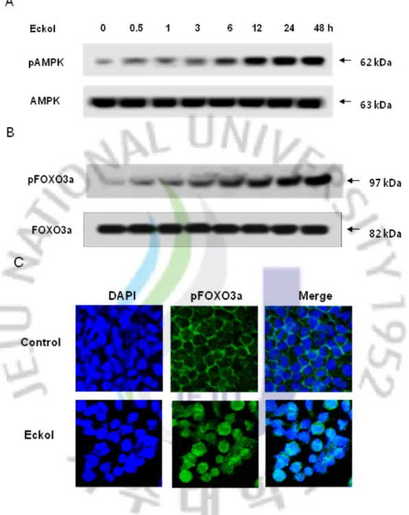
We examined whether eckol is able to activate AMPK-mediated with phosphorylation of Foxo3a up-regulation. As shown in Figs. 6A and B, exposure to eckol caused an increase in the phosphorylation of AMPK and FOXO3a in a time- dependent manner and confirmed by the level of phosphorylation of ACC, which downstream of AMPK. The phosphorylated FOXO3a was clearly increased in the eluted fraction of cells treated with eckol, and resulted in a translocalization of FOXO3a protein from the cytosol to the nucleus. (Fig. 6C) These results suggesting of the eckol mediated the involvement of the AMPK/FOXO3a pathway.
27
Fig. 6. Eckol mediated on AMPK/FOXO3a signaling pathway and FOXO3a
translocalization into nucleus. (A) Cell lysates treated by a variety of time courses of eckol at 10 μg/ml was electrophoresed and the expression of phospho AMPK was detected by the respective specific antibodies. (B) Nuclear extracts from Chang liver cells were prepared after treatment with 10 μg/ml of eckol for the indicated times. Western blot for nuclear lysates were detected with on phosphorylation of FOXO3a specific antibody. (C).Confocal
28
imaging using FITC-conjugated secondary antibody staining indicates the location of phospho FOXO3a (green) by phospho FOXO3a antibody, DAPI staining indicates the location of the nucleus (blue), and the merged image in eckol-treated cells and the merged image in eckol-treated cells indicates the nuclear translocation of phospho FOXO3a translocated into the nucleus (D) from eckol-treated cells was detected using immunocytochemical analysis.
29 3.6. Eckol activated Mn SOD via of AMPK/FOXO3a
To further elucidate the signaling pathway involved in eckol-mediated Mn SOD activation and induction of FOXO3a. We analyzed whether FOXO3a pathways is involved in the induction of Mn SOD expression. Cells were transfected with siFOXO3a for 24 h prior to eckol treatment. As shown in Fig. 7A, the both constitutive and eckol-induced expression of Mn SOD was markedly inhibited by siRNA knock down of FOXO3a gene. We then analyzed whether AMPK pathways were involved in the induction of phospho-FOXO3a activation and Mn SOD expression. Cells were pre-incubated for 30 min with inhibitors of compound C (an AMPK inhibitor) and then treated with eckol for additional 24 h. The effectiveness of these inhibitors was confirmed in eckol-treated cells with antibodies specific for phospho-FOXO3a and Mn SOD (data not shown). Increase in nuclear phospho-FOXO3a accumulation and Mn SOD protein expression occurred following treatment with eckol as expected. Inhibition of the AMPK pathways dramatically reduced the capacity of eckol to increase FOXO3a and Mn SOD protein levels (Fig. 7B). To further confirm these observations, cells were transfected with siAMPK. As shown in Fig. 7C, the expression of nuclear phospho-FOXO3a and Mn SOD were markedly inhibited in siAMPK-transfected cells regardless of eckol treatment. These results indicate that AMPK is required to induce Mn SOD expression as well as nuclear accumulation of FOXO3a.
31
Fig.7. Induction of Mn SOD by eckol via AMPK/FOXO3a. (A) Cells were transfected in
10–50nM siControl and siFOXO3a. At 24 h after transfection, the cells were treated with 10 μg/ml of eckol for 24 h and the expression of Mn SOD protein was examined by Western blot analysis. (B) Cells were incubated with eckol in presence of compound C. Next Cell lysates were electrophoresed, and phospho-Foxo3a and Mn SOD were detected using their respective specific antibodies.(C) Cells were transfected in 10–50nM siControl and siAMPK for 24 h after transfection, the cells were treated with 10 μg/ml of eckol for 24 h and the expression of phospho-FoxO3a and Mn SOD protein were examined by Western blot analysis.
32
3.7. Involvement of Mn SOD in cell damage induced by oxidative stress
As shown in Fig. 8, the protein expression of Mn SOD was markedly inhibited in siMn SOD-transfected cells regardless of eckol treatment. To determine whether the increased level of Mn SOD activity enhanced by eckol confers cytoprotection against oxidative stress, Chang liver cells were pretreated with the Mn SOD inhibitor (DEDTC). DEDTC attenuated the protective effect of eckol on H2O2-induced cytotoxicity and siMn SOD-transfected cells exhibited similar results Therefore, the cytoprotective effect of eckol is likely to be mediated through Mn SOD induction.
33
Fig.8. The cytoprotective effect of eckol against H2O2-induced cell death. Cells were transfected in 10–50nM siControl and siMn SOD. At 24 h after transfection, the cells were pre-incubated with DEDTC at 10 μM for 30min, followed by 1 h of incubation with eckol and exposure to 600 μM of H2O2 for 24 h. Cell viability was measured using the MTT assay. *Significantly different from H2O2 treated cells (p < 0.05). **Significantly different from eckol plus H2O2treated cells (p < 0.05).
34
DISCUSSION
Marine algae have been well-known as an important source to produce natural bioactive secondary metabolites including phenols and polyphenols with unique linkages (48) Phlorotannin components, which are marine algal polyphenols and especially found in brown algae, are polymers of phloroglucinol (49). Eckol is a polymer of phloroglucinol with a polyphenol structure (Fig.1). We have previously shown that eckol protected cells against H2O2 induced cell damage via the cellular antioxidant activation, and ERK-NFkB activation pathway (50). In additional , that it protected V79-4 lung fibroblast cells against gamma-ray radiation-induced apoptosis by scavenging of ROS and inhibiting of the JNK pathway (51). Mitochondria contributes to a number of different processes in living cells, such as ATP synthesis by oxidative phosphorylation, the production of ROS, and Ca2+ uptake and release (52), of which the most important is ATP synthesis by oxidative phosphorylation (53). Oxidative stress may be induced by increasing generation of ROS and other free radicals. ROS production by mitochondria can lead to oxidative damage to mitochondrial proteins, membranes and DNA, impairing the ability of mitochondria to synthesize ATP and to carry out their wide range of metabolic functions (54). In our result, H2O2 increased the Ca2+ levels, and eckol inhibited the overloading of Ca2+ in mitochondria (Fig.3). For Ca2+ and ROS, a delicate balance exists between beneficial and detrimental effects on mitochondria. The ATP production and succinate dehydrogenase activity in the respiratory chain was decreased by H2O2 treatment. Impaired mitochondrial function exposed to H2O2 was restored by eckol (Fig.4). Mitochondria are the major source of superoxide production and are subject to direct attack by reactive oxygen species (ROS) (55,56). Superoxides in mitochondria are conveyed through a series of electron carriers arranged spatially according to their redox potentials, but superoxides are quickly dismutated to hydrogen peroxide (H2O2) by the mitochondrial
35
manganese superoxide dismutase (Mn SOD) (57,58). To determine the mechanism of eckol recovering H2O2-induced the attenuation of MnSOD mRNA level and protein expression, as well as Mn SOD activity (Fig .5). In our study, we demonstrate that cytoprotective effect of eckol is likely to mediate through Mn SOD induction via AMPK/FOXO3a dependent pathway.
Recent studies are providing further insights into the complexity of forkhead box (FOXO) regulatory pathways. The FOXO family of proteins is a large family of transcription factors with diverse physiological functions. FOXO factors have been shown to be regulated by a variety of additional stress stimuli, including DNA damage, nutrient deprivation, cytokines, and hypoxia (59-63). Additionally, FOXO3a transcription factors protect against oxidative stress by increasing the levels of the antioxidant enzymes Mn SOD and catalase and the DNA repair enzyme GADD45 (64-66).
Activation of FOXO3a factors by AMPK promotes the preferential expression of a gene expression program that enhances cellular stress resistance (63,64). AMPK is a heterotrimeric protein, comprising two catalytic (α1 and α2) and five different regulatory (β1–2 and γ 1–3) subunits, and functions as a protein serine/threonine kinase. AMPK is activated by a mechanism independent of energy changes, but dependent on mitochondrial ROS generated in response to the interaction of NO with the cytochrome c oxidase (69,70). As a further mechanism of regulation, it has been recently shown that the activation of the energy sensor pathway, which is triggered by an increased AMP/ATP ratio, leads to the AMPK-mediated phosphorylation of FOXO3a (69). Increasing evidence suggests that AMPK can directly phosphorylate FOXO3, which mediates AMPKs ability to reduce cell stress and increase cell survival (71.72).
We examined effect of eckol clearly induced the expression of phosphorylated AMPK and FOXO3a in Chang liver cells. The increased FOXO3a might play a role in its activation or
36
nuclear translocation (Fig 6). Our results demonstrated that the increase of Mn SOD protein level induced by eckol was dependent on the activation AMPK/FOXO3a, since as well as AMPK and FOXO3a siRNA decreased the eckol-induced accumulation of Mn SOD protein through inhibition of AMPK/FOXO3a. These results show that AMPK/FOXO3a pathways are regulated by eckol, and they participate in the induction of Mn SOD (Fig 7). In addition, we showed that DEDTC (a potent inhibitor of Mn SOD), and Mn SOD siRNA can partially reverse the protective effects of eckol, thus providing further evidence for MnSOD as a possible cytoprotective pathway for eckol (Fig. 8).
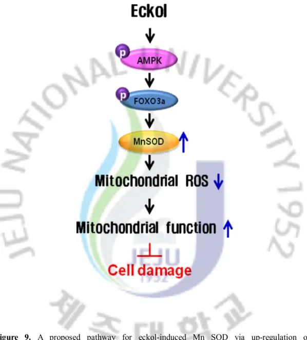
In conclusion, eckol attenuated mitochondrial oxidative stress by activating AMPK/FOXO3a-mediated Mn SOD induction, and this suggests its possible application in the amelioration of mitochondrial ROS related pathological conditions (Fig.9).
37
Figure 9. A proposed pathway for eckol-induced Mn SOD via up-regulation of
AMPK/FOXO3a, explaining the cytoprotective effect against oxidative stress in Chang liver cells.
38
REFERENCES
1. Dröge, W. 2002. Free radicals in the physiological control of cell function.Physiol.Rev. 82:47–95.
2. Balaban RS, Nemoto S, Finkel T. Mitochondria, oxidants, aging. Cell120: 483–495, 2005. 3. Richter, C., 1993. Pro-oxidants and mitochondrial Ca2+: their relationship toapoptosis and
oncogenesis. FEBS Lett. 325, 104–107.
4. Lowell, B.B., Shulman, G.I., 2005. Mitochondrial dysfunction and type 2 diabetes.Science 307, 384–387
5. Wei, Y.Z., Rector, R.S., Thyfault, J.P., Ibdah, J.A., 2008. Nonalcoholic fatty liver disease and mitochondrial dysfunction. World J. Gastroenterol. 14, 193–199.
6. Mattson, M.P., Gleichmann, M., Cheng, A.W., 2008. Mitochondria in neuroplasticity and neurological disorders. Neuron 60, 748–766.
7. Castellani, R., Hirai, K., Aliev, G., Drew, K.L., Nunomura, A., Takeda, A., Cash, A.D., Obrenovich, M.E., Perry, G., Smith, M.A., 2002. Role of mitochondrial dysfunction in Alzheimer’s disease. J. Neurosci. Res. 70, 357–360.
8. Weisiger RA, Fridovich I (1973) Superoxide dismutase. Organelle specificity.J BiolChem 248:3582–3592
9. Marcus, D.L., Strafaci, J.A., Freedman, M.L., 2006. Differential neuronal expression of manganese superoxide dismutase in Alzheimer's disease. Med. Sci. Monit. 12, BR8– BR14.
10. Oberley, L.W., Buettner, G.R., 1979. Role of superoxide dismutase in cancer: a review. Cancer Res. 39, 1141–1149.
11. Kiningham KK, Oberley TD, Lin SM, Mattingly CA, St. Clair DK (1999) Overexpression of manganese superoxide dismutase protects
againstmitochondrial-39
initiated poly (ADP-ribose) polymerase mediated celldeath. FASEB J13:1601–1610 12. Greer, E.L., and Brunet, A. (2005). FOXO transcription factors at the interfacebetween
longevity and tumor suppression. Oncogene 24,7410–7425.
13. Medema RH, Kops GJ, Bos JL, Burgering BM (2000). "AFX-like Forkhead transcription factors mediate cell-cycle regulation by Ras and PKB through p27kip1.". Nature 404 (6779): 782–7.
14. Akt promotes cell survival by phosphorylating and inhibiting a Forkhead transcription factor. Brunet A, Bonni A, Zigmond MJ, Lin MZ, Juo P, Hu LS, Anderson MJ, Arden KC, Blenis J, Greenberg ME. Cell. 1999 Mar 19;96(6):857-68.
15. The forkhead transcription factor Foxo1 links insulin signaling to Pdx1 regulation of pancreatic β cell growth. Nakae, J., Kitamura, T., Silver, D. L., and Accili, D. (2001) J. Clin. Invest.108, 1359–1367
16. Dijkers, P.F., et al. 2000. Forkheadtranscriptionfactor FKHR-L1 modulates cytokine-dependent transcriptional regulation of p27(KIP1). Mol. Cell.Biol. 20:9138–9148. 17. Martinez-Gac, L., Marques, M., Garcia, Z., Campanero,M.R., and Carrera, A.C. 2004.
Control of cyclin G2 mRNA expression by forkheadtranscriptionfactors: novel mechanism for cell cycle controlbyphosphoinositide 3-kinase and forkhead. Mol.Cell. Biol. 24:2181–2189.
18. Tran, H., Brunet, A., Grenier, J. M., Datta, S. R., Fornace, A. J., Jr., DiStefano, P. S. et al. DNA repair pathway stimulated by the forkhead transcription factor FOXO3a through the Gadd45 protein. Science 296, 5567:530-534
19. Brunet, A., et al. 1999. Akt promotes cell survival byphosphorylating and inhibiting a Forkheadtranscriptionfactor. Cell. 96:857–868.
20. Ghaffari, S., Jagani, Z., Kitidis, C., Lodish, H.F., andKhosravi-Far, R. 2003. Cytokines and BCR-ABL mediate suppression of TRAIL-induced apoptosisthrough inhibition of
40
forkhead FOXO3atranscription factor. Proc. Natl. Acad. Sci. U. S. A.100:6523–6528. 21. Kops, G.J., et al. 2002. Forkhead transcription factorFOXO3a protects quiescent cells
from oxidativestress. Nature. 419:316–321.
22. Nemoto, S., and Finkel, T. 2002. Redox regulationofforkhead proteins through a p66shc-dependentsignaling pathway.Science. 295:2450–2452
23. Hardie, D. G. and Carling, D. (1997) The AMP-activated protein kinase: fuel gauge of themammalian cell? Eur. J. Biochem. 246, 259–273
24. Salt, I. P., Johnson, G., Ashcroft, S. J. and Hardie, D. G. (1998) AMP-activated proteinkinase is activated by low glucose in cell lines derived from pancreatic β cells, and mayregulate insulin release. Biochem. J. 335, 533–539
25. Marsin, A. S., Bouzin, C., Bertrand, L. and Hue, L. (2002) The stimulation of glycolysis byhypoxia in activated monocytes is mediated by AMP-activated protein kinase andinducible 6-phosphofructo-2-kinase. J. Biol. Chem. 277, 30778–30783
26. Oliveira, R. L., Ueno, M., de Souza, C. T., Pereira-da-Silva, M., Gasparetti, A. L., Bezzera,R. M., Alberici, L. C., Vercesi, A. E., Saad, M. J. and Velloso, L. A. (2004) Cold-inducedPGC-1α expression modulates muscle glucose uptake through an insulinreceptor/Akt-independent, AMPK-dependent pathway. Am. J. Physiol. Endocrinol. Metab.287, E686–E695
27. Greer EL, Oskoui PR, Banko MR, Maniar JM, Gygi MP, Gygi SP, et al. The energy sensor AMPactivated protein kinase directly regulates the mammalian FOXO3 transcription factor. J Biol Chem 2007; 282:30107-19.
28. Li XN, Song J, Zhang L, LeMaire SA, Hou X, Zhang C, et al. Activation of the AMPK-FOXO3 pathway reduces fatty acid-induced increase in intracellular reactive oxygen species by upregulating thioredoxin. Diabetes 2009; 58:2246-57.
41
endothelial cells. Biochem J 2009; 421:163-9.
30. Kang, K., Park, Y., Hwang, H.J., Kim, S.H., Lee, J.G., Shin, H.C., 2003. Antioxidativeproperties of brown algae polyphenolics and their perspectives as chemopreventiveagents against vascular risk factors. Arch. Pharm. Res. 26, 286–293. 31. Kang, H.S., Chung, H.Y., Kim, J.Y., Son, B.W., Jung, H.A., Choi, J.S., 2004a. Inhibitory
phlorotannins from the edible brown alga Eckloniastolonifera on total reactive oxygen species (ROS) generation.Arch. Pharm. Res. 27, 194–198.
32. Kang, H.S., Kim, H.R., Byun, D.S., Son, B.W., Nam, T.J., Choi, J.S., 2004b. Tyrosinaseinhibitorsisolated from the edible brown alga Eckloniastolonifera. Arch. Pharm. Res. 27,1226–1232.
33. Kang, K.A., Lee, K.H., Chae, S., Koh, Y.S., Yoo, B.S., Kim, J.H., Ham, Y.M., Baik, J.S., Lee, N.H.,Hyun, J.W., 2005a. Triphlorethol-A from Ecklonia cava protects V79-4 lungfibroblast against hydrogen peroxide induced cell damage. Free Radic. Res. 39,883– 892.
34. Kang, K.A., Lee, K.H., Chae, S., Zhang, R., Jung, M.S., Lee, Y., Kim, S.Y., Kim, H.S., Joo, H.G.,Park, J.W., Ham, Y.M., Lee, N.H., Hyun, J.W., 2005b. Eckol isolated from Eckloniacavaattenuates oxidative stress induced cell damage in lung fibroblast cells. FEBS Lett.579, 6295–6304.
35. Kang, K.A., Lee, K.H., Chae, S., Zhang, R., Jung,M.S.,Ham, Y.M., Baik, J.S., Lee,N.H., Hyun, J.W.,2006a. Cytoprotective effect of phloroglucinol on oxidative stress induced cell damagevia catalase activation. J. Cell. Biochem. 97, 609–620.
36. Kang, K.A., Zhang, R., Lee, K.H., Chae, S., Kim, B.J., Kwak, Y.S., Park, J.W., Lee, N.H., Hyun, J.W., 2006b. Protective effect of triphlorethol-A from Ecklonia cava against ionizingradiation in vitro. J. Radiat. Res. (Tokyo) 47, 61–68.
42
Protectiveeffect of phlorotannin components phloroglucinol and eckol on radiation-inducedintestinal injury in mice.Phytother. Res. 21, 15–24.
38. Kang, K.A., Lee, K.H., Park, J.W., Lee,N.H.,Na,H.K., Surh,Y.J.,You, H.J., Chung,M.H.,Hyun, J.W.,2007. Triphlorethol-A induces heme oxygenase-1 via activation of ERK and NF-E2related factor 2 transcription factor. FEBS Lett. 581, 2000–2008. 39. Brain Res Dev Brain Res. 2002 Mar 31;134(1-2):93-102. Role of mitochondria in serum
withdrawal-induced apoptosis of immortalized neuronal precursors. L. Colombaioni, L. Colombini and M. Garcia-Gil
40. Connop, B. P., Thies, R. L., Beyreuther, K., Ida, N., and Reiner, P. B. 1999. Novel effects of FCCP [carbonyl cyanide p-(trifluoromethoxy)phenylhydrazone] on amyloid precursor protein processing. J. Neurochem. 72: 1457–1465.
41. Pearse AG (1972). Histochemistry, theoretical and applied. Baltimore: Williams and Wilkins, pp. 921-961.
42. Alley MC, Scudiero DA, Monks A, Hursey ML, Czerwinski MS, Fine DL, et al. (1988). Feasibility of drug screening with panels of human tumor cell lines using a microculture tetrazolium assay. Cancer Res 48:589-601.
43. Wataha JC, Hanks CT, Craig RG (1991). The in vitro effects of metal cations on eukaryotic cell metabolism. J Biomed Mater Res 25:1133-1149.
44. Carmichael, J., Degraff, W.G., Gazdar, A.F., Minna, J.D., Mitchell, J.B., 1987. Evaluation of a tetrazolium-based semiautomated colorimetric assay: assessment of chemosensitivity testing. Cancer Res. 47, 936–942.
45. Misra, H. P., and Fridovich, I. 1972. The role of superoxide anion in the autoxidation of epinephrine and a simple assay for superoxide dismutase. J. Biol.Chem. 247:3170 3175. 46. Casano, L. M., Martin, M., and Sabater, B. 1994. Sensitivity of superoxide dismutase
43
young barley leaves. Plant Physiol. 106: 1033–1039.
47. Fridovich, I. 1995. Superoxide radical and superoxide dismutases. Annu. Rev. Biochem. 64:97–112.
48. Shibata, T., Yamaguchi, K., Nagamura, K., Kawaguchi, S. and Nagamura, T. (2002) Inhibitory activity of brown algae phlorotannins against glycosidases from the viscera of the turban shell Turbo cornutus. Eur. J. Phycol. 37, 493–500
49. Torres, M.A., Barros, M.P., Campos, S.C., Pinto, E., Rajamani, S., Sayre, R.T., Colepicolo,P., 2008. Biochemical biomarkers in algae and marine pollution: a review. Ecotoxicology and Environmental Safety 71, 1–15.
50. Kang, K.A., Lee, K.H., Chae, S., Koh, Y.S., Yoo, B.S., Kim, J.H., Ham, Y.M., Baik, J.S., Lee, N.H., Hyun, J.W., 2005. Triphlorethol-A from Ecklonia cava protects V79-4 lung fibroblast against hydrogen peroxide induced cell damage. Free Radic. Res. 39, 883-892. 51. Zhang R, Kang KA, Piao MJ, Ko DO, Wang ZH, Lee IK, et al. Eckol protects V79-4
lungfibroblast cells against gamma-ray radiation-induced apoptosis via the scavenging of reactive oxygen species and inhibiting of the c-Jun NH(2)-terminal kinase pathway. Eur J Pharmacol 2008;591:114–23.
52. Pedersen, P.L., 1999. Mitochondrial events in the life and death of animal cells: a brief overview. J. Bioenerg. Biomembr. 31, 291–304.
53. Saraste, M., 1999. Oxidative phosphorylation at the fin de siecle. Science 283, 1488– 1493.
54. Madamanchi NR, Runge MS. Mitochondrial dysfunction in atherosclerosis. Circ Res 2007;100:460–473
55. Harman, D., 1972. The biological clock: the mitochondria? J. Am. Geriatr. Soc. 20,145-147
44
Metab. Rev. 39, 443-455.
57. Sutteon, A., Khoury, H., Prip-Buus, C., Cepanec, C., Pessayre, D., Degoul, F., 2003. The Ala16Val genetic dimorphism modulates the import of human manganese superoxide dismutase into rat liver mitochondria. Pharmacogenetics 13, 145-157.
58. Shimoda-Matsubayashi, S., Matsumine, H., Kobayashi, T., Nakagawa-Hattori, Y.,Shimizu, Y., Mizuno, Y., 1996. Structural dimorphism in the mitochondrial targeting sequence in the human manganese superoxide dismutase gene. Biochem. Biophys. Res. Commun. 226, 561-565
59. Seoane J, Le HV, Shen L, Anderson SA, Massague J: Integrationof Smad and forkhead pathways in the control of neuroepithelial and glioblastoma cell proliferation. Cell 2004, 117:211-223
60. Gomis RR, Alarcon C, Nadal C, Van Poznak C, Massague J: C/EBPbeta at the core of the TGFbeta cytostatic response and its evasion in metastatic breast cancer cells. Cancer Cell 2006, 10:203-214.
61. Gomis RR, Alarcon C, He W, Wang Q, Seoane J, Lash A,Massague J: A FoxO-Smad synexpression group in human keratinocytes. Proc Natl Acad Sci U S A 2006, 103:12747-12752.
62. Huang H, Regan KM, Lou Z, Chen J, Tindall DJ: CDK2-dependent phosphorylation of FOXO1 as an apoptotic response to DNA damage. Science 2006, 314:294-297.
63. Greer EL, Dowlatshahi D, Banko MR, Villen J, Hoang K, Blanchard D, Gygi SP, Brunet A: An AMPK-FOXO pathway mediates longevity induced by a novel method of dietary restriction in C. elegans. Curr Biol 2007, 17:1646-1656.
64. Greer EL, Oskoui PR, Banko MR, Maniar JM, Gygi MP, Gygi SP, Brunet A: The energy sensor AMP-activated protein kinase directly regulates the mammalian FOXO3 transcription factor. J Biol Chem 2007, 282:30107-30119.
45
65. Bakker WJ, Harris IS, Mak TW: FOXO3a is activated in response to hypoxic stress and inhibits HIF1-induced apoptosis via regulation of CITED2. Mol Cell 2007, 28:941-953. 66. Kops, G. J., T. B. Dansen, P. E. Polderman, I. Saarloos, K. W. Wirtz, P. J. Coffer, T. T.
Huang, J. L. Bos, R. H. Medema, and B. M. Burgering. 2002. Forkhead transcription factor FOXO3a protects quiescent cells from oxidative stress. Nature. 419: 316?321. 67. Tran, H., A. Brunet, J. M. Grenier, S. R. Datta, A. J. Fornace, Jr., P. S. DiStefano, L. W.
Chiang, and M. E. Greenberg. 2002. DNA repair pathway stimulated by the forkhead transcription factor FOXO3a through the Gadd45 protein. Science. 296: 530?534. 68. Nemoto, S., and T. Finkel. 2002. Redox regulation of forkhead proteins through a
p66shc-dependent signaling pathway. Science. 295: 2450?2452.
69. Quintero, M., Colombo, S. L., Godfrey, A. and Moncada, S. (2006) Mitochondria as signaling organelles in the vascular endothelium. Proc. Natl. Acad. Sci. U.S.A. 103,5379-5384.
70. Palacios-Callender, M., Quintero, M., Hollis, V. S., Springett, R. J. and Moncada, S. (2004) Endogenous NO regulates superoxide production at low oxygen concentrations by modifying the redox state of cytochrome c oxidase. Proc. Natl. Acad. Sci. U.S.A. 101, 7630-7635
71. Greer EL, Oskoui PR, Banko MR, Maniar JM, Gygi MP, Gygi SP, et al. The energy sensor AMPactivated protein kinase directly regulates the mammalian FOXO3 transcription factor. J Biol Chem 2007; 282:30107-19.
72. Greer EL, Dowlatshahi D, Banko MR, Villen J, Hoang K, Blanchard D, Gygi SP, Brunet A. An AMPK-FOXO pathway mediates longevity induced by a novel method
46
ABSTRACT IN KOREAN
이 연구에서는 감태(Ecklonia cava)로부터 분리 정제한 Phlorotannin 화합물인 eckol의 산화적 스트레스에 의한 미토콘드리아 손상에 대한 세포 보호효과와 그 기작에 대해 밝히고자 하였다. 세포 보호기작 연구에 있어서 중요한 항산화효소로 알려진 Mn SOD의 산화적 손상으로부터의 보호 작용을 검색하였다. Eckol은 The human hepatocyte-derived cell line인 Chang liver 세포에서 hydrogen peroxide로 미토콘드리아 ROS와 세포내 칼슘농도를 증가시켰으나 eckol에 의해 감소하는 것을 확인할 수 있었다. 게다가 H2O2에 의해 낮아진 ATP의 양을 eckol을 처리함에 따라 ATP양이 증가하는 것도 확인하였다. 따라서 우리는 eckol이 미토콘드리아 ROS의 연관성과 세포내 칼슘농도의 균형을 유지하고 미토콘드리아내의 에너지를 생산하고 조절하는 것을 알 수 있었다. Mn SOD는 산화적 스트레스에 의해 유도되는 미토콘드리아의 세포손상을 저해하는 항산화 효소로서 중요한 역할을 한다. 따라서 Eckol을 Mn SOD 단백질량과 mRNA의 수준을 증가 시켰고, Mn SOD의 효소활성도 증가 시켰다. 에너지항상성을 조절하는 AMP-activated protein kinase (AMPK)와 수명을 연장하는데 주요한 유전자인 Forkhead box O3a (FOXO3a)의 기작은 산화적 스트레스와 연관대어 다양한 세포내 역할을 하는 것으로 보고되고 있다. 따라서 Eckol을 처리하였을 때 AMPK와 FOXO3a의 시간 의존적으로 단백질 발현량을 증가시켰고 FOXO3a의 핵 내로의 이동을 유도하였다. Eckol은 AMPK와 FOXO3a 둘 다의 활성을 증가 시켰으며 AMPK의 활성 억제재인 compound C를 처리 하였을 FOXO3a와 Mn SOD의 발현을 억제하였으며, AMPK와 AMPK를 억제하기 위해 siAMPK과 siAMPK를 이식하였을 때 역시 같은 결과를 얻을 수 있었다. 마지막으로 Mn SOD의 산화적 손상으로부터의 보호효과를 확인하기 위해 hydrogen peroxide로 세포 손상을 유도하였으며 Mn SOD특이적 억제재인 DEDTC 처리와 siMn SOD
47
이식으로 eckol에 의해 유도된 세포보호효과가 상쇄되었다. 이 연구는 eckol이 AMPK와 FOXO3a를 통하여 항산화 효소인 Mn SOD의 증가에 의해 미토콘드리아내의 산화적 손상을 억제시킨다는 내용을 증명하였다.
검색어:
Manganese superoxide dismutase · Forkhead box O3a, AMP-activated
48
감사의
글
저에게는 2년이라는 짧지만 길었던 석사과정 동안 더 많은 것을 배우고 경험하였기에 너무 뿌듯하고 즐거웠습니다. 저를 걱정해주시고 버팀목이 되어 주셨던 모든 분들에게 지면으로 나마 감사의 마음을 전하고 싶습니다. 먼저, 학부생활부터 지금까지 부족한 저에게 가르침을 주시고 바르게 지도해주신 영원한 스승님이신 전유진 교수님, 2년전 처음 만나서 부족한 저를 안아주시고 이끌어주신 현진원 교수님, 항상 같은 모습으로 제가 힘들 때 격려해 주신 김기영 교수님, 언제나 인자하시고 고요하게 그림자처럼 저에게 힘을 불어넣어 주신 이기완 교수님께 진심으로 감사하고 사랑한다는 말을 전합니다. 또한 학부생활부터 저에게 많은 사랑과 관심을 주셨던 송춘복 교수님, 허문수 교수님, 이제희 교수님, 여인규 교수님, 최광식 교수님, 이영돈 교수님, 이경준 교수님, 정준범 교수님께 진심으로 감사드립니다. 또한 항상 자기학생처럼 아껴주시고 보듬어주신 식품공학과의 김수현교수님, 하진환 교수님, 화학과의 이남호 교수님, 수의학과 지영흔 교수님, 의과대 강희경 교수님, 유은숙 교수님, 은수용 교수님, 정성철 교수님, 김수영 교수님, 고영상 교수님, 조문제 교수님, 이근화 교수님, 박덕배 교수님 항상 저에게 힘을 주시는 정원교 교수님께도 진심으로 감사드립니다. 지난 2년간 가장 많이 나를 아껴준 실험실 사람들 수진오빠, 길남오빠, 선희언니, 긴내언니, 승홍오빠, 석천오빠, 원우오빠, 지혁오빠, 창익오빠, 재영이, 수동오빠, 자나카오빠와 사랑스런 후배들인 혜미, 은아, 나래 항상 고맙고 미안하고 사랑한다고 전하고 싶습니다. 또한 항상 저에게 관심과 사랑을 주시는 의대 실험실의 경아언니, 미경언니, 장예언니, 기천오빠, 영미선생님, 희경선생님, 진영선생님에게도 진심으로 감사드립니다. 그리고 항상 저에게 격려를 아끼지49 않으셨던 김필연 연구사님, 강호정사장님, 영건오빠, 만철오빠, 문휴오빠, 주상오빠, 철홍오빠, 맹진오빠, 상규오빠, 현실언니, 현성오빠, 태원오빠, 송헌오빠, 경주오빠, 민주언니, 혜영언니, 봉근오빠, 대승오빠, 윤범오빠, 영득오빠, 치훈오빠, 상우오빠, 성표오빠, 상혁오빠, 경용오빠, 지웅오빠, 현기오빠, 희중오빠, 희도오빠, 세진오빠, 창희오빠, 창영오빠, 동민오빠, 준상오빠, 익수오빠, 영민오빠와 항상 웃음과 힘을 불어넣어주는 병훈오빠, 정훈오빠, 승민오빠, 상민오빠에게 고맙다는 말을 전합니다. 지난 6년간 옆에서 다독여주고 힘이 되어주었던 은기오빠, 태혁오빠, 제관오빠, 권우오빠, 현국오빠께 진심으로 감사드립니다. 그리고 너무나 보고 싶은 현식오빠, 봉규오빠에게도 고맙다는 말을 전합니다. 2년동안 서로서로의 힘이 되주었던 동기인 경필오빠, 용재오빠, 유철오빠, 진우오빠, 민철오빠, 숙경이, 주영언니께 고맙다는 말을 전합니다. 나의 곁에서 나를 격려해주고 버팀목이 되어준 나의 서울 친구들 자윤이, 범신이, 정욱이, 성용이, 세엽이, 세나, 한희와 장난꾸러기 친구들인 진호, 우영이, 지훈이, 윤호한테 너무너무 사랑하고 고맙다는 말을 전합니다. 니쳐의 탄생을 축하하며 내가 가장 힘들 때 나를 가장 많이 안아준 멋지고 시크한 나의 친구들 명순이, 연수, 수희, 우리한테 엄청 많이 사랑한다고 전합니다. 그리고 항상 만나면 유쾌하고 사랑스런 나의 친언니 역할을 해준 우리언니한테도 고맙고 사랑한다는 말을 전합니다. 사랑하는 나의 가족들.. 자상하시고 항상 배려가 깊으신 아빠, 너무나 이쁘고 사랑스런 엄마, 항상 최선을 다해 노력하는 내동생 하나에게 항상 건강하고 행복하길 바라고 깊이 사랑한다고 전합니다. 마지막으로 늘 곁에서 나를 지켜주고 격려해주고 다독여 주며 나에게 가장 큰 힘이 되는 사랑하는 우리 성명이 오빠에게 고맙고 사랑한다는 말을 전합니다. 모든 분들에게 좋은 일만 가득하시길 바라며 항상 행복하셨으면 좋겠습니다 다시 한 번 진심으로 감사드립니다.