저작자표시-비영리-변경금지 2.0 대한민국 이용자는 아래의 조건을 따르는 경우에 한하여 자유롭게 l 이 저작물을 복제, 배포, 전송, 전시, 공연 및 방송할 수 있습니다. 다음과 같은 조건을 따라야 합니다: l 귀하는, 이 저작물의 재이용이나 배포의 경우, 이 저작물에 적용된 이용허락조건 을 명확하게 나타내어야 합니다. l 저작권자로부터 별도의 허가를 받으면 이러한 조건들은 적용되지 않습니다. 저작권법에 따른 이용자의 권리는 위의 내용에 의하여 영향을 받지 않습니다. 이것은 이용허락규약(Legal Code)을 이해하기 쉽게 요약한 것입니다. Disclaimer 저작자표시. 귀하는 원저작자를 표시하여야 합니다. 비영리. 귀하는 이 저작물을 영리 목적으로 이용할 수 없습니다. 변경금지. 귀하는 이 저작물을 개작, 변형 또는 가공할 수 없습니다.
A Thesis for the Degree of Master of Philosophy
Juniperus chinensis extract induces
apoptosis via ROS generation in human
pancreatic cancer cell lines
Boram Go
(Supervised by Professor Jae-Hoon Kim)
Department of Biotechnolohy
GRADUATE SCHOOL
JEJU NATIONAL UNIVERSITY
I
Content
LIST OF FIGURES ... III
LIST OF TABLES ... V
ABSTRACT ... VI
1. Introduction... 1
2.
Materials and Methods ... 3
2.1. Sample preparation and antibodies ... 3
2.2. Cell culture ... 3
2.3. Cell viability assay ... 3
2.4. Scratch wound healing assay ... 4
2.5. Western blot analysis ... 4
2.6. Flow cytometry analysis ... 5
2.7. GC-MS MS analysis ... 5
2.8. Statistical analysis ... 6
3. Results... 7
3.1. JCX inhibits cell viability in human pancreatic cancer
cell lines. ... 7
3.2. JCX inhibits cell migration in human pancreatic cancer
cells lines. ... 10
II
3.3. JCX
downregulates
the
tyrosine-phosphorylated
proteins in human pancreatic cancer cell lines. ... 12
3.4. JCX induces apoptosis through activation of caspase-3
in human pancreatic cancer cells lines. ... 15
3.5. JCX induces ROS generation in PANC-1 and SNU-213 ... 18
3.6. GC-MS MS analysis ... 21
4. Discussion ... 25
REFERENCES... 27
ACKNOWLEDGMENT
III
LIST OF FIGURES
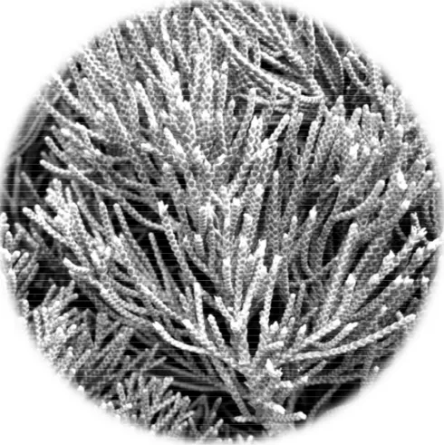
Figure. 1 The picture of Juniperus chinensis leaf. ... 8
Figure. 2 JCX inhibited cell viability ... 9
Figure. 3 JCX inhibited cell migration ... 11
Figure. 4 JCX downregulated the tyrosine-phosphorylated proteins .... 13
Figure. 5 JCX downregulated the p-FAK and p-ERK ... 14
Figure. 6 JCX induced apoptosis ... 16
Figure. 7 JCX downregulated the apoptosis-related proteins ... 17
Figure. 8 JCX induced ROS generation ... 19
IV
V
LIST OF TABLES
VI
ABSTRACT
Pancreatic cancer is one of the dreadful tumors. Less symptoms or signs at early stage cause over a half of patients has distant metastases, which leads resistance to cancer therapies. In this study, we found Juniperus chinensis extract (JCX) inhibited the cell viability and migration activity of PANC-1 and SNU-213 in a dose-dependent manner. JCX increased capase-3 activation, and reactive oxygen species (ROS) generation. N-acetylcysteine (NAC) treatment blocked anti-viability activity of JCX and JCX -induced ROS generation. In addition, JCX downregulated the levels of p-FAK/FAK and p-ERK/ERK.
Altogether, these results indicated that JCX induced apoptosis in human pancreatic cancer cell lines via ROS generation and downregulation of FAK/ERK signaling and activation of caspase-3. We suggest the JCX-derived compounds could be an alternative medicine candidate for pancreatic cancer treatment.
1
1. Introduction
Pancreatic cancer is one of the most dreadful tumors. Its 5- year survival ratio is around 8%. [1] There are few symptoms or signs at early stage, which leads over 50 % of patients have distant metastases when their diagnosis [1]. Invasive pancreatic cancer has resistance against common cancer therapies as chemotherapy and radiation therapy [2]. It’s necessary to improve the treatment strategies for pancreatic cancer.
Juniperus chinensis is native to East Asian countries such as Korea, China, Japan, and
Mongolia. Extract of Juniperus genus exhibits various activities as antioxidants, antimicrobiotics, and antitumor agents. [3, 4, 5] The Juniperus–derived compounds have been reported inducing apoptosis in cancer cells, for example, nardosinen from J. foetidissim, deoxypodophyllotoxin from J. communis, and widdrol from J. lucayana. [6, 7, 8].
Apoptosis is a type of cell deaths. It takes place without inflammation, which is a distinct feature from other cell deaths. It occurs under control remarkably and is a important biological process such as ageing, embryogenesis, and diseases. Reactive oxygen species (ROS) is a byproduct of metabolisms, and acts in cell signaling and homeostasis. Oxidative stress arises between the production of ROS and the biological ROS removal system. Excessively high levels of ROS induce cell apoptosis or necrosis [9, 10]. ROS or oxidative stress involved in regulation of signal transductions such as FAK/ERK, which are related in tumorigenesis. Many therapeutic strategies are devised to elevate intracellular ROS levels with the goal to induce irreparable damages, subsequently resulting in tumor cell apoptosis [11] .
In this study, we report that Juniperus chinensis extract (JCX) inhibits cellviability and -migration in human pancreatic cancer cells. JCX increased intracellular ROS levels, subsequently regulates FAK/ERK pathway and the apoptosis-relating proteins, revealing the
2
3
2. Materials and Methods
2.1. Sample preparation and antibodies
The dried extract of J. chinensis leaf was obtained from Jeju Biodiversity Research Institute (JBRI), dissolved in 70% ethanol, and filtered with a 0.2 μm syringe filter. Antibodies against p-tyrosine, and GAPDH were purchased from Santa Cruz Bio Technology. Antibodies against p-FAK, FAK, p-ERK, ERK, caspase-3, cleaved caspase-3, and proliferating cell nuclear antigen (PCNA) were purchased from Cell Signaling Technology.
2.2. Cell culture
PANC-1, SNU-213, and 293T cells were obtained from Korean Cell Line Bank (Seoul, Korea). 293T and PANC-1 cells were maintained in Dulbbeco’s modified Eagle’s medium supplemented with 10 % fetal bovine serum, 100 U/mL penicillin, and 100 μg/mL streptomycin. SNU-213 cells were maintained in RPMI-1640 medium containing 10 % fetal bovine serum, 25 mM HEPES, 100 U/mL penicillin, and 100 μg/mL streptomycin. All cells
were cultured in a humidified incubator at 37 ℃, and 5% CO2.
2.3. Cell viability assay
PANC-1, SNU-213, and 293T cells were seeded at a density of 1 × 10⁴ cells/well in
24-well plates overnight, and then treated with different concentrations (0, 2.5, 5, 7.5, 10, 15 μg/mL) of JCX for 72 h. Pretreatments of N-acetylcysteine (NAC) was implemented 1.5 h prior to treatments of JCX when performed. Cell viability was assessed by WST assay (EZ-cytox, DoGenbio). Each well was added WST solution 50μL, and incubated for 30 min at 37 ℃. The absorbance was measured by a microplate reader (ScanIt, BD) at 450 nm.
4
Cell viability = OD (sample)
OD (average of control)
2.4. Scratch wound healing assay
PANC-1 and SNU-213 cells were plated at a 5 × 10⁵ cells/well in 6-well plates overnight,
and medium were changed into 0.5% FBS medium (PANC-1) or 0.75% FBS medium (SNU-213) for 24 h. Confluent monolayer cells were manually wounded by scratchers
(SPLScarTM, SPL), washed with PBS, and incubated with fresh medium containing JCX
(0, 10 μg/mL) for 24 h. At 0 and 24 h of incubation, the wounded area were photographed with a microscopy, and measured by imageJ.
Wound healing rate = 1 − remained area (24 h)
ounded area (0 h)
2.5. Western blot analysis
PANC-1, SNU-213, and 293T cells, were treated with JCX (0, 10 μg/mL) for 48 h. The total proteins were extracted with M-PER lysis buffer (Thermo science) containing protease inhibitors such as 1X complete protease inhibitor cocktail (Roche), 2 mM sodium vanadate, 30 mM sodium pyrophosphate, and 100 mM sodium fluoride . After heated at 95 ℃ for 5min, the same amounts of proteins were separated by 12% SDS-PAGE and transferred onto a nitrocellulose blotting membrane (GE healthcare life science). Membranes were blocked with 5% BSA in TBST, incubated with primary antibodies (1:1,000) overnight at 4 ℃. After washing with TBST, membranes were incubated with the corresponding secondary antibodies (1:4,000) room temperature for 3 h. The protein bands were detected by ECL kit (biosesang) on X-ray film (AGFA).
5
2.6. Flow cytometry analysis
For apoptosis analysis, PANC-1, SNU-213, and 293T cells (1 × 10⁵ cells/well) were
seeded in 6-well plates overnight, and treated JCX (0, 10 μg/mL) for 72 h. Cells were collected through tripsinization, and washed with PBS. Then, the cells were incubated with PI and Annexin V-FITC (FITC Annexin V apoptosis detection kit, BD pharmigen) in the dark for 15 min. The apoptotic cells were analyzed by flow cytometry (LSRFortessa, BD)
For intercellular ROS measurement, PANC-1 and SNU-213 cells (1.5× 10⁵ cells/well)
were plated in 12-well plates overnight, pretreated NAC (0, 10 mM) prior 1.5 h to treat JCX, and treated JCX (0, 10 μg/mL) for 6 h. Cells were harvested, and stained with 10 μΜ
H2DCF-DA (ROS detection reagent, Invitrogen.) in the dark for 15 min. The intracellular
ROS measurement was conducted by flow cytometry.
2.7. GC-MS MS analysis
The sample was eluted in methanol. The analysis was performed using a gas chromatograph (GC2010, Shimadzu Corporation). The GC was equipped with an AOC-20i auto-injector and a triple quadruple tandem mass spectrometer (TQ8040, Shimadzu Corporation). 1 μL volume of the sample was injected in splitless mode on Rtx-5MS (30 m × 0.25 mm, 0.25-µm film thickness) column (restek). The carrier gas and the collision gas were helium and argon, respectively. Oven temperature started from 80 ℃ (3-min hold time) to 310 ℃ (10-min hold time). The interface and ion source temperature were 280 °C and 250 °C, respectively. The mass spectrometer was operated in multiple reaction monitoring (MRM) mode. The data acquisition was started at 3.5 min.
6
2.8. Statistical analysis
Data were presented as average ± standard deviation (SD). The differences between multiple groups were analyzed using tukey’s post hoc method. P < 0.5 was regarded statistically significant.
7
3. Results
3.1. JCX inhibits cell viability in human pancreatic cancer cell lines.
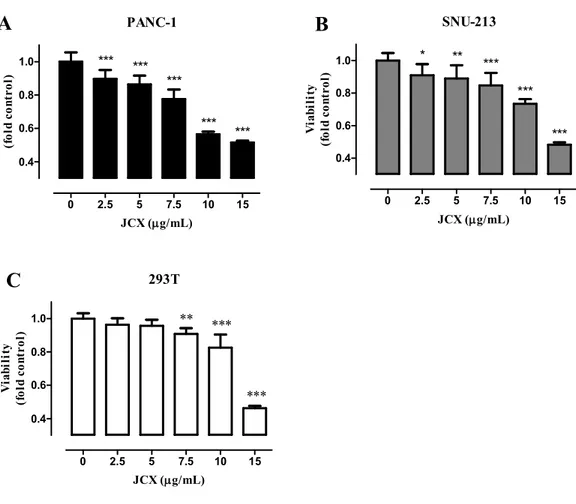
The picture of J. chinensis leaf is shown in Fig. 1. The WST assay was conducted to examine whether the JCX mediates the inhibition effects on the cell viability of PANC-1, SNU-213, and 293T. Cells were treated with different concentrations (0, 2.5, 5, 7.5, 10, 15 μg/mL) of JCX for 72 h. The results demonstrated the JCX inhibited the cell viability in a dose-dependent manner. (Fig. 2) We found JCX has higher inhibition in PANC-1 and SNU-213 human pancreatic cancer cells than 293T, derived from human embryonic kidney cells. Compared with the control, the cell viabilities were inhibited approximately 44% in PANC-1, 36% in SNU-213, and 13% in 293T at treatment of 10 μg/mL JCX.
8
9 293T 0 2.5 5 7.5 10 15 0.4 0.6 0.8 1.0 ** *** *** JCX (g/mL) V ia b il it y (f o ld c o n tr o l) PANC-1 0 2.5 5 7.5 10 15 0.4 0.6 0.8 1.0 *** *** *** *** *** JCX (g/mL) V ia b il it y (f o ld c o n tr o l) SNU-213 0 2.5 5 7.5 10 15 0.4 0.6 0.8 1.0 * ** *** *** *** JCX (g/mL) V ia b il it y (f o ld c o n tr o l)
Fig. 2 JCX inhibited cell viability PANC-1 (A), SNU-213 (B), and 293T (C) cells were
treated with various concentrations (0, 2.5, 5, 7.5, 10, 15 μg/mL) of JCX for 72 h. Cell viabilities were measured using WST assay. Each point is the mean ± SD. *P < 0.05, **P < 0.01, ***P < 0.001
A
C
10
3.2. JCX inhibits cell migration in human pancreatic cancer cells lines.
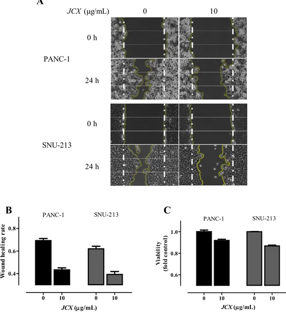
The scratch wound healing assay was performed to assess the JCX-mediated inhibition on the cell migration of PANC-1, and SNU-213. Cells were treated with JCX (0,10 μg/mL) for 24 h, after scratchiness. The quantification was carried out by photoshoot and measuring the wounded area after 0 and 24 h treatment. As shown in Fig. 3, the wounded areas were reduced approximately 27% (PANC-1) and 23% (SNU-213), respectively.
11 0 10 0 10 0.6 0.8 1.0 PANC-1 SNU-213 JCX (g/mL) V ia b il it y (f o ld c o n tr o l) 0 10 0 10 0.4 0.6 0.8 PANC-1 SNU-213 JCX (g/mL) W o u n d h e a li n g r a te
Fig. 3 JCX inhibited cell migration Pancreatic cancer cells were treated with JCX (0, 10
μg/mL) for 24 h. The wounded area pictures at 0, 24 h (A), the numeric results (B), and the viabilities of the PANC-1 and SNU-213 (C).
0 10 0 h 24 h 0 h 24 h JCX (μg/mL) PANC-1 SNU-213
A
B
C
12
3.3. JCX downregulates the tyrosine-phosphorylated proteins in human pancreatic
cancer cell lines.
Western blot analysis was performed to determine JCX mediated molecular mechanisms
of JCX suchas the phosphorylation of tyrosine, FAK and ERK in PANC-1, SNU-213, and
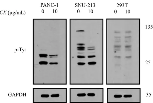
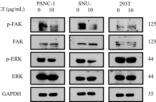
293T. Cells were treated with JCX (0, 10 μg/mL) for 48 h, and then lysates were prepared. As shown in Fig. 4, JCX treatment downregulated the levels of tyrosine-phosphorylated proteins, which are sized between approximately 25 and 135 kDa in PANC-1 and SNU-213, but not in 293T. Furthermore, the results indicated the treatment of JCX decreased the levels of p-FAK and p-EKR in PANC-1 and SNU-213, whereas the expression of ERK and FAK were not decreased. (Fig. 5)
13
Fig. 4 JCX downregulated the tyrosine-phosphorylated proteins PANC-1, SNU-213, and
293T cells were treated with JCX for 48 h. The expression of tyrosine-phosphorylated proteins.
GAPDH
293T
0 10
PANC-1
0 10
SNU-213
0 10
135
25
p-Tyr
JCX (μg/mL)
35
14
Fig. 5 JCX downregulated the p-FAK and p-ERK PANC-1, SNU-213, and 293T cells
were treated with JCX for 48 h. The expression of p-FAK, FAK, p-ERK, ERK, and GAPDH.
293T
0 10
PANC-1
0 10
SNU-0 1SNU-0
125
p-FAK
44
p-ERK
35
GAPDH
125
FAK
44
ERK
JCX (μg/mL)
15
3.4. JCX induces apoptosis through activation of caspase-3 in human pancreatic cancer
cells lines.
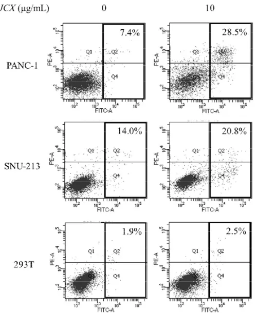
To investigate whether JCX induces anti-viability due to apoptosis, PI and Annexin V-FITC staining was performed after treatment of JCX (0, 10 μg/mL) in PANC-1, SNU-213, and 293T for 72 h. Compared to untreated counterparts, the percentage of the apoptotic cells were increased 21.1% (PANC-1), 6.8% (SNU-213), and 0.6% (293T), respectively (Fig. 6).
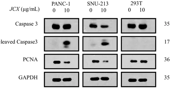
To determine whether J. chinesis extract regulates the levels of caspase-3, cleaved caspase-3, and PCNA, which are related to apoptosis, in PANC-1, SNU-213, and 293T cells by Western blot analysis. As shown in Fig. 7, treatment of JCX increased the expression of cleaved csaspase-3, and decreased the expression of PCNA in PANC-1 and SNU-213. However, treatments of JCX in 293T seemed not causing the activation and expression of cleaved Caspase-3 and PCNA significantly as PANC-1 and SNU-213.
16
Fig. 6 JCX induced apoptosis PANC-1, SNU-213, and 293T cells were treated with JCX
17
Fig. 7 JCX downregulated the apoptosis-related proteins PANC-1, SNU-213, and 293T
cells were treated with JCX for 48 h. The expression of caspase3, cleaved caspase-3, PCNA, and GAPDH. DCF-DA staining, and then flow cytometry analysis
PANC-1
0 10
SNU-213
0 10
GAPDH
35
293T
0 10
17
36
35
PCNA
cleaved Caspase3
Caspase 3
JCX (μg/mL)
18
3.5. JCX induces ROS generation in PANC-1 and SNU-213
To determine whether JCX -induced anti-viability and apoptosis is coupled with oxidative
stress level, H2DCF-DA staining was performed after co-treatment of NAC and JCX in
PANC-1 and SNU-213 for 6 h. As shown as Fig. 8, treatments of JCX increased intracellular ROS levels in PANC-1 (17.1%) and SNU-213 (14.2%), respectively, and 10 mM NAC pretreated cells showed less intracellular ROS levels.
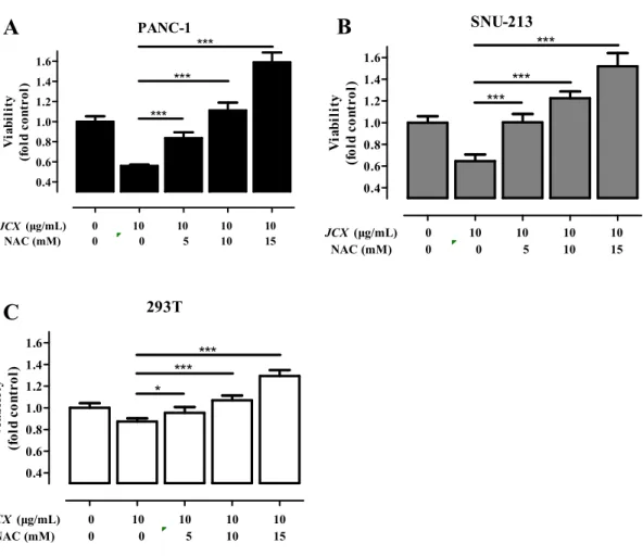
To investigate whether JCX-induced anti-viability and apoptosis is related in ROS generation, the cells were co-treated with JCX and NAC. The pretreatment of NAC leaded to a inhibition of JCX-mediated anti-viability effect in a dose-dependent manner (Fig. 9). Compared to control, the viabilities of 10 mM NAC pretreatments and 10 μg/mL JCX treatment were 111 % (PANC-1), 122 % (SNU-213), and 107 % (293T), respectively. These results demonstrate that pretreatment of 10 mM NAC offset the anti-viability effect of JCX in PANC-1, SNU-213, and 293T.
19 J C X ( μ g /m L ) 0 0 1 0 1 0 N A C ( m M ) 0 1 0 0 1 0 S N U-2 13 P A NC-1 5 0 .4 4 2 .7 6 7 .5 5 1 .9 4 8 .2 4 5 .7 6 2 .4 5 7 .0 F ig . 8 JC X i n d u ce d R O S ge n er at ion P re tr ea tm ent of N A C or ve hi cl e pr ior t o tr ea tm ent o f J. c hi ne ns is e xt ra ct . A ft er 6 hour -tr ea tm ent of J . chi ne ns is e xt ra ct . H2 D C F -D A st ai ni ng, a nd the n fl ow c yt om et ry a na lys is .
20 PANC-1 0.4 0.6 0.8 1.0 1.2 1.4 1.6 JCX (μg/mL) 0 10 10 10 10 NAC (mM) 0 0 5 10 15 *** *** *** V ia b il it y (f o ld c o n tr o l) SNU-213 0.4 0.6 0.8 1.0 1.2 1.4 1.6 JCX (μg/mL) 0 10 10 10 10 NAC (mM) 0 0 5 10 15 *** *** *** V ia b il it y (f o ld c o n tr o l) 293T 0.4 0.6 0.8 1.0 1.2 1.4 1.6 JCX (μg/mL) 0 10 10 10 10 NAC (mM) 0 0 5 10 15 * *** *** V ia b il it y (f o ld c o n tr o l)
Fig. 9 NAC offset the anti-viability effect of JCX Pretreatment of NAC or vehicle prior to
treatment of JCX. PANC-1, SNU-213, and 293T cells were incubated with JCX for 72 h. WST assay. Each point is the mean ± SD. *P < 0.05, **P < 0.01, ***P < 0.001
A
B
21
3.6. GC-MS MS analysis
We performed GC-MS MS analysis to choose candidates among the compounds in JCX. The identified chemicals were shown in Fig. 10 and Table. 1.
22 5 .0 7 .5 1 0 .0 1 2 .5 1 5 .0 1 7 .5 2 0 .0 2 2 .5 2 5 .0 2 7 .5 3 0 .0 3 2 .5 3 5 .0 3 7 .5 4 0 .0 4 2 .5 4 5 .0 4 7 .5 5 0 .0 5 2 .5 5 5 .0 5 7 .5 0 .5 1 .0 1 .5 2 .0 2 .5 3 .0 3 .5 4 .0 (x 1 0 ,0 0 0 ,0 0 0 ) 1 2 3 4 5 6 7 8 9 1 0 1 1 1 2 1 3 1 4 1 5 1 6 1 7 1 8 1 9 2 0 2 1 2 2 2 3 2 4 2 5 2 6 2 7 2 8 2 9 3 0 3 1 3 2 3 3 3 4 3 5 3 6 3 7 3 8 3 9 4 0 4 1 4 2 4 3 4 4 F ig. 10 G as c h rom at ogr ap h y ta n d em m as s sp ec tr om et er an al ys is T h e dr ie d J. c hi ne ns is l ea f ext ra ct w as di lut ed in m et ha nol . 1 μ L of s am pl e w as i n je ct ed . T he c ar ri er ga s and the c ol li si on ga s w er e he li um a nd ar gon , re spe ct ive ly . O ve n te m pe ra tu re s ta rt ed fr om 80 ℃ ( 3 -m in hol d ti m e) t o 310 ℃ ( 10 -m in h ol d ti m e) .
23
Table. 1 Gas chromatography tandem mass spectrometer analysis
Peak
No. AREA % NAME
1 1.36 2-Methoxy-4-vinylphenol 2 5.35 Elemol 3 0.53 2-Butanone, 4-(2,6,6-trimethyl-2-cyclohexen-1-ylidene)- 4 1.70 beta-Eudesmol 5 2.02 10-epi-.gamma.-eudesmol 6 0.64 AROMADENDRENEPOXIDE-(I) 7 0.63 .delta.-Cadinol 8 2.68 (-)-Caryophyllene oxide
9 0.28 Tetradecanoic acid, ethyl ester
10 3.01 Bicyclo[5.3.0]decane, 2-methylene-5-(1-methylvinyl)-8-methyl-
11 0.67 alpha-Eudesmol
12 2.65 B-Norcholestan-3-one, cyclic 1,2-ethanediyl acetal, (5.alpha.)-
13 1.80 Ethanol, 2-(9-octadecenyloxy)-, (Z)-
14 1.10 LONGIFOLENALDEHYDE
15 5.63 Hexadecanoic acid, ethyl ester
16 3.58 3,7,11,15-Tetramethyl-2-hexadecene
17 2.50 Ethyl linoleate
18 4.76 ETHYL LINOLEOLATE
19 0.53 Octadecanoic acid, ethyl ester
20 1.06 5-Hydroxymethyl-1,1,4a-trimethyl-6-methylenedecahydronaphthalen-2-ol 21 0.52 Cyclohexane, hexaethylidene- 22 0.82 1-Phenanthrenecarboxaldehyde, 1,2,3,4,4a,9,10,10a-octahydro-1,4a-dimethyl-7-(1-methylethyl)-, [1S-(1.alpha.,4a.alpha.,10a.beta. 23 1.78 Kaur-16-en-19-ol 24 4.79 Totarol 25 5.26 Cycloisolongifolene, 9,10-dehydro-
26 1.40 Podocarpa-6,8,11,13-tetraen-12-ol, 13-isopropyl-, acetate
27 0.99 Ferruginol
28 0.58 (Albicanol) Decahydro-2-methylene-5,5,8a-trimethyl-1-naphthalenemethanol
29 1.78 Vitamin A alcohol
30 2.89 METHYL TRANS-COMMUNATE
31 0.62 Methane, phenyl(2,2,3-trimethylcyclopentylidene)-
32 2.83 1-Phenanthrenecarboxylic acid,
1,2,3,4,4a,9,10,10a-octahydro-1,4a-dimethyl-7-(1-methylethyl)-, [1R-(1.alpha.,4a.beta.,10a.alpha
33 3.83 1-Phenanthrenecarboxylic acid,
1,2,3,4,4a,10a-hexahydro-1,4a-dimethyl-7-(1-methylethyl)-, methyl ester, [1R-(1.alpha.,4a.beta.,
34 2.12 Bicyclo[3.1.1]hept-2-ene, 2,2'-(1,2-ethanediyl)bis[6,6-dimethyl-
24
36 0.72 Hexadecanoic acid, 2-hydroxy-1-(hydroxymethyl)ethyl ester
37 1.24 BORNYL ESTER OF 3-ISOPROPYLIDENE-CYCLOPENTANECARBOXYLIC ACID
38 2.25 9,12-Octadecadienoic acid (Z,Z)-, 2,3-dihydroxypropyl ester
39 3.13 (-)HINOKININ
40 0.64 Sesamin
41 0.98 Bicyclo[2.2.1]hept-2-en-7-ol, 7-(4-methoxyphenyl)-, anti-
42 1.15 Racemic Savinin [
2-[(3,4-Methylenedioxy)benzylidene]-3-[(3,4-methylenedioxy)benzyl]-.gamma.-butyrolactone isomer]
43 3.35 Stigmast-5-en-3-ol, (3.beta.)-
25
4. Discussion
Cancer cells are having acquired capabilities unlike normal cells. Resistance to apoptosis is an important index for cancer cells. Cancer cells are avoiding apoptosis with encouraging the anti-apoptotic mechanisms and/or downregulating the pro-apoptotic program. [12, 13] In this study, we found JCX induces anti-viability in a dose-dependent manner. The upregulation of cleaved (active) caspase-3 and downregulation of PCNA is proving JCX-mediated apoptosis in human pancreatic cancer cells. Caspase-3 plays a centrol role in the execution of apoptosis and PCNA is essential for DNA replication. Likewise, the Juniperus-derived compounds reported inducing caspase-dependent apoptosis in cancer cells, for
examples, nardosinen from J. foetidissim in MCF-7 breast cancer cell,
deoxypodophyllotoxin from J. communis in breast cancer cells, and widdrol from J.
lucayana in HT-29 colon cancer cells. [6, 7, 8] In addition, the crude leaf extract from Juniperus chinensis was also suggested as a antitumor-promoting and antitumor candidate.
[5]
Focal adhension kinase (FAK) is a non-receptor kinase localized to focal adhension. [14] Extracellular signal-regulated protein kinases 1/2 (ERK1/2) are members of the mitogen-activated protein kinase super family. The regulation of FAK/ERK modulates cell proliferation, migration, adhension, apoptosis, and differentication. [15, 16, 17] The increased FAK complex increases Extracellular signal Regulated Kinase 1/2 (ERK1/2) signal pathway, subsequently boosting the ability of cancer cell survival, and growth in a cell detached condition [18]. Overexpression of FAK has been shown to block the caspase-3-mediated apoptosis [19]. In this study, we found JCX inhibits cell viability and migration via downregulations of p-FAK and p-ERK in pancreatic cancer cells.
ROS is known as a mediator regulation of cell signaling. However, exorbitant ROS levels lead to extreme oxidative stress, and eventually apoptosis in pancreatic cancer cells. [20] For
26
example, for pancreatic cancer, to date only few treatment strategies have been discovered as effective for therapy. Like the combinations of gemcitabine with trichostatin A, epigallocate-3-gallatae (EGCG), capsaicin and benzyl isothiocyanate (BITC), these share the same mechanism to elevate intracellular ROS levels to trigger apoptosis [20, 21, 22, 23] 30,31,32,33]. In this study, JCX -mediated anti-viability and -migration is coupled with ROS generation. The pretreatment of NAC, a ROS scavenger, offset the inhibition effect of JCX.
We suggest JCX as an alternative anticancer medicine. JCX leads to a caspase-related apoptosis in human pancreatic cancer cells via ROS-mediated FAK/EKR signaling. Furthermore, to find the major compounds having anticancer activity in JCX, the next study is in progress.
27
References
[1] Siegel, Rebecca L., Kimberly D. Miller, and Ahmedin Jemal. "Cancer Statistics, 2017." CA: A Cancer Journal for Clinicians 67.1 (2017): 7-30.
[2] Samulitis, Betty K., et al. "Gemcitabine resistant pancreatic cancer cell lines acquire an invasive phenotype with collateral hypersensitivity to histone deacetylase inhibitors." Cancer biology & therapy 16.1 (2015): 43-51.
[3] Lim, Jong Pil, et al. "Free radical scavengers from the heartwood ofJuniperus chinensis." Archives of pharmacal research 25.4 (2002): 449-452.
[4] Yu, M. J., et al. "Antimicrobial activity and antioxidative effects of Juniperus chinensis and protective effects on human HaCaT keratinocyte." Korean J. Aesthetics and Cosmetology 8.2 (2010): 107.
[5] Ali, A. M., et al. "Antitumour-promoting and antitumour activities of the crude extract from the leaves of Juniperus chinensis." Journal of ethnopharmacology 53.3 (1996): 165-169.
[6] Shahali, Amirhosein, et al. "Mitochondrial and caspase pathways are involved in the induction of apoptosis by nardosinen in MCF-7 breast cancer cell line." Research in Pharmaceutical Sciences 13.1 (2018): 12.
[7] Benzina, Sami, et al. "Deoxypodophyllotoxin isolated from Juniperus communis induces apoptosis in breast cancer cells." Anti-Cancer Agents in Medicinal Chemistry (Formerly Current Medicinal Chemistry-Anti-Cancer Agents) 15.1 (2015): 79-88. [8] Kang, Moo Rim, et al. "Widdrol induces apoptosis via activation of AMP-activated
protein kinase in colon cancer cells." Oncology reports 27.5 (2012): 1407-1412.
[9] Finkel, Toren, and Nikki J. Holbrook. "Oxidants, oxidative stress and the biology of ageing." Nature 408.6809 (2000): 239.
28
stress: signaling for suicide and survival." Journal of cellular physiology 192.1 (2002): 1-15.
[11] "Diehn, Maximilian, et al. ""Association of reactive oxygen species levels and
radioresistance in cancer stem cells."" nature 458.7239 (2009): 780.
[12] Hanahan, Douglas, and Robert A. Weinberg. "The hallmarks of cancer." cell 100.1
(2000): 57-70.
[13] Fernald, Kaleigh, and Manabu Kurokawa. "Evading apoptosis in cancer." Trends in
cell biology 23.12 (2013): 620-633.
[14] Zachary, Ian. "Focal adhesion kinase." The international journal of biochemistry &
cell biology 29.7 (1997): 929-934.
[15] Zhao, Jihe, and Jun-Lin Guan. "Signal transduction by focal adhesion kinase in
cancer." Cancer and Metastasis Reviews 28.1-2 (2009): 35-49.
[16] McLean, Gordon W., et al. "The role of focal-adhesion kinase in cancer—a new
therapeutic opportunity." Nature Reviews Cancer 5.7 (2005): 505.
[17] Mebratu, Yohannes, and Yohannes Tesfaigzi. "How ERK1/2 activation controls cell
proliferation and cell death: Is subcellular localization the answer?." Cell cycle 8.8 (2009): 1168-1175.
[18] Bouchard, Véronique, et al. "Fak/Src signaling in human intestinal epithelial cell
survival and anoikis: differentiation state‐specific uncoupling with the PI3‐K/Akt‐1 and MEK/Erk pathways." Journal of cellular physiology 212.3 (2007): 717-728.
[19] Kamarajan, Pachiyappan, and Yvonne L. Kapila. "An altered fibronectin matrix
induces anoikis of human squamous cell carcinoma cells by suppressing integrin alpha v levels and phosphorylation of FAK and ERK." Apoptosis 12.12 (2007): 2221-2231.
[20] "Zhang, Ruifen, et al. ""In vitro and in vivo induction of apoptosis by capsaicin in
pancreatic cancer cells is mediated through ROS generation and mitochondrial death pathway."" Apoptosis 13.12 (2008): 1465-1478.
29
[21] Qanungo, Suparna, et al. "Epigallocatechin-3-gallate induces mitochondrial
membrane depolarization and caspase-dependent apoptosis in pancreatic cancer cells." Carcinogenesis 26.5 (2005): 958-967.
[22] Donadelli, Massimo, et al. "Synergistic inhibition of pancreatic adenocarcinoma
cell growth by trichostatin A and gemcitabine." Biochimica et Biophysica Acta (BBA)-Molecular Cell Research 1773.7 (2007): 1095-1106.
[23] Sahu, Ravi P., et al. "Benzyl isothiocyanate-mediated generation of reactive oxygen
species causes cell cycle arrest and induces apoptosis via activation of MAPK in human pancreatic cancer cells." Carcinogenesis 30.10 (2009): 1744-1753.