- 국문요약 -
기미병변에서
기미병변에서
기미병변에서
기미병변에서 섬유아
섬유아
섬유아 세포
섬유아
세포
세포 기원
세포
기원
기원 사이토카인의
기원
사이토카인의
사이토카인의 발현
사이토카인의
발현
발현
발현
기미는 자외선, 여성호르몬 및 유전적 소인 등에 의해 유발 또는 악화된다고 알려져 있으나 아직까지 그 기전은 확실히 밝혀져 있지 않다. 최근 보고에 의하면 진피의 섬유아 세포가 표피 색소성 질환의 발생에 중요한 영향을 미치는 것으로 밝혀지고 있다. 따라서 본 연구에서는 기미 병변에서 섬유아 세포 기원 사이토카인의 발현양상을 관찰하였다. 기미환자 50 명의 안면 병변 및 인접정상조직을 2 mm 펀치 생검하여 hematoxylin-eosin, Fontana-Masson 염색, Masson trichrome 염색, Verhoeff-van Gieson 염색 및 NKI/beteb, hepatocyte growth factor (HGF), stem cell factor (SCF), tumor necrosis factor (TNF)-α, interleukin (IL)-1α 및 c-kit 에 대한 면역조직화학염색을 실시하고 육안평가 및 컴퓨터 영상분석법 (computerized image analysis)으로 결과를 분석하였다. 기미 병변의 표피는 인접 정상에 비해 얇아져 있었고 상부 진피에 일광탄력변성(so- lar elastosis)소견이 증가되어 있었다. HGF 와 SCF 의 발현은 병변의 진피에서 인접 정상 에 비해 유의하게 증가되어 있었고 c-kit 의 발현은 병변의 표피에서 유의하게 증가되어 있었다. 반면 TNF-α와 IL-1α의 발현은 인접 정상에 비해 병변의 표피에서 각각 약간 감소 또는 증가되어 있었으나 유의한 수준은 아니었다. 이러한 연구 결과 진피의 섬유아 세포로부터 생성된 사이토카인인 HGF 와 SCF 가 기미의 발생에 중요한 역할을 담당할 것으로 생각되었다. 핵심 핵심 핵심 핵심어어어:::: 기미, 섬유아 세포, 면역조직화학염색, HGF, SCF, c-kit 어차례
차례
차례
차례
국문요약--- ---i 차례 ---ii 그림 차례` ---iv 표 차례---v I. 서론---1 II. 연구 대상 및 방법 ---3 A. 연구 대상 ---3 B. 방법 ---3 1. 기미환자의 안면조직수집---3 2. 일반 및 조직화학염색--- 33. 면역조직화학염색 : NKI/beteb, HGF, SCF, TNF-α, IL-1α, c-kit -4 4. 염색결과 분석 ---6
5. 통계학적 분석 ---7
III. 결과 ---8
A. General histologic features---8
B. Fontana-Masson 염색 : 멜라닌 양---9 C. NKI/beteb 면역조직화학염색 : 멜라닌 세포 수---10 D. Masson trichrome 염색 : 콜라겐 섬유---11 E. Verhoeff-van Gieson 염색 : 탄력섬유---12 F. HGF 의 면역조직화학염색---13 G. SCF 의 면역조직화학염색---15 H. TNF-α의 면역조직화학염색---17
J. c-kit 의 면역조직화학염색---21
IV. 고찰 ---23
V. 결론---26
참고문헌 ---27
그림
그림
그림
그림 차례
차례
차례
차례
Fig. 1. hematoxylin-eosin stains---8
Fig. 2. Fontana-Masson stains---9
Fig. 3. NKI/beteb immunostains---10
Fig. 4. Masson trichrome stains---11
Fig. 5. Verhoeff-van Gieson stains---12
Fig. 6. HGF immunostains---13
Fig. 7. The comparision of dermal reactivity about HGF---14
Fig. 8. SCF immunostains---15
Fig. 9. The comparision of dermal reactivity about SCF---16
Fig. 10. TNF-α immunostains---17
Fig. 11. The comparision of epidermal reactivity about TNF-α---18
Fig. 12. IL-1α immunostains---19
Fig. 13. The comparision of epidermal reactivity about IL-1α---20
Fig. 14. c-kit immunostains---21
Fig. 15. The comparision of epidermal reactivity about c-kit---22
표
표
표
표 차례
차례
차례
차례
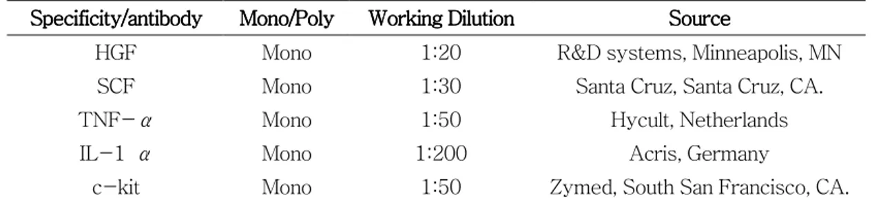
Table 1. List of primary antibodies used in this study---6
Table 2. The comparision of reactivity about HGF---14
Table 3. The comparision of reactivity about SCF---16
Table 4. The comparision of reactivity about TNF-α---18
Table 5. The comparision of reactivity about IL-1α---20
Table 6. The comparision of epidermal reactivity about c-kit---22
I.
I.
I.
I. 서
서
서 론
서
론
론
론
기미는 자외선 노출부위에 갈색 반 및 판을 특징으로 하는 후천적 과색소 질환이다. 기미의 발생원인으로는 자외선, 여성호르몬 및 유전적 소인에 의해 유발 또는 악화된다고 알려져 있으나 아직까지 그 기전은 확실히 밝혀져 있지 않다(Ortonne 등, 2003). 피부의 색소침착은 표피에 존재하는 멜라닌 세포의 활성의 결과이다. 피부는 표피와 진피로 구성되어 있고, 표피에는 다수의 각질형성세포와 멜라닌 세포가 있다. 멜라닌 세포 는 스스로 어떠한 물질을 분비하기 보다는 주변 세포들이 분비한 성장인자 또는 사이토카 인에 반응하는 세포이다. 지금까지 멜라닌 색소형성에 관한 연구는 멜라닌 세포와 가장 가 까이 인접해 있는 각질형성세포가 분비하는 물질에 의한 멜라닌 세포의 활성에 초점이 맟 춰져 왔다(Halaban R, 2000). 피부에 자외선을 조사하면 배양된 각질형성세포에서 α- melanocyte stimulating hormone (MSH)와 endothelin-1 (ET-1)이 분비되어 이들 인 자들에 의해 멜라닌 세포가 증식하고 멜라닌 형성이 증가한다고 알려져 있다. 또한, 실제 로 기미 조직의 각질형성세포에서 α-MSH 의 발현이 증가되어 있음이 보고되었고(Im S 등, 2002), 자외선 관련 과색소 질환인 노인성 흑자와 지루 각화증의 병변에서 ET-1, 표 피 stem cell factor (SCF) 및 tumor necrosis factor (TNF)-α의 발현이 증가되어 있음이 밝혀져 각질형성세포가 분비하는 멜라닌 세포 성장자극인자가 이들 질환의 병인에 중요한 역할을 할 것으로 생각되고 있다(Teraki 등, 1996; Kadono 등, 2001; Hattori 등, 2004). 그러나 기미의 조직학적 소견은 다른 자외선에 의한 과색소질환인 지루성 각화증 이나 노인성 흑자의 조직소견과는 아주 다르다. 지루성 각화증이나 노인성 흑자는 각질형 성세포의 과형성에 의한 표피의 증식이 특징적인 반면 기미에서는 표피가 정상이거나 오 히려 납작해져 있는 것을 알 수 있다(Kang 등, 2002). 또 하나의 특징적인 소견은 진피 의 일광탄력변성(solar elastosis)이다. 기미 병변부 진피에서 인접한 주변 정상피부에 비변성된 섬유아 세포가 변성된 탄력섬유를 생성하기 때문이라고 알려져 있다(Kang 등, 2002).
최근 연구결과에 의하면 진피에 존재하는 섬유아 세포가 표피 멜라닌 세포에 작용하 여 멜라닌 세포를 활성시키는 것으로 생각되고 있다. 섬유아 세포는 hepatocyte growth factor (HGF), SCF, TNF-α, interleukin(IL)-1α, platelet derived growth factor, in- sulin like growth factor, transforming growth factor-β, vascular endothelial growth factor, keratinocyte growth factor, IL-6, IL-8 및 granulocyte-colony stimulating factor 등의 사이토카인을 분비한다(Mansbridge 등, 1999). Imokawa 등은 HGF 와 SCF 가 표피에 존재하는 멜라닌 세포를 활성시켜 멜라닌 합성을 증가시킨다는 사실을 보고한 바 있다(Imokawa 등, 1998; Hedley SJ, 2002). HGF 형질 전환 쥐를 이용한 실험결과 HGF 는 멜라닌 세포의 증식과 분화에 관여한다고 밝혀졌다(Kunisada 등, 2000). 또한 섬 유아 세포가 만드는 콜라겐I, 콜라겐IV, fibronectin 등의 세포간질은 멜라닌 세포 내 티로 로시나제 활성을 증가시키고 섬유아 세포가 멜라닌 세포를 화학 유도하는 작용도 있음이 밝혀졌다(Buffey 등, 1994; Archambault 등, 1995). SCF 와 fibronectin 은 멜라닌 세포 의 증식, 분화 및 이동에 중요한 역할을 한다(Takano 등, 2002).
따라서 진피 변화가 특징적인 기미의 발생에 섬유아 세포가 중요한 역할을 하리라고 생각되는 바, 본 연구에서는 기미환자의 조직에서 섬유아 세포에서 유도되는 사이토카인의 발현양상을 관찰하였다.
II.
II.
II.
II. 연구대상
연구대상
연구대상
연구대상 및
및
및
및 방법
방법
방법
방법
A. A. A. A. 연구연구연구 대상연구대상대상 대상 임상 및 조직학적 소견으로 기미에 합당한 환자 50 명을 대상으로 하였다. B. B. B. B. 방법방법방법방법 1. 기미환자의 안면조직 수집 기미환자 50 명으로부터 2 mm punch 를 이용하여 병변 및 인접 정상에서 각각 피부 조직을 얻었다. 2. 일반 및 조직화학염색 hematoxylin-eosin 염색을 시행하여 인접 정상과 기미 조직의 일반적인 조직소견을 관찰하였으며, 멜라닌 색소에 대해 Fontana-Masson 염색을 실시하였다. 또한 진피의 변 화는 콜라겐에 대해 Masson trichrome 염색을, 그리고 탄력섬유에 대해 Verhoeff-van Gieson 염색을 실시하여 알아보았다. hematoxylin-eosin 염색은 파라핀 절편을 탈파라핀 시킨 후 크실렌, 증류수에 차례 로 수세한 후, 헤마톡실린 액으로 옮겨 4 내지 6 분간 염색하였다. 증류수로 충분히 수세한 후 에오진 액으로 옮겨 1 내지 2 분간 염색하였다. 가볍게 수세한 후 70%, 80%, 90%, 95 %, 무수알코올의 순서로 각 1 분씩 탈수한 후 봉입하여 현미경 하에서 관찰하였다. Fontana-Masson 염색은 파라핀 포매 조직으로 부터 얻은 4.5 ㎛ 두께의 절편을 탈 파라핀 및 수세과정을 거친 후 10% 은용액(silver nitrate)에 옮겨 하룻밤 동안 침투시킨 후, 증류수로 2 내지3 회 수세하고 5% sodium thiosulfate 용액에 2 분간 정착시킨 후 1% neutral red 수용액에 3 분간 대조염색 후 탈수시켜 염색하였다.
Masson trichrome 염색은 파라핀 절편을 탈파라핀 시킨 후 크실렌, 증류수에 차례로 수세하고 포르말린에 1 차 고정 시킨 뒤, Bouin 액에 옮겨 56℃에서 1 시간 동안 고정하였 다. 절편에 착색된 피크르산을 제 거하고 Weigert 철 헤마톡실린 액에 10 분간 염색하였 다. 흐르는 증류수에 10 분간 수세한 후 0.9% Biebrich scarlet-acid fuchsin 액에 15 분 간 염색하였다. 증류수로 1 내지 2 회 수세한 후 phosphomolybdic-phosphotungstic acid (phosphomolybdic 2.5g and phosphotungstic acid 2.5g in distiled water 100ml)액으로 15 분간 염색하였다. 수세과정 없이 2% light green 수용액에 2 내지 5 분 염색한 후 1% acetic acid 에 2 내지 5 분간 수세 후 탈수, 봉입과정을 거쳐 현미경 하에서 관찰하였다. Verhoeff-van Gieson 염색은 파라핀 절편을 탈파라핀 시킨 후 크실렌, 증류수에 차 례로 수세하고 Verhoeff 용액에 옮겨 15 내지30 분간 염색하였다. 흐르는 증류수에 2 내 지3 회 수세한 후 2% ferrick chloride 수용액으로 탈색한 후 현미경으로 보아가며 회색배 경에 탄력섬유가 흑색으로 보일 때 탈색을 멈췄다. 그 후 증류수로 수세 후 5% sodium thiosulfate 에 1 분간 두어 착색된 iodine 을 제거한 후 흐르는 수도물에 5 분간 수세하였 다. 그 후 van Gieson 용액에 옮겨 2 내지 3 분간 대조염색을 하였다. 3. 면역조직화학염색 멜라닌 세포에 대한 염색은 GP-100 항원에 대한 면역화학염색인 NKI/beteb 염색을 시행하였다. NKI/beteb 염색은 파라핀 포매 조직으로부터 얻은 3 ㎛ 두께의 절편을 탈파 라핀 및 phosphate-buffered saline(PBS)세척 후 단백질 비특이 흡수를 저지하였다. 이 후 NKI/beteb 항체(Monoclonal antibodies to NKI/beteb, melanoma-associated anti-gen, Monosan, the Netherlands)로 15 분간 적용시킨 후에 PBS 로 10 분에 걸쳐서 5 회 세척하고, 제 2 항체(IgG type, biotinylated anti-mouse immunoglobulin, Monosan, the Netherlands)를 15 분간 적용시키고 나서 PBS 로 10 분에 걸쳐서 5 회 세척하였다. 그
이후에 3-amino-9-ethylcarbazol (Thermo, U.S.)로 발색시킨 후 흐르는 물에 수세 후 탈수하여 시행하였다.
섬유아 세포 기원 사이토카인인 HGF, SCF, TNF-α, IL-1α및 c-kit 에 대한 면역 조직화학염색은 4.5 ㎛ 두께의 파라핀 절편을 탈파라핀 시킨 후 크실렌, 에탄올, 증류수에 차례로 수세한 후 avidine biotin peroxidase complex 방법을 이용해서 면역조직화학염색 을 시행하였다. 3% hydrogen peroxide 용액에 옮겨 실온에서 10 분간 두어 quenching 한 후 0.1 M tris-buffered saline (DAKO, Carpinteria, CA)로 3 회 세척하였다. 그 후 0.05% tripsin 에 옮겨 37℃에서 10 분간 방치하고 나서 tris-buffered saline 으로 3 회 세척한 후 protein blocking agent (Immunon, Pittsburgh, PA)에 10 분간 실온에서 방치 하여 non-specific protein binding site 에 포화되도록 하였다. 상기 절편들을 PBS 로 세 척 후 일차항원을 4℃에서 하룻밤 동안 침투시켰다. 사용한 일차항원을 Table 1 에 정리 하였다. 5 분간 PBS 로 세척 후 biotinylated universal secondary antibody reagent (I- mmunon, Pittsburgh, PA)에 30 분간 두고 나서 다시 10 분간 PBS 로 세척하였다. Hor- se radish peroxidase 를 30 분 실온에서 반응시킨 뒤 주의하여 5 분간 PBS 로 6 회 세척 후 10 분간 실온에서 10 mmol L-1 hydrogen peroxide 가 포함된 0.05 mol L-1 Tris b-
uffer(pH 7.6)로 희석시킨 3-amino-9-ethylcarbazol 에 반응시켰다. 이후 Mayer’s Hematoxylin (Sigma, St Louis, MO)으로 대조염색 후 현미경으로 관찰하였다.
Table 1. Lis Table 1. Lis Table 1. Lis
Table 1. List of primary antibodies used in this studyt of primary antibodies used in this studyt of primary antibodies used in this studyt of primary antibodies used in this study
Specificity/antibody Specificity/antibodySpecificity/antibody
Specificity/antibody Mono/PolyMono/Poly Mono/PolyMono/Poly Working DilutionWorking DilutionWorking DilutionWorking Dilution SourceSourceSourceSource
HGF Mono 1:20 R&D systems, Minneapolis, MN SCF Mono 1:30 Santa Cruz, Santa Cruz, CA. TNF-α Mono 1:50 Hycult, Netherlands IL-1 α Mono 1:200 Acris, Germany
4. 염색결과 분석
4-1. 피부과 의사에 의한 면역조직화학염색 강도 평가
HGF, SCF, TNF-α, IL-1α 및 c-kit 의 면역조직화학염색에 대한 결과 분석은 인 접 정상과 병변에 염색된 정도를 3 명의 피부과 의사가 독립적으로 평가하였다. 즉 면역조 직화학염색의 강도를 0 부터 4 까지의 grade (0 : no staining, 1 : weak staining, 2 : mild staining, 3 : moderate staining, 4 : strong staining)로 나누어 점수를 매기고, 결과의 평균값을 구해 각 조직의 대표 값으로 하였다.
4-2. 컴퓨터 이미지 분석(Computerized image analysis)
앞서 실시한 면역조직화학염색 결과 중 c-kit 의 결과를 가지고 이미지 분석을 시행 하였다. 영상분석은 현미경(Zeiss, Axioplan, Germany)상에서의 조직을 현미경에 부착 된 디지탈 카메라(Olympus DP11, Japan)로 불러들여 아날로그 형태의 영상을 IBM PC 내에 장착된 영상전환기(image converter)를 통하여 디지털 형태의 영상으로 전환한 후 software 인 Image pro plus V4.5 (Media Cybertics Co. Georgia)를 이용하여 분석하였다. c-kit 의 염색 강도는 현미경 배율 200 배상에서 표피의 일정한 면 적(Epidermal area : EA)을 먼저 측정한 후 동일면적 내의 염색을 영상 분석하여, 정상과 병변의 표피의 측정 넓이(EA)당 착색밀도(density)와 착색부위의 넓이(stained area : S A)를 측정한 값(SA/EA)을 서로 비교하였다.
5. 통계학적 분석
비 모수적 평균치 분석법인 Wilcoxon t-test 를 이용하여 분석하였고, 모든 통계결과 는 p 값이 0.05 이하일 때 유의한 차이가 있는 것으로 간주하였다.
III.
III.
III.
III. 결
결
결 과
결
과
과
과
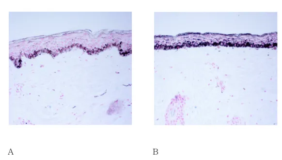
A. General histologic features
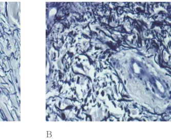
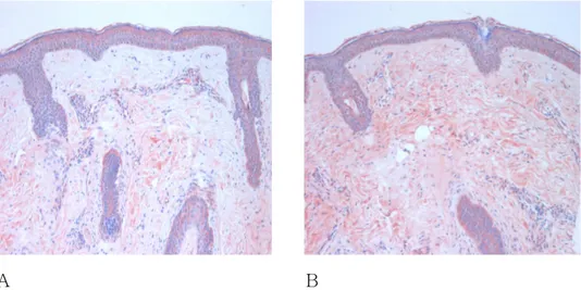
기미 병변의 표피는 인접 정상에 비해 다소 얇아져 있었고 표피능이 소실되어 편평한 모습을 보였으며, 진피는 인접 정상에 비해 일광 탄력변성이 증가되어 있었고 섬유아 세포 의 증식소견을 보였다(Fig. 1). A B Fig. Fig.Fig.
Fig. 1. Hematoxylin1. Hematoxylin1. Hematoxylin1. Hematoxylin----eosin stainseosin stainseosin stains.... ((((A) Perilesional normal skin, (B) Melasma skin. eosin stains Melasma skin showed flattened epidermis, loss of rete ridge, and increased solar elastosis in the dermis compared to perilesional normal skin (original magnification x 200).
B. Fontana-Masson 염색 : 멜라닌 양 병변의 표피부 멜라닌 색소는 인접 정상에 비해 주로 기저층 부위에 현저하게 증가되 어 있었다(Fig. 2). A B Fig. 2. Fontana Fig. 2. Fontana Fig. 2. Fontana
Fig. 2. Fontana---Masson stains-Masson stainsMasson stainsMasson stains.... ((((A) Perilesional normal skin, (B) Melasma skin. The melanin pigment on lesional epidermis increased compared to those of perilesional normal skin (original magnification x 200).
C. NKI/beteb 면역조직화학염색 : 멜라닌 세포 수
병변의 표피부 멜라닌 세포는 기저층을 따라 인접 정상보다 증가되어 있었다(Fig. 3).
A B
Fig. 3. NKI/beteb immunostains Fig. 3. NKI/beteb immunostainsFig. 3. NKI/beteb immunostains
Fig. 3. NKI/beteb immunostains. . . . ((((A) Perilesional normal skin, (B) Melasma skin. The number of epidermal melanocyte on lesional skin increased compared to perilesional normal skin (original magnification x 200).
D. Masson trichrome염색 : 콜라겐 섬유
콜라겐 섬유는 병변과 인접정상 진피에서 별다른 차이가 없었다(Fig. 4).
A B
Fig. 4. Masson Trichrome stain Fig. 4. Masson Trichrome stainFig. 4. Masson Trichrome stain
Fig. 4. Masson Trichrome stainssss. . . . ((((A) Perilesional normal skin, (B) Melasma skin. The collagen fibers(blue color) appeared normal feature in both perilesional normal skin and melasma skin (original magnification x 200).
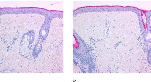
E. Verhoeff-van Gieson 염색 : 탄력섬유 탄력섬유는 인접정상에 비해 병변부의 진피에서 더 두껍고 분절되어 있는 소견을 보 였다(Fig. 5). A B Fig. Fig. Fig.
Fig. 555. 5. . . VerhoeffVerhoeffVerhoeffVerhoeff--van Gieson stain--van Gieson stainvan Gieson stainvan Gieson stainssss. . . ((((A) Perilesional normal skin, (B) Melasma skin. . Melasma skin showed thick and more fragmented elastic fibers than in perilesional normal skin (original magnification x 200).
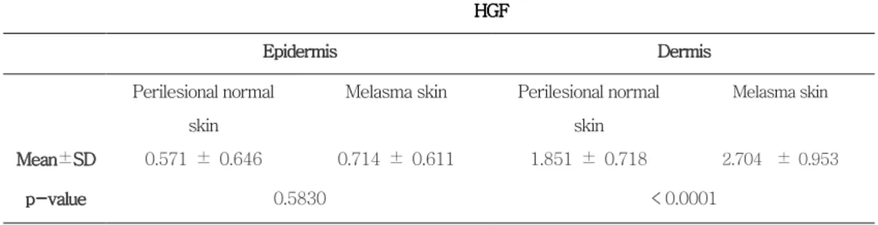
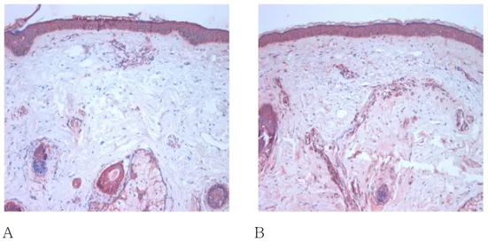
F. HGF 의 면역조직화학염색
표피의 HGF염색 정도는 인접 정상(0.571±0.646)과 병변(0.714±0.611)간 별 차 이를 보이지 않았으나(p=0.583), 진피에서는 인접 정상(1.851±0.718)에 비해 병변 (2.704±0.953)에서 현저하게 증가되어 있었다(p<0.0001).(Fig. 6, Fig. 7, Table 2).
A B
Fig. 6 Fig. 6 Fig. 6
Fig. 6. HGF immunostains. HGF immunostains. HGF immunostains. . HGF immunostains. . . ((((A) Perilesional normal skin, (B) Melasma skin. Melasma skin showed stronger intensity of HGF in the dermis than perilesional normal skin (original magnification x 200).
Tab TabTab
Table 2. The comparision of le 2. The comparision of le 2. The comparision of le 2. The comparision of reactivity about HGF.reactivity about HGF.reactivity about HGF.reactivity about HGF.
HGF HGF HGF HGF Epidermis Epidermis Epidermis
Epidermis DermisDermisDermisDermis Perilesional normal
skin
Melasma skin Perilesional normal skin Melasma skin Mean Mean Mean Mean±SDSDSD SD 0.571 ± 0.646 0.714 ± 0.611 1.851 ± 0.718 2.704 ± 0.953 p p p
p---value-valuevaluevalue 0.5830 < 0.0001 0 : no staining, 1 : weak staining, 2 : mild staining, 3 : moderate staining, 4 : strong staining
Fig. 7 Fig. 7Fig. 7
Fig. 7. The comparision of dermal reactivity about HGF. The comparision of dermal reactivity about HGF. The comparision of dermal reactivity about HGF. . The comparision of dermal reactivity about HGF. . . The expression of HGF was significantly increased in the melasma skin compared to perilesional normal skin.
normal melasma 0 1 2 3 4 *
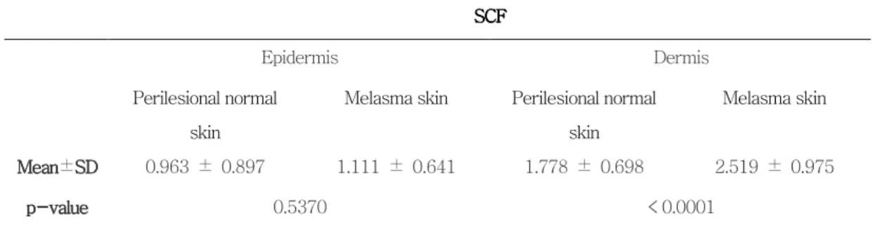
G. SCF 의 면역조직화학염색
표피의 SCF 염색 정도 역시 인접 정상(0.963±0.897)과 병변(1.111±0.641)간 별 차 이를 보이지 않았으나(p=0.537), 진피에서는 인접 정상(1.778±0.698)에 비해 병변
(2.519±0.975)에서 현저하게 증가되었다(p<0.0001)(Fig. 8, Fig. 9, Table 3).
A B
Fig. 8 Fig. 8 Fig. 8
Fig. 8. SCF immunostains. SCF immunostains. SCF immunostains. . SCF immunostains. . . ((((A) Perilesional normal skin, (B) Melasma skin. Melasma skin showed stronger intensity of SCF in the dermis than perilesional normal skin (original magnification x 200).
Table 3 Table 3Table 3
Table 3. The comparisi. The comparisi. The comparisi. The comparision of on of on of on of reactivity about SCreactivity about SCreactivity about SCreactivity about SCF.F.F. F.
SCF SCF SCF SCF Epidermis Dermis Perilesional normal skin
Melasma skin Perilesional normal skin Melasma skin Mean Mean Mean Mean±SDSDSDSD 0.963 ± 0.897 1.111 ± 0.641 1.778 ± 0.698 2.519 ± 0.975 p pp
p----valuevaluevalue value 0.5370 < 0.0001 0 : no staining, 1 : weak staining, 2 : mild staining, 3 : moderate staining, 4 : strong staining
Fig. 9. The comparision of derma Fig. 9. The comparision of dermaFig. 9. The comparision of derma
Fig. 9. The comparision of dermal reactivity about SCF.l reactivity about SCF.l reactivity about SCF.l reactivity about SCF. The expression of SCF was significantly increased in the melasma skin compared to perilesional normal skin.
1 4 3 2 1 0 0 * normal melasma
G. TNF-α의 면역조직화학염색
TNF-α는 진피에서 염색이 되지 않았으며 표피에서는 인접 정상(1.630±1.006)에
비해 병변(1.481±1.014)에서 약간 감소한 소견을 보였으나, 유의한 수준은 아니었다(p= 0.3430)(Fig. 10, Fig. 11, Table 4).
A B
Fig. 10 Fig. 10Fig. 10
Fig. 10. TNF. TNF. TNF. TNF----ααα immunostainsα immunostains immunostains immunostains. . . . ((((A) Perilesional normal skin, (B) Melasma skin. Melasma skin showed slightly weaker intensity of TNF-α in the epidermis than perilesional normal skin (original magnification x 200).
Table 4 Table 4Table 4
Table 4. The comparisi. The comparisi. The comparisi. The comparision ofon ofon ofon of reactivity about TN reactivity about TNF reactivity about TN reactivity about TNFFF----ααα....α
TNF TNF TNF TNF----αααα
Epidermis Dermis
Perilesional normal skin Melasma skin Perilesional normal skin Melasma skin Mean Mean Mean Mean±SDSDSDSD 1.630 ± 1.006 1.481 ± 1.014 0 0 p pp
p----valuevaluevalue value 0.3430 0 0 : no staining, 1 : weak staining, 2 : mild staining, 3 : moderate staining, 4 : strong staining
Fig. 11. The comparision of epidermal reactivity about TNF Fig. 11. The comparision of epidermal reactivity about TNF Fig. 11. The comparision of epidermal reactivity about TNF
Fig. 11. The comparision of epidermal reactivity about TNF---α-ααα.... The expression of
TNF-α was slightly decreased in the melasma skin compared to perilesional normal skin. 0 1 2 3 4 normal melasma
H. IL-1α의 면역조직화학염색
IL-1α는 진피에서 염색 강도가 매우 낮았고 인접 정상(0.357±0.497)과 병변
(0.538±0.518)간 별 차이가 없었으며(p=0.3940), 표피에서는 인접 정상(1.379±
0.885)에 비해 병변(1.437±0.964)쪽이 약간 증가된 소견을 보였으나 유의한 수준은
아니었다(p=0.5020)(Fig. 12, Fig. 13, Table 5).
A B
Fig. 12 Fig. 12Fig. 12
Fig. 12. IL. IL. IL. IL----11α11ααα immunostains immunostains immunostains. immunostains. . . ((((A) Perilesional normal skin, (B) Melasma skin. Melasma skin showed slightly stronger intensity of IL-1α in the epidermis than perilesional normal skin (original magnification x 200).
Table 5 Table 5Table 5
Table 5. The comparisi. The comparisi. The comparision. The comparisiononon of of of of reactivity about IL reactivity about IL- reactivity about IL reactivity about IL---1111αααα....
IL IL IL IL----1111αααα
Epidermis Dermis
Perilesional normal skin Melasma skin Perilesional normal skin Melasma skin Mean Mean Mean Mean±SDSDSDSD 1.379 ± 0.885 1.437 ± 0.964 0.357 ± 0.497 0.538 ± 0.518 p pp
p----valuevaluevalue value 0.3940 0.5020 0 : no staining, 1 : weak staining, 2 : mild staining, 3 : moderate staining, 4 : strong staining
Fig. 13. The comparision of epidermal reactivity about IL Fig. 13. The comparision of epidermal reactivity about IL Fig. 13. The comparision of epidermal reactivity about IL
Fig. 13. The comparision of epidermal reactivity about IL----111α1ααα.... The expression of IL-1α was slightly increased in the melasma skin compared to perilesional normal skin.
normal melasma 0 1 2 3 4
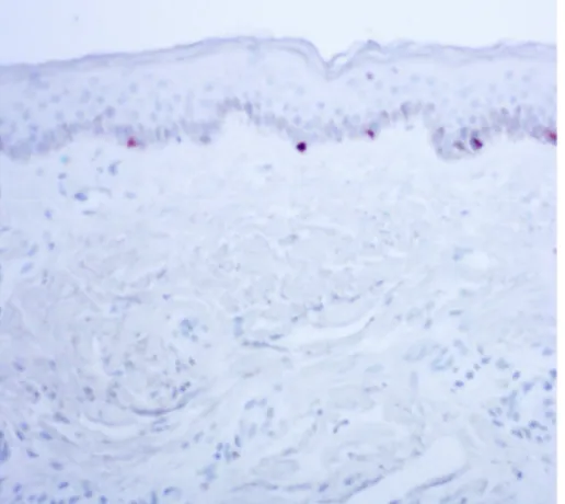
I. c-kit 의 면역조직화학염색
c-kit 은 인접 정상에 비해 병변의 멜라닌 세포에서 보다 더 증가되었다(Fig. 14). 컴
퓨터 영상분석 결과 착색 밀도는 인접 정상(18124,42±10443.82)에 비해 병변(25375.05
±10230.47)의 표피에서 유의한 수준으로 증가되어 있었다(p=0.0146). SA/EA 측정값 역
시 인접 정상(0.0807±0.0594)에 비해 병변(0.1365±0.0749)의 표피에서 유의한 수준으 로 증가되어 있었다(p=0.0086) (Fig. 14, Fig. 15, Table 6).
A B
Fig. 14 Fig. 14Fig. 14
Fig. 14. c. c. c. c----kit immunostainskit immunostainskit immunostains. kit immunostains. . . ((((A) Perilesional normal skin, (B) Melasma skin. The melanocytes of melasma skin showed stronger intensity of c-kit than those of perilesional normal skin (original magnification x 400).
Table 6 Table 6Table 6
Table 6. The comparisi. The comparisi. The comparisi. The comparision of epidermal reactivity about on of epidermal reactivity about on of epidermal reactivity about on of epidermal reactivity about cc-cc---kitkitkit. kit. . .
c cc c----kitkitkitkit O
OO
Optical densityptical densityptical densityptical density SA/EASA/EASA/EASA/EA Perilesional
normal skin
Melasma skin Perilesional normal skin Melasma skin Mean±SD 18124,42 ± 10443.82 25375.05 ± 10230.47 0.0807 ± 0.0594 0.1365 ± 0.0749 p-value 0.0146 0.0086 0 0.05 0.1 0.15 0.2 1 Fig. 15 Fig. 15Fig. 15
Fig. 15.... The comparision of The comparision of The comparision of The comparision of epidermal reactivityepidermal reactivityepidermal reactivity aboutepidermal reactivity about c about about c c- c--kit.-kit.kit. A : Optical density, B : kit. SA/EA. The density and stained area per epidermal area of c-kit was significantly
0 5000 10000 15000 20000 25000 30000 35000 * normal melasma normal melasma * A B
IV.
IV.
IV.
IV. 고
고
고
고 찰
찰
찰
찰
섬유아 세포는 진피의 주된 세포로 진피의 구성 성분인 교원섬유, 탄력섬유 및 기질을 생산하는 역할을 담당하는 세포이다. 따라서 그 동안 섬유아 세포는 노화와 관련한 진피의 콜라겐 remodeling 에서의 역할을 중심으로 연구되어 왔다. 그러나 최근 연구에 따르면 섬유아 세포가 분비하는 사이토카인이 표피 색소형성에 역할을 미치는 것으로 나타나 섬 유아 세포가 표피 색소형성에도 중대한 역할을 담당할 것으로 생각되고 있다. 실제 임상에 서는 섬유아 세포에 이상이 있는 질환에서 과색소침착이 동반되는 것을 볼 수 있다. 즉 loose skin 과 과색소침착을 보이는 Costello syndrome 의 조직소견에서 표피의 과색소침 착과 진피의 탄력섬유 손상이 동시에 나타나는 것이 확인되었다(Hataochi 등, 2000). 섬 유아 세포 기원 종양의 경우 표피의 과색소침착이 특징적으로 나타나는데, 제 1 형 신경 섬유종증 환자의 밀크커피 반점으로부터 분리한 섬유아 세포에서 HGF 와 SCF 의 발현이 증가되어 있었고(Okazaki 등, 2003), 피부섬유종(dermatofibroma)에서 표피의 과색소침 착 역시 진피의 섬유아 세포가 분비하는 HGF 와 SCF 때문인 것으로 밝혀졌다(Shishido 등, 2001). 또한 전신경화증(systemic sclerosis)의 색소침착이 섬유아 세포가 분비하는 SCF 에 의한 것이라는 보고도 있다(Kihira 등, 1998). Yaar 와 Gilchrest 는 자외선에 의 한 멜라닌 합성의 증가가 각질형성세포 단독의 monolayer culture 에서는 나타나지 않으 며 dermal equivalent 가 같이 있어야 색소침착이 가능하다고 하여 색소형성에서 섬유아 세포의 중요성을 증명하였다(Yaar M 와 Gilchrest, 1991). 본 연구에서는 각질형성세포나 멜라닌 세포의 배양을 통한 in vitro 실험이 아닌, 실제 기미환자의 인접 정상과 병변 부위의 조직으로 시행한 in vivo 실험을 통해 기미의 발생기 전을 밝히고자 하였다. 그 결과, 인접 정상에 비해 기미 병변의 진피에서 섬유아 세포 기 원 사이토카인인 HGF 와SCF 의 발현이 섬유아 세포를 중심으로 증가되어 있음을 면역조 직화학염색을 통해 알 수 있었다. 각질형성세포는 membrane bound form SCF 을 분비하나 soluble form SCF 는 자외선에 노출되는 조건 하 에서도 분비되지 않는다(Hachiya 등, 2001), 노인성 흑자의 표피에서도 soluble form SCF 는 발견되지 않았다 (Hattori 등, 20 04). 반면 진피의 섬유아 세포는 soluble form SCF 를 분비할 수 있으며, 밀크 커피반점 이나 피부섬유종에서 증가되어 있는 것도 soluble form SCF 으로서 진피와 기저막을 통해 분산되어 표피 멜라닌 세포를 활성시키게 된다고 하였다(Okazaki 등, 2003; Shishido 등, 2001). 그러므로 기미 병변 진피에서도 증가되어 있는 SCF 는 섬유아 세포에 의해 분비 된 soluble form SCF 로서 이것이 표피의 멜라닌 세포에 영향을 준 것이라고 생각해 볼 수 있었다. 반면, 표피의 경우 염색강도가 인접 정상과 기미 병변 간에 유의한 차이가 없 었으므로 각질형성세포가 분비한 membrane bound form SCF 는 기미의 병인에 큰 영향 을 주었을 것으로 생각되지 않는다.
c-kit 은 SCF 의 수용체로서, 자외선에 노출된 멜라닌 세포와 노인성 흑자의 표피에 서 SCF 와 c-kit 이 증가되었다는 보고가 있으며(Hachiya 등, 2001; Hattori 등, 2004), c-kit 의 억제항체(inhibitory antibody)를 표피 하 주입시 자외선에 노출시켜도 색소침착 이 야기되지 않았다는 연구 결과도 있다(Imokawa 등, 1986). 본 연구 결과도 c-kit 의 발현이 인접 정상에 비해 기미 병변의 멜라닌 세포에서 증가되어 있다는 것을 이미지 분 석을 통해 알 수 있었다. 즉 진피의 섬유아 세포로부터 기원한 SCF 의 증가에 의해 표피 기저층의 멜라닌 세포의 c-kit 이 활성화되어 멜라닌 세포의 활성을 유도하였을 것으로 추측할 수 있다. TNF-α는 인접정상에 비해 기미 병변의 각질형성세포에서 약간 감소되어 있었는데 통계학적으로 의의가 없었고, IL-1α의 경우는 그와 반대로 인접 정상에 비해 기미 병변 의 각질형성세포에서 약간 증가되어 있었는데 역시 통계학적으로 의의가 없었다. TNF-α 와IL-1α가 색소형성에 미치는 영향은 보고자 마다 차이가 있어 아직 확실히 밝혀져 있 지 않다. TNF-α와 IL-1α은 섬유아 세포에서 HGF 와 SCF 의 발현을 증가시킨다고 알
가 ET-1 의 발현을 증가시켜 색소형성에 중요한 역할을 한다고 보고되었다(Imokawa 등, 1992). 반면 지루성 각화증이나 노인성 흑자 병변에서는 각질형성 세포에서 IL-1α의 발현은 감소되어 있었고TNF-α의 발현은 ET-1 과 함께 증가되어 있음이 밝혀져 있다 (Kadono 등, 2001; Manaka 등, 2001). 본 연구 결과 자외선이 병인의 하나라고 여겨지 는 기미 조직의 경우 표피 각질형성세포에서 TNF-α나 IL-1α의 발현이 인접 정상과 유의한 차이가 없는 것을 보아 기미의 병인에는 각질형성세포 기원 TNF-α나 IL-1α의 역할이 중요한 것으로 생각되지 않았다. 또한 기미조직의 진피에서 TNF-α가 염색되지 않았고 IL-1α는 인접 정상과 별 차이가 없는 것으로 보아 섬유아 세포 기원 TNF-α와 IL-1α이 기미에서HGF 와 SCF 의 발현 증가 및 기미 병변 유발에 큰 영향을 미치지 않 을 것으로 생각되었다. 본 연구 결과, 자외선에 의해 변성된 진피의 섬유아 세포로부터 생성된 사이토카인인 HGF 와 SCF 가 표피의 멜라닌 세포의 활성을 유도하여 기미를 유발하였을 것으로 생각 되었다. 이는 기미 발생에서 진피의 효과를 밝혀서, 지금까지 밝혀져 있지 않은 기미 유발 의 새로운 이론적 근거를 제시하는 결과이다. 또한, 피부 색소질환의 병인을 각질형성세포 와 멜라닌 세포에 국한시키지 않고 진피 구성세포인 섬유아 세포와 표피 멜라닌 세포와의 유기적 관계에 의한 현상으로 해석함으로써 많은 다른 색소질환의 병인을 밝히는데도 기 여할 것으로 기대된다.
V.
V.
V.
V. 결
결
결 론
결
론
론
론
1. 기미 병변의 진피에서 인접 정상에 비해 섬유아 세포 기원 사이토카인인 HGF 와 SCF 의 발현이 유의하게 증가되어 있었다. 2. SCF 의 수용체인 c-kit 의 발현도 기미 병변의 멜라닌 세포에서 인접 정상에 비해 의 의있게 증가되어 있었다. 3. TNF-α와 IL-1α는 인접정상에 비해 기미 병변의 각질형성세포에서 약간 감소 또는 증가되어 있었으나, 유의한 수준은 아니었다. 이상의 결과로 자외선에 의해 변성된 기미 병변 진피의 섬유아 세포로부터 생성된 사이토카인인 HGF 와 SCF 가 멜라닌 세포의 활성을 유도하여 기미를 유발하였을 것으로 생각되었다.참
참
참
참 고
고
고
고 문
문
문
문 헌
헌
헌
헌
1. Archambault M, Yaar M, Gilchrest BA: Keratinocytes and fibroblasts in a human skin equivalent model enhance melanocyte survival and melanin synthesis after ultraviolet irradiation. J Invest Dermatol 104:859-867, 1995
2. Buffey JA, Messenger AG, Taylor M, Ashcroft AT, Westgate GE, MacNeil S: Extracellular matrix derived from hair and skin fibroblasts stimulates human skin melanocyte tyrosinase activity. Br J Dermatol 131:836-842, 1994
3. Hachiya A, Kobayashi A, Ohuchi A, Takema Y, Imokawa G: The paracrine role of stem cell factor / c-kit signaling in the activation of human melanocytes in
ultraviolet-B-induced pigmentation. J Invest Dermatol 116:578-586, 2001
4. Halaban R: The regulation of normal melanocyte proliferation. Pigment Cell Res 13:4-14, 2000
5. Hataochi A, Nagayama H, Kuroda K, Shinkai H, Ishikiriyama S, Kobayashi M, Kobayashi K: Costello syndrome with decreased gene expression of elastin in cultured dermal fibroblasts. Dermatology 201:366-369, 2000
is over-expressed in lentigo senilis: implication for the mechanism of hyperpigmentation. J Invest Dermatol 122: 1256-1265, 2004
7. Hedley SJ, Layton C, Heaton M, Chakrabarty KH, Dawson RA, Gawkrodger DJ, MacNeil S: Fibroblasts play a regulatory role in the control of pigmentation in reconstructed human skin from skin types I and II. Pigment Cell Res 15:49-56, 2002
8. Imokawa G, Kawai M, Mishima Y, Motegi I: Differential analysis of experimental hypermelanosis induced by UVB, PUVA and allergic contact dermatitis using brownish guinea pig model. Arch Dermatol Res 278:352-362, 1986
9. Imokawa G, Yada Y, Miyagishi M: Endothelins secreted from human keratinocytes are intrinsic mitogens for human melanocytes. J Biol Chem 267: 24675-24680, 1992
10. Imokawa G, Yada Y, Morisaki N, Kimura M: Biological characterization of human fibroblast-derived mitogenic factors for human melanocytes. Biochem J
15:1235-1239, 1998
11. Im S, Kim J, On WY, Kang WH: Increased expression of alpha-melanocyte-stimulating hormone in the lesional skin of melasma. Br J Dermatol
146:165-12. Kadono S, Manaka I, Kawashima M, Kobayashi T, Imokawa G: The role of the epidermal endothelin cascade in the hyperpigmentation mechanism of lentigo senilis. J Invest Dermatol 116:571-577, 2001
13. Kang WH, Yoon KH, Lee ES, Kim J, Lee KB, Yim H, Sohn S, Im S: Melasma: histopathological characteristics in 56 Korean patients. Br J Dermatol 146:228-237, 2002
14. Kihira C, Mizutani H, Asahi K, Hamanaka H, Shimizu M: Increased cutaneous immunoreactive stem cell factor expression and serum stem cell factor level in systemic scleroderma. J Dermatol Sci 20:72-78, 1998
15. Kunisada T, Yamazaki H, Hirobe T, Kamei S, Omoteno M, Tagaya H, Hemmi H, Koshimizu U, Nakamura T, Hayashi SI: Keratinocyte expression of transgenic hepatocyte growth factor affects melanocyte development, leading to dermal melanocytosis. Mech Dev 94:67-78, 2000
16. Manaka I, Kadono S, Kawashima M, Kobayashi T, Imokawa G: The mechanism of hyperpigmentation in seborrhoeic keratosis involves the high expression of endothelin-converting enzyme-1α and TNF-α, which stimulate secretion of endothelin 1. Br J Dermatol 145:895-903, 2001
factors secreted by fibroblasts: role in healing diabetic foot ulcers. Diabetes, obesity, and metabolism 1:265-279, 1999
18. Okazaki M, Yoshimura K, Suzuki Y, Uchida G, Kitano Y, Harii K, Imokawa G: The mechanism of epidermal hyperpigmentation in cafe-au-lait macules of neurofibromatosis type 1 (von Recklinghausen's disease) may be associated with dermal fibroblast-derived stem cell factor and hepatocyte growth factor. Br J Dermatol 148:689-697, 2003
19. Ortonne JP, Bahadoran P, Fitzpatrick TB, Mosher DB, Hori Y: Hypomelanosis and hypermelanosis. In Fitzpatrick’s Dermatology in general medicine. 6th ed, New
York, McGrow-Hill, pp868-869, 2003
20. Shishido E, Kadono S, Manaka I, Kawashima M, Imokawa G: The mechanism of epidermal hyperpigmentation in dermatofibroma is associated with stem cell factor and hepatocyte growth factor expression. J Invest Dermatol 117:627-633, 2001
21. Takano N, Kawakami T, Kawa Y, Asano M, Watabe H, Ito M, Soma Y, Kubota Y, Mizoguchi M: Fibronectin combined with stem cell factor plays an important role in melanocyte proliferation, differentiation and migration in cultured mouse neural crest cells. Pigment Cell Res 15:192-200, 2002
22. Teraki E, Tajima S, Manaka I, Kawashima M, Imokawa G: Role of endothelin-1 in hyperpigmentation in seborrhoeic keratosis. Br J Dermatol 135:918-923, 1996
23. Yaar M, Gilchrest BA: Human melanocyte growth and differentiation: a decade of new data. J Invest Dermatol 97:611-617, 1991
- ABSTRACT -
The role of fibroblast
The role of fibroblast
The role of fibroblast
The role of fibroblast-
-
-derived cytokines on melasma
-
derived cytokines on melasma
derived cytokines on melasma
derived cytokines on melasma
Ji Yeon Kim
Department of Medical Sciences The Graduate School, Ajou University
(Supervised by Assistant Professor Hee Young Kang)
Melasma is developed or aggravated by UV radiation, sex hormone, and genetic predisposing factors, however, the exact mechanism of pathogenesis is not clearly demonstrated. Previous studies have been bringing focus into the pigmentation formed by interaction between keratinocytes and melanocytes. Recently, it was reported that dermal fibroblasts played an important roles on the cutaneous pigmentary disorders. Therefore, we studied the role of fibroblast-derived cytokines on the development of melasma. We collected the specimens of perilesional normal site and lesional site by 2 mm punch biopsy technique on the faces of 50 melasma patients. We performed on the specimens to stain of hematoxylin-eosin, Fontana-Masson, Masson trichrome, Verhoeff-van Gieson and immunohistochemical stain of NKI/beteb, hepatocyte growth factor (HGF), stem cell factor (SCF), tumor necrosis
analyzed by estimation of three dermatologists and computerized image analysis. There were marked epidermal thinning of the lesional sites than those of perilesional normal sites, and solar elastosis at lesional dermis were significantly increased. The expression of HGF and SCF significantly increased at lesional dermis and the expression of c-kit was considerably increased at melanocytes of lesional epidermis. However, TNF-α was slightly decreased and IL-1α increased in expression at epidermal melasma skin, but no significant differences as compared to perilesional normal skin. These results may indicate that the dermal fibroblast-derived cytokines, such as HGF and SCF induced activation of melanocytes and subsequently was brought about the development of the melasma.
Key Words: melasma, fibroblast, immunohistochemistry, HGF, SCF, c-kit