Clinical Study
Medial Subluxation or Dislocation of the Biceps on Magnetic
Resonance Arthrography Is Reliably Correlated with Concurrent
Subscapularis Full-Thickness Tears Confirmed Arthroscopically
Ji-Sang Yoon, Sung-Jae Kim, Yun-Rak Choi, Wonyong Lee,
Sang Ho Kim, and Yong-Min Chun
The Department of Orthopaedic Surgery, Arthroscopy and Joint Research Institute, Severance Hospital, Yonsei University College of Medicine, Seoul, Republic of Korea
Correspondence should be addressed to Yong-Min Chun; min1201@hanmail.net
Received 15 June 2018; Revised 17 August 2018; Accepted 28 August 2018; Published 9 September 2018 Academic Editor: Sae Hoon Kim
Copyright © 2018 Ji-Sang Yoon et al. This is an open access article distributed under the Creative Commons Attribution License, which permits unrestricted use, distribution, and reproduction in any medium, provided the original work is properly cited.
Background. The purpose of this study was to investigate the relationship between biceps medial subluxation/dislocation on
the magnetic resonance arthrography (MRA) imaging and subscapularis full-thickness tear confirmed arthroscopically. We hypothesized that presence of a biceps medial subluxation or dislocation would strongly indicate a subscapularis full-thickness tear.
Methods. A total of 432 consecutive patients who underwent arthroscopic repair for rotator cuff tears with/without subscapularis
tears at our institute were retrospectively reviewed. The inclusion criterion of this study was preoperative MRA images taken within 6 months of arthroscopic repair. The presence of medial subluxation/dislocation was evaluated on the preoperative MRA images, and subscapularis tear was confirmed on arthroscopic examination. Results. Biceps subluxation/dislocation was identified in 46 of the 432 patients on MRA. Forty-five of these 46 patients also had a subscapularis full-thickness tear identified in arthroscopic examination. Among the 386 patients who did not have biceps subluxation or dislocation, 54 patients had a subscapularis thickness tear diagnosed arthroscopically. The presence of a biceps subluxation/dislocation could predict a subscapularis full-thickness tear with sensitivity of 45% (45/99), specificity of 99% (332/333), positive predictive value of 98% (45/46), negative predictive value of 86% (332/386), and accuracy of 87% (377(45 +332)/432). Conclusion. Medial subluxation/dislocation of the biceps on MRA images was highly associated with a concurrent subscapularis full-thickness tear which was confirmed arthroscopically. This association had 99% specificity and 98% positive predictive value. Therefore, if a biceps subluxation/dislocation is identified on MRA images, there is a high chance that a concurrent subscapularis full-thickness tear exists.
1. Introduction
Various imaging modalities can be used to detect rotator cuff tears. In particular, magnetic resonance imaging (MRI) is an effective tool with high sensitivity and specificity for diagnosing supraspinatus and infraspinatus tears. However, several studies have indicated that MRI and even magnetic resonance arthrography (MRA) do not reliably predict sub-scapularis tears [1–4]. There are several physical examinations used to detect subscapularis tears, including the lift-off test, belly press test, and bear hug test; however, these maneuvers have relatively low sensitivity [5, 6]. Unfortunately, many subscapularis tears may be overlooked clinically.
At an early stage of a subscapularis full-thickness tear without retraction, it is often difficult to differentiate between a normal insertion and tear. This is particularly true if the tear is limited to the upper portion of the tendon. If the MR image does not exactly capture the torn part of the tendon, it may appear to attach well to the lesser tuberosity. Thorough and meticulous investigation of MR images may identify a subtle change in signal intensity (Figure 1).
The superolateral border of the subscapularis tendon is connected to the biceps pulley, which precludes medial displacement of the biceps. Considering that subscapularis tears are likely to involve the biceps pulley and lead to medial displacement of the biceps subsequently, a biceps subluxation
Volume 2018, Article ID 2674061, 5 pages https://doi.org/10.1155/2018/2674061
Figure 1: A biceps medial dislocation protrudes out of bicipital groove on an axial oblique image of MR arthrography in the right shoulder. Although the insertion of the subscapularis tendon on the footprint seems to be intact, a full-thickness tear at the upper portion of the subscapularis tendon was identified arthroscopically. There is increased signal intensity (arrow head) at the tear site.
or dislocation can be a reliable clue of an underlying sub-scapularis tear [1, 2, 7–9].
The purpose of this study was to investigate the relation-ship between biceps medial subluxation/dislocation on the MRA images and subscapularis full-thickness tear which was confirmed arthroscopically. We hypothesized that presence of the biceps medial subluxation/dislocation would strongly indicate a concurrent subscapularis full-thickness tear.
2. Materials and Methods
We retrospectively reviewed a consecutive series of patients who underwent arthroscopic repair for rotator cuff tears either with or without subscapularis full-thickness tears at our institute between January 2010 and August 2015. The inclusion criterion of this study was preoperative MR arthrography (MRA) (3.0T MR imager, Magnetom Tim Trio, Siemens, Erlangen, Germany) taken in neutral position of the affected shoulder at our institute, within 6 months prior to arthroscopic repair [1, 7]. The exclusion criteria were MRA images obtained at other institutes and history of previous shoulder surgery. Biceps subluxation and dislocation were defined on axial images as biceps displacement over the lesser tuberosity and bicipital groove (subluxation) (Figure 2(a)) or completely out of the bicipital groove (dislocation) (Fig-ure 2(b)). An experienced shoulder specialist (one of senior authors) who was blinded to arthroscopic findings reviewed MRA images of the included patients and determined the presence of the biceps subluxation/dislocation on the axial images. This study was approved by our institutional review board, and the need for informed consent was waived.
Arthroscopic evaluation was performed by a single sur-geon in a beach-chair position under general anesthesia. Both 30∘and 70∘arthroscopes were used to investigate the articular and bursal side of the subscapularis insertion. With use of the 70∘arthroscope, as indicated by several studies, the footprint of the subscapularis as well as tear within the bicipital groove can be more clearly visualized [10–12]. Through the standard
Subluxation/Dislocation (+) 45 1 46 Subluxation/Dislocation (-) 54 332 386
Total 99 333 432
posterior portal, the footprint of the subscapularis tendon, location of the biceps, their relation to the biceps pulley, and structural integrity of the biceps pulley itself were investigated (Figure 3).
We defined the full-thickness tear of the subscapularis as complete detachment of the subscapularis from its foot-print on the lesser tuberosity, which was confirmed under arthroscopic examination. Despite a full-thickness tear, in many cases, the torn edge of the subscapularis tendon was connected to the transverse ligament over the bicipital groove and, subsequently, the torn subscapularis tendon remained in place without any medial retraction. These were also regarded as full-thickness tears.
3. Results
This study included 432 patients (189 male and 243 female), and their mean age at the time of surgery was 63.2 years (range, 47 to 80 years). Among them, biceps subluxa-tion/dislocation was identified in 46 patients on preopera-tive MRA (intraclass correlation coefficient of intraobserver reliability = 0.857). Of these 46 patients, 45 patients also had a subscapularis full-thickness tear identified on arthroscopic examination. Of 386 patients who did not have biceps sub-luxation/dislocation on preoperative MRA, 54 patients had subscapularis full-thickness tears diagnosed arthroscopically (Table 1). The presence of a biceps subluxation/dislocation could predict the subscapularis full-thickness tear with sen-sitivity of 45% (45/99), specificity of 99% (332/333), positive predictive value of 98% (45/46), negative predictive value of 86% (332/386), and accuracy of 87% (377(45 +332)/432).
4. Discussion
Subscapularis tears seem to be slightly different than supraspinatus tears. A supraspinatus full-thickness tear can be easily identified because, regardless of the degree of retraction, complete detachment of the cuff from its footprint is detected on MR imaging. However, even in full-thickness subscapularis tears, the subscapularis tendon often appears to attach to its footprint in the setting of biceps sublux-ation/dislocation. This may be attributed to the transverse ligament over the bicipital groove. The transverse ligament seems to hold the subscapularis tendon in place, preventing it from retracting medially even after a full-thickness tear [13]. Therefore, unlike other supraspinatus or infraspinatus tears, it is often difficult to clearly identify subscapularis tears on the MR images. Consistent with our original hypothesis, biceps subluxation/dislocation was highly associated with concur-rent subscapularis full-thickness tear. This association has
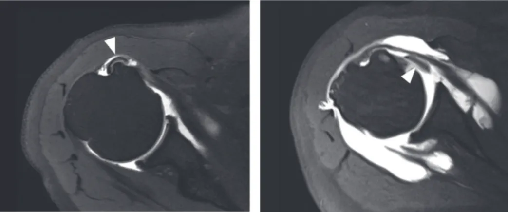
Figure 2: (a) A biceps medial subluxation (white arrow head) over the bicipital groove and lesser tuberosity, right shoulder. (b) A biceps medial dislocation (white arrow head) in the setting of a subscapularis full-thickness tear, right shoulder.
Figure 3: (a) Biceps medial subluxation. The biceps is located behind the torn subscapularis tendon, viewed from the posterior portal, right shoulder. (b) Biceps medial dislocation. The biceps is placed behind the torn subscapularis tendon and completely out of the bicipital groove, viewed from the posterior portal, right shoulder.
both a high positive predictive value and specificity. On the other hand, there were also many subscapularis full-thickness tears in the absence of biceps subluxation/dislocation. There-fore, the sensitivity of this association was low in this study. Nevertheless, the negative predictive value and accuracy were relatively high.
Interestingly, Shi et al. recently reported that the diagnos-tic value of biceps subluxation lies in the negative predictive value; if there is no biceps subluxation, then it is unlikely that there is a subscapularis full-thickness tear [7]. In contrast to the current study, the group reported a positive predictive value of 35% and negative predictive value of 97%; among 26 patients diagnosed with biceps long head subluxation, only 9 patients had full-thickness subscapularis tears confirmed arthroscopically. In our data, biceps subluxation/dislocation in the absence of subscapularis full-thickness tear was only one case and it seems to be rare, although there are previous case reports that describe biceps dislocation without rotator cuff tears [14, 15]. Nevertheless, the incidence of biceps subluxation without subscapularis tear in their study was relatively high. Based on their study and current study, the accuracy itself for the diagnosis of the biceps subluxation or dislocation based on MR axial image seems to be important for predicting the subscapularis full-thickness tear. In the current study, its accuracy was 96.7 % when employing the 3.0
T MRA, although direct comparison was not feasible because there was no comment on this issue in other studies.
Lafosse et al. used arthroscopic static and dynamic evaluation to classify biceps instability in rotator cuff tears as anterior or posterior instability [16]. In their study, anterior instability of the biceps tendon, which is consistent with cur-rent biceps subluxation/dislocation over the bicipital groove, was often related to a subscapularis tear. In contrast, posterior instability was more related to a supraspinatus tear. Among 200 consecutive cases, anterior and anteroposterior biceps instability were identified in as many as 26% of cases, which is a higher rate than that in the current study, and in others [7, 9, 17]. This finding is attributable to the fact that our study and the others mentioned exclusively addressed anterior static instability and not posterior or dynamic instability.
Walch et al. reported several types of biceps subluxa-tions/dislocations that were associated with the status of the adjacent subscapularis tear [9]. Among the various types, there was a biceps dislocation inside a subscapularis tear that resulted in an interstitial subscapularis partial-thickness tear. In the current study, there were similar cases, although there was no biceps subluxation/dislocation; the tear existed within the bicipital groove, which could be only identified using a 70∘ arthroscope into the bicipital groove (Figure 4). It was very difficult to identify this lesion on preoperative MR image,
Figure 4: A subscapularis interstitial tear identified within the bicipital groove, viewed from the posterior portal using a 70∘ arthroscope, right shoulder.
Figure 5: A biceps medial subluxation without a subscapularis full-thickness tear, right shoulder. A subscapularis partial-full-thickness tear (white arrow head) within the bicipital groove was identified.
including MRA. In addition, there were no instances of biceps subluxation/dislocation in interstitial subscapularis partial-thickness tears in the current study.
The vast majority (90%) of subscapularis tears started from the articular side and cephalad portion of the tendon [1, 2, 18, 19]. Therefore, biceps medial subluxation/dislocation appears to be a result of subscapularis full-thickness tears, even though interstitial or bursal side partial-thickness sub-scapularis tears can exist. It is likely that the subsub-scapularis tear starts from the articular side as a partial-thickness tear and extends toward the lateral side. Finally, the tear may disrupt the medial sling of the biceps, connected with the bicipital groove. Eventually, the biceps can be medially subluxated or dislocated through an unstable biceps sling. Therefore, when biceps medial subluxation/dislocation is identified, causative subscapularis full-thickness tear seems to be an inevitable consequence. In the current study, there was only one case of biceps subluxation diagnosed on preoperative MR image without a concomitant subscapularis full-thickness tear (Figure 5). Arthroscopic examination revealed subscapu-laris partial-thickness tear within the bicipital groove with an intact articular portion of the subscapularis tendon.
This study has several limitations. First, this study evalu-ated just static anterior biceps instability, rather than assessing
limited 3.0 T MRA image evaluated in our institute to get the uniformity and accuracy of the images. However, in contrast, in a clinical setting, it might have been more realistic to include MR images from various sources.
5. Conclusion
Medial subluxation or dislocation of the biceps on MRA images was highly associated with a concurrent subscapularis full-thickness tear confirmed arthroscopically. This associa-tion had 99% specificity and 98% positive predictive value. Therefore, if a biceps subluxation/dislocation is identified on MRA images, there is a high chance that a concurrent subscapularis full-thickness tear exists.
Data Availability
The data used to support the findings of this study are available from the corresponding author upon request.
Ethical Approval
This study was conducted following the approval of Insti-tutional Review Board of Severance Hospital, Yonsei 180 University (IRB no. 2015-3041-001).
Conflicts of Interest
The authors declare that there are no conflicts of interest to report regarding the work and publication of this study.
Acknowledgments
The authors are grateful to Man-Seo Cho (research assistant and medial illustrator) for his help with the figures.
References
[1] C. R. Adams, P. C. Brady, S. S. Koo et al., “A systematic approach for diagnosing subscapularis tendon tears with preoperative magnetic resonance imaging scans,” Arthroscopy - Journal of
Arthroscopic and Related Surgery, vol. 28, no. 11, pp. 1592–1600,
2012.
[2] C. R. Adams, J. D. Schoolfield, and S. S. Burkhart, “Accuracy of preoperative magnetic resonance imaging in predicting a subscapularis tendon tear based on arthroscopy,” Arthroscopy
- Journal of Arthroscopic and Related Surgery, vol. 26, no. 11, pp.
1427–1433, 2010.
[3] A. Foad and C. A. Wijdicks, “The accuracy of magnetic resonance imaging and magnetic resonance arthrogram versus arthroscopy in the diagnosis of subscapularis tendon injury,”
Arthroscopy - Journal of Arthroscopic and Related Surgery, vol.
[4] G. Garavaglia, H. Ufenast, and E. Taverna, “The frequency of subscapularis tears in arthroscopic rotator cuff repairs: A retrospective study comparing magnetic resonance imaging and arthroscopic findings,” International Journal of Shoulder
Surgery, vol. 5, no. 4, pp. 90–94, 2011.
[5] J. R. H. Barth, S. S. Burkhart, and J. F. De Beer, “The Bear-Hug Test: A New and Sensitive Test for Diagnosing a Subscapularis Tear,” Arthroscopy - Journal of Arthroscopic and Related Surgery, vol. 22, no. 10, pp. 1076–1084, 2006.
[6] J. P. Yoon, S. W. Chung, S. H. Kim, and J. H. Oh, “Diagnostic value of four clinical tests for the evaluation of subscapularis integrity,” Journal of Shoulder and Elbow Surgery, vol. 22, no. 9, pp. 1186–1192, 2013.
[7] L. L. Shi, M. G. Mullen, M. T. Freehill, A. Lin, J. J. P. Warner, and L. D. Higgins, “Accuracy of long head of the biceps subluxation as a predictor for subscapularis tears,” Arthroscopy - Journal of
Arthroscopic and Related Surgery, vol. 31, no. 4, pp. 615–619, 2015.
[8] Y. Morag, J. A. Jacobson, G. Shields et al., “MR arthrography of rotator interval, long head of the biceps brachii, and biceps pulley of the shoulder,” Radiology, vol. 235, no. 1, pp. 21–30, 2005. [9] G. Walch, L. Nov´e-Josserand, P. Boileau, and C. Levigne, “Subluxations and dislocations of the tendon of the long head of the biceps,” Journal of Shoulder and Elbow Surgery, vol. 7, no. 2, pp. 100–108, 1998.
[10] S. J. Kim, M. Jung, J. H. Lee, C. Kim, and Y. M. Chun, “Arthro-scopic repair of anterosuperior rotator cuff tears: InContinu-ity technique Vs. disruption of subscapularis supraspinatus tear margin comparison of clinical outcomes and structural integrity between the two techniques,” Journal of Bone and Joint
Surgery - American Volume, vol. 96, no. 24, pp. 2056–2061, 2014.
[11] A. J. Sheean, R. U. Hartzler, P. J. Denard, A. L¨adermann, B. T. Hanypsiak, and S. S. Burkhart, “A 70∘Arthroscope Significantly Improves Visualization of the Bicipital Groove in the Lateral Decubitus Position,” Arthroscopy - Journal of Arthroscopic and
Related Surgery, vol. 32, no. 9, pp. 1745–1749, 2016.
[12] P. J. Denard and S. S. Burkhart, “Arthroscopic Recognition and Repair of the Torn Subscapularis Tendon,” Arthroscopy
Techniques, vol. 2, no. 4, pp. e373–e379, 2013.
[13] S.-J. Kim, Y.-R. Choi, M. Jung, Y.-K. Yoon, and Y.-M. Chun, “Concomitant coracoplasty during arthroscopic subscapularis repair does not yield better clinical outcomes and structural integrity,” Knee Surgery, Sports Traumatology, Arthroscopy, vol. 26, no. 1, pp. 56–62, 2018.
[14] M. L. Gambill, T. S. Mologne, and M. T. Provencher, “Dis-location of the long head of the biceps tendon with intact subscapularis and supraspinatus tendons,” Journal of Shoulder
and Elbow Surgery, vol. 15, no. 6, pp. e20–e22, 2006.
[15] D. H. O’Donoghue, “Subluxing biceps tendon in the athlete,”
Clinical Orthopaedics and Related Research, vol. 164, pp. 26–29,
1982.
[16] L. Lafosse, Y. Reiland, G. P. Baier, B. Toussaint, and B. Jost, “Anterior and Posterior Instability of the Long Head of the Biceps Tendon in Rotator Cuff Tears: A New Classification Based on Arthroscopic Observations,” Arthroscopy - Journal of
Arthroscopic and Related Surgery, vol. 23, no. 1, pp. 73–80, 2007.
[17] A. Deutsch, D. W. Altchek, D. M. Veltri, H. G. Potter, and R. F. Warren, “Traumatic tears of the subscapularis tendon: Clinical diagnosis, magnetic resonance imaging findings, and operative treatment,” The American Journal of Sports Medicine, vol. 25, no. 1, pp. 13–22, 1997.
[18] G. Sakurai, J. Ozaki, Y. Tomita, T. Kondo, and S. Tamai, “Incom-plete tears of the subscapularis tendon associated with tears
of the supraspinatus tendon: Cadaveric and clinical studies,”
Journal of Shoulder and Elbow Surgery, vol. 7, no. 5, pp. 510–515,
1998.
[19] S. S. Burkhart and A. M. Tehrany, “Arthroscopic subscapularis tendon repair: Technique and preliminary results,” Arthroscopy:
The Journal of Arthroscopic and Related Surgery, vol. 18, no. 5, pp.