Detection of MYD88 L265P in patients with lymphoplasmacytic
lymphoma/Waldenstrom macroglobulinemia and other B-cell
non-Hodgkin lymphomas
Sang-Yong Shin
1, Seung-Tae Lee
4, Hyun-Young Kim
2, Chang-Hun Park
2, Hee-Jin Kim
2,
Jong-Won Kim
2, Seok Jin Kim
3, Won Seog Kim
3, Sun-Hee Kim
21
Department of Laboratory Medicine, Center for Diagnostic Oncology, Hospital and Research Institute, National Cancer Center, Goyang, 2Department of Laboratory Medicine & Genetics, 3Internal Medicine, Samsung Medical Center, Sungkyunkwan University School of Medicine, 4Department of Laboratory Medicine, Yonsei University College of Medicine, Seoul, Korea
p-ISSN 2287-979X / e-ISSN 2288-0011 http://dx.doi.org/10.5045/br.2016.51.3.181 Blood Res 2016;51:181-6. Received on April 2, 2016 Revised on April 24, 2016 Accepted on May 31, 2016 Background
Recent studies have identified a high prevalence of the MYD88 L265P mutation in lympho-plasmacytic lymphoma (LPL)/Waldenstrom macroglobulinemia (WM) cases, whereas low frequencies have been observed in other B cell non-Hodgkin lymphomas (NHLs).
Methods
We evaluated the sensitivity of the mutant enrichment 3’-modified oligonucleotide (MEMO)-PCR technique, a new detection method. We examined the MYD88 L265P mu-tation in a series of Korean patients with LPL/WM and other B cell NHLs in bone marrow aspirates, using the MEMO-PCR technique.
Results
The sensitivity of MEMO-PCR was estimated to be approximately 10‒16.7%. MYD88 L265P was detected in 21 of 28 LPL cases (75%) and only three of 69 B cell NHL cases (4.3%).
Conclusion
Although MEMO-PCR had relatively low sensitivity, we confirmed the high prevalence of the MYD88 L265P mutation in Korean LPL patients. Our study suggests the diagnostic value of MYD88 L265P for differentiating B-cell NHLs.
Key Words Lymphoplasmacytic lymphoma, MYD88 L265P, Aspirate, MEMO-PCR
*This research was supported by the Basic Science Research Program through the National Research Foundation of Korea (NRF) funded by the Ministry of Education, Science and Technology (NRF-2012R1A1A2043879). Correspondence to Seung-Tae Lee, M.D., Ph.D. Sun-Hee Kim, M.D., Ph.D.
Department of Laboratory Medicine, Yonsei University College of Medicine, 50, Yonsei-ro, Seodaemun-gu, Seoul 03722, Korea (S.T.L.)
Department of Laboratory Medicine & Genetics, Samsung Medical Center, Sungkyunkwan University School of Medicine, 81, Irwon-ro, Gangnam-gu, Seoul 06351, Korea (S.H.K.)
E-mail: S.T.L., LEE.ST@yuhs.ac S.H.K., sunnyhk@skku.edu Ⓒ 2016 Korean Society of Hematology
INTRODUCTION
Lymphoplasmacytic lymphoma/Waldenstrom macro-globulinemia (LPL/WM) is a relatively rare subgroup of non-Hodgkin lymphoma (NHL). Differential diagnosis of LPL/WM from other NHLs is somewhat difficult, especially from marginal zone lymphoma (MZL) or B cell lymphoma with plasmacytic differentiation [1, 2]. Recent studies have reported a high prevalence (80-100%) of the MYD88 L265P
mutation in LPL/WM compared to a low prevalence in other NHLs; therefore, testing for this mutation could be of diag-nostic value [3-5]. The incidence of LPL in Korea has been suggested to be low (0.8%) compared to that in western countries [6]. One recent study on Korean patients with LPL/WM also showed a relatively high prevalence (69%) of MYD88 L265P [7], whereas studies on other NHLs showed a low prevalence [8, 9].
The detection methods for MYD88 L265P differed among different studies, and included conventional Sanger
sequenc-Fig. 1. Principles of MEMO-PCR for MYD88 L265P detection. Blocking primers, complementary to the normal sequence, anneal to normal DNA, hampering PCR amplifica-tion. In the presence of a missense mutation, the binding affinity of the blocking primers to the mutant DNA is decreased due to the mis-match, and therefore, amplifica-tion of the mutant DNA is markedly enhanced.
ing, allele-specific PCR (AS-PCR), and real-time PCR assays [4, 10]. The specimens for molecular testing can be obtained from the lymph node, bone marrow aspirate, or peripheral blood samples. Since there is a high chance of inclusion of stromal and normal hematopoietic cells in such specimens, more sensitive methods will be required for accurate testing. In this study, we investigated the MYD88 L265P prevalence in Korean patients with LPL/WM and other B-cell NHLs using mutant enrichment 3ˊ-modified oligonucleotide (MEMO)-PCR and sequencing, a sensitive technique with low false-positive rates [11-13]. We observed a relatively high frequency of MYD88 L265P and determined the clinical utility of testing for this mutation.
MATERIALS AND METHODS
Patients and specimens
From a review of LPL/WL and other B-cell NHL cases diagnosed at the Samsung Medical Center between 2001 and 2014, we selected 97 cases with bone marrow involve-ment, including 28 LPL/WL and 69 other B-cell NHL cases. Patient demographics, clinical features, treatment histories, and hematologic, immunophenotypic, and cytogenetic find-ings were all comprehensively analyzed. To identify the deletion of 6q, we additionally performed fluorescence in situ hybridization (FISH) using the XL 6q21/6q23 probe (Metasystem, Altlussheim, Germany). For the evaluation of MEMO-PCR, samples were diluted with equimolar normal DNA to produce 1:1, 1:3, 1:5, 1:9, 1:17, 1:21, and 1:41 ratios of mutant DNA.
Mutant enrichment 3ˊ-modified oligonucleotide–PCR (MEMO-PCR) and sequencing
DNA was extracted from bone marrow aspirate slides using the QIAamp DNA Blood Mini Kit (Qiagen, Foster City, CA,
USA) after treating with proteinase K. To detect the MYD88
L265P by MEMO-PCR, we designed generic forward (5ˊ- CAGGTGCCCATCAGAAGC-3ˊ) and reverse (5ˊ-GAAGTTG GCATCTCCAGGAA-3ˊ) primers along with a blocking pri-mer (5ˊ-AAGCGACTGATCCCCATCAA-[C3 spacer]-3ˊ) em-ploying a C3 spacer at the 3ˊ end. PCR amplification was performed using these three primers (10, 10, and 50 pmol each, respectively), with the following amplification cycle: 94oC for 5 min, 40 cycles of the main reaction (94oC for 30 s, 59oC for 30 s, and 72oC for 60 s), and 72oC for 7 min. Downstream sequencing analysis was performed using the BigDye Terminator Cycle Sequencing Ready Reaction Kit on the ABI Prism 3130 Genetic Analyzer (Applied Biosystems, Foster City, CA, USA). Principles of MEMO-PCR for MYD88 L265P detection are shown in Fig. 1 [12].
Statistical analysis
Statistical analysis was performed using PASW Statistics 20.0 software (IBM, Armonk, New York, USA). To compare the clinical parameters between MYD88 L265P positive and negative groups, we performed a chi-square test for catego-rical variables and a Mann-Whitney test or student’s t-test for continuous variables. A P-value<0.05 was considered statistically significant.
RESULTS
Clinical characteristics of LPL/WM
Patient demographics and clinical characteristics of cases are summarized in Table 1. The mean age was 65 years and the male/female ratio was 1.8:1. Patients generally had mild anemia (mean 9.7 g/dL) with variable white blood cell and platelet counts. All patients had IgM-type monoclonal gammopathies, except for one patient with IgG-type. Chromosomal aberration was detected in three of 25 patients
Fig. 2. Sensitivity of MEMO-PCR (estimated to be approximately 10–16.7%) and sequencing analysis. Table 1. Demographics and clinical characteristics of LPL/WM.
LPL/WM
(N=28) Other NHL (N=69)
Age, years (mean±SD) 65±13.0 56±15.9
Male/female 18/10 54/15
Treated/untreated (initial diagnosis) 1/27 8/61 Hemoglobin, g/dL (mean±SD) 9.7±2.3 11.8±2.0 White blood cell, ×109/L (mean±SD) 5.9±2.2 14.9±15.7
Platelet, ×109/L (mean±SD) 220±102 158±83
BUN (mean±SD) 17.2±5.8 16.8±12.2
Serum creatinine (mean±SD) 1.3± 1.2 1.1±1.9 Lymphoma cell, % (mean±SD) 50.6±23.0 46.2±29.5
Monoclonal gammopathy 28 11
IgG/IgA/IgM/not available 1/0/27/0 0/0/5/6 6q deletion, confirmed by FISH 5/13 (38.5%) NA Chromosome aberration 3/25 (12%) 36 (52.2%) Abbreviations: FISH, fluorescence in situ hybridization; BUN, blood urea nitrogen; LPL/WM, lymphoplasmacytic lymphoma/ Waldenstrom macroglobulinemia; NA, not available; NHL, non-Hodgkin lymphoma; SD, standard deviation.
analyzed (12%), including two complex karyotypes and one single balanced translocation. By FISH analysis, a 6q deletion was detected in 38.5% (5/13) of LPL/WM cases, suggesting that the deletion is cryptic. One case was CD5-positive (1/19,
5.3%). The positivity rate of CD23, CD25, and FMC-7 was 29.4% (5/17), 22.2% (2/9), and 50% (9/18), respectively.
Clinical characteristics of NHL
Demographic and clinical characteristics of the enrolled patients are summarized in Table 1. The mean age was 56 years and male/female ratio was 3.6:1. Patients generally had mild anemia (mean 11.8 g/dL) with variable white blood cell and platelet counts. Eleven patients had monoclonal gammopathies, which were most commonly of the IgM-type. Chromosome aberration was detected in 36 patients (52.2%).
Detection of MYD88 L265P
Using diluted samples, the sensitivity of MEMO-PCR was estimated to be approximately 10–16.7% (Fig. 2). By MEMO- PCR and sequencing of bone marrow aspirate specimens,
MYD88 L265P was detected in 21 (75%) of 28 LPL/WM cases. In other B-cell NHLs, this mutation was detected in only three cases (4.3%), including B-cell prolymphocytic leukemia, marginal zone lymphoma, and follicular lympho-ma (Table 2).
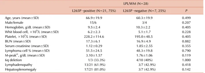
None of the clinical and cytogenetic parameters were sig-nificantly different between MYD88 L265P positive and neg-ative groups (Table 3). Nonetheless, we observed relatively high serum paraprotein levels in the mutation-positive group compared to the wild type group (3.10±1.57 g/dL vs.
Table 2. Prevalence of MYD88 L265P in LPL/WM and other B-cell NHLs.
MYD88 L265P N of positive/total patients
B-cell prolymphocytic leukemia 1/1
Marginal zone lymphoma 1/8
Follicular lymphoma 1/13
Lymphoplasmacytic lymphoma/Waldenstrom macroglobulinemia 21/28
B-cell lymphoma, unclassifiable, with features intermediate between DLBCL and Burkitt lymphoma 0/2
IgM-type myeloma 0/2
Burkitt lymphoma 0/9
Diffuse large B-cell lymphoma 0/11
Mantle cell lymphoma 0/11
Chronic lymphocytic leukemia/small lymphocytic lymphoma 0/12
Table 3. Clinical characteristics of LPL/WM according to MYD88 L265P status.
LPL/WM (N=28)
L265P –positive (N=21, 75%) L265P –negative (N=7, 25%) P
Age, years (mean±SD) 66.9±19.9 60.3±19.9 0.499
Male/female 15/6 3/4 0.207
Hemoglobin, g/dL (mean±SD) 9.5±2.4 10.3±2.2 0.405
Whit blood cell, ×109/L (mean±SD) 6.2±2.3 5.1±1.7 0.228
Platelet, ×109/L (mean±SD) 228.2±114.6 195.0±48.5 0.405
BUN (mean±SD) 17.3±6.1 16.9±4.9 0.882
Serum creatinine (mean±SD) 1.12±0.29 1.85±2.55 0.355
Lymphoma cell % (mean±SD) 51.5±24.5 48.3±19.8 0.790
M-peaka), g/dL (mean±SD) 3.10±1.57 1.76±1.06 0.119
6q deletion 1/3 (33.3%) 4/10 (40%) 1.000
Lymphadenopathy 13/21 (61.9%) 3/7 (42.9%) 0.418
Hepatosplenomegaly 17/21 (81.0%) 3/7 (42.9%) 0.142
a)17 cases and 6 cases were available for serum paraprotein levels.
Abbreviations: BUN, blood urea nitrogen; LPL/WM, lymphoplasmacytic lymphoma/Waldenstrom macroglobulinemia; SD, standard deviation.
1.76±1.06 g/dL, respectively; P=0.119).
DISCUSSION
We observed a high prevalence of the MYD88 L265P mutation in Korean LPL/WM patients, which was similar to results of previous studies [7, 10, 14]. In 2012, Treon
et al. initially described a high prevalence of MYD88 L265P in LPL/WM (91%) using whole genome sequencing [14]. They also developed real-time AS-PCR assays for sensitive detection and quantification of MYD88 L265P [10]. Other subsequent studies confirmed this high prevalence (over 90%) in LPL/WM and in IgM monoclonal gammopathy of undetermined significance (MGUS; up to 87% of cases) [5, 15]. In addition, MYD88 L265P was considered a risk factor for progression to LPL/WM or other lymphomas in patients with IgM MGUS [15-17].
MYD88 L265P can be detected in other B-cell NHLs; for example, approximately 30% of cases of the activated
B-cell-like (ABC) subtype of diffuse large B-cell lymphoma (DLBCL) had the MYD88 L265P mutation [18]. Bonzheim
et al. [19] have reported a high prevalence of MYD88 L265P (69%) in vitreoretinal diffuse large B-cell lymphomas. Cases of splenic marginal zone lymphomas and chronic lympho-cytic lymphomas have also been reported to have MYD88
L265P although the frequencies are relatively low (10% and 4%, respectively) [10]. We also observed MYD88 L265P in three case of B-cell NHL, including B prolymphocytic leuke-mia, marginal zone lymphoma, and follicular lymphoma. These observations indicate that MYD88 L265P should not be considered an exclusive diagnostic marker of WM/LPL. Nonetheless, MYD88 L265P is relatively rare in marginal zone lymphomas, IgM myelomas, or chronic lymphocytic leu-kemia, of which the discrimination from LPL/WM is difficult. Therefore, the diagnostic utility of MYD88 L265P testing for differential diagnosis will be still valuable [1, 5, 15].
Several studies have shown the association of the MYD88
L265 mutation with higher bone marrow tumor burden [3, 10, 20, 21], serum paraprotein levels [15, 20], and probability
of progression [15, 16, 20]. In contrast, we could not identify any significant differences in laboratory and clinical parame-ters between MYD88 L265P positive and negative cases. This might be because of the small number of cases enrolled and the heterogeneity of clinical and pathological charac-teristics. Regarding cytogenetic abnormalities, no karyotype is known to be specific to LPL/WM. Loss of 13q14, 11q21-q22, and 6q23–q24 and gain of 3, 12, and 18 are reported to be common in LPL/WM [22] but these abnormalities are also common in other low-grade lymphomas such as MZL and CLL [22, 23]. We observed only three LPL/WM cases with chromosomal abnormalities by conventional chromo-some analysis; however, a high frequency of 6q deletion (38.5%) was detected with FISH, which is in accordance with another study on Korean LPL/WM patients [7]. This report described an association between 6q deletion and
MYD88 L265P mutation [7]. However, we failed to find a similar association; cases with a 6q deletion also had a high frequency of MYD88 L265P mutation.
For molecular testing, we used bone marrow aspirate slides instead of biopsy specimens from primary sites such as lymph nodes. Given that the detection method is sensitive, obtaining samples from bone marrow or peripheral blood specimens will be more practical and easier than performing biopsies on primary sites. One popular method for this purpose is AS-PCR, used in many studies for MYD88 L265P detection [4, 10, 16, 24, 25]. AS-PCR shows good sensitivity but some-times suffers the drawback of high false-positive rates [13, 26]; therefore, we adopted a MEMO-PCR method with high specificity [11, 13, 27] and confirmed that this technique could be efficiently used for the detection of MYD88 L265P mutations in LPL/WM and other B-cell NHLs.
Several limitations were encountered in this study. We did not perform extensive clinical and morphologic review of selected cases to differentiate LPL/WM from other lym-phoma diagnoses. In addition, only a few cases of MZL or B cell lymphoma with plasmatic differentiation were en-rolled in this study. The sensitivity of MEMO-PCR was in-sufficient to detect minimal mutant levels of MYD88 L265P. This low sensitivity could have caused the relatively low mutation prevalence (75%), when this parameter was com-pared to that in other studies (∼90%). Therefore, clinical and laboratory differences, according to mutation status, might not have yielded statistical significance in this study. Further optimization of MEMO-PCR and sequencing might be required to increase detection sensitivity.
In conclusion, we observed a high prevalence of MYD88
L265P in LPL/WM and a low prevalence in other NHLs based on a series of Korean patients. Although several limi-tations were encountered in this study, our results demon-strate the diagnostic value of MYD88 L265P for differ-entiating B-cell NHLs.
AuthorsÊ Disclosures of Potential Conflicts of Interest
No potential conflicts of interest relevant to this article
were reported.
REFERENCES
1. Martinez-Lopez A, Curiel-Olmo S, Mollejo M, et al. MYD88 (L265P) somatic mutation in marginal zone B-cell lymphoma. Am J Surg Pathol 2015;39:644-51.
2. Chong Y, Kang CS, Oh WJ, Kim TJ, Lee EJ. Nodal involvement of extranodal marginal zone lymphoma with extreme plasmacytic differentiation (Mott cell formation) simulating plasma cell neoplasm and lymphoplasmacytic lymphoma. Blood Res 2014;49:275-85.
3. Xu L, Hunter ZR, Yang G, et al. Detection of MYD88 L265P in peripheral blood of patients with Waldenström's Macroglo-bulinemia and IgM monoclonal gammopathy of undetermined significance. Leukemia 2014;28:1698-704.
4. Staiger AM, Ott MM, Parmentier S, et al. Allele-specific PCR is a powerful tool for the detection of the MYD88 L265P mutation in diffuse large B cell lymphoma and decalcified bone marrow samples. Br J Haematol 2015;171:145-8.
5. Jiménez C, Sebastián E, Chillón MC, et al. MYD88 L265P is a marker highly characteristic of, but not restricted to, Waldenström's macroglobulinemia. Leukemia 2013;27:1722-8.
6. Ko YH, Kim CW, Park CS, et al. REAL classification of malignant lymphomas in the Republic of Korea: incidence of recently recognized entities and changes in clinicopathologic features. Hematolymphoreticular Study Group of the Korean Society of Pathologists. Revised European-American lymphoma. Cancer 1998;83:806-12.
7. Kim JA, Im K, Park SN, et al. MYD88 L265P mutations are correlated with 6q deletion in Korean patients with Waldenström macroglobulinemia. Biomed Res Int 2014;2014:363540. 8. Kim Y, Ju H, Kim DH, et al. CD79B and MYD88 mutations in
diffuse large B-cell lymphoma. Hum Pathol 2014;45:556-64. 9. Choi JW, Kim Y, Lee JH, Kim YS. MYD88 expression and L265P
mutation in diffuse large B-cell lymphoma. Hum Pathol 2013;44:1375-81.
10. Xu L, Hunter ZR, Yang G, et al. MYD88 L265P in Waldenström macroglobulinemia, immunoglobulin M monoclonal gammo-pathy, and other B-cell lymphoproliferative disorders using conventional and quantitative allele-specific polymerase chain reaction. Blood 2013;121:2051-8.
11. Lee ST, Kim JY, Kown MJ, et al. Mutant enrichment with 3'-modified oligonucleotides a practical PCR method for detecting trace mutant DNAs. J Mol Diagn 2011;13:657-68. 12. Shin SY, Ki CS, Kim HJ, Kim JW, Kim SH, Lee ST. Mutant
enrichment with 3'-modified oligonucleotides (MEMO)- quantitative PCR for detection of NPM1 mutations in acute myeloid leukemia. J Clin Lab Anal 2015;29:361-5.
13. Lee ST, Kim SW, Ki CS, et al. Clinical implication of highly sensitive detection of the BRAF V600E mutation in fine-needle aspirations of thyroid nodules: a comparative analysis of three molecular assays in 4585 consecutive cases in a BRAF V600E mutation-prevalent area. J Clin Endocrinol Metab 2012;97: 2299-306.
in Waldenström's macroglobulinemia. N Engl J Med 2012;367: 826-33.
15. Varettoni M, Arcaini L, Zibellini S, et al. Prevalence and clinical significance of the MYD88 (L265P) somatic mutation in Waldenstrom's macroglobulinemia and related lymphoid neoplasms. Blood 2013;121:2522-8.
16. Varettoni M, Zibellini S, Arcaini L, et al. MYD88 (L265P) mutation is an independent risk factor for progression in patients with IgM monoclonal gammopathy of undetermined signifi-cance. Blood 2013;122:2284-5.
17. Kyle RA, Therneau TM, Rajkumar SV, et al. Long-term follow-up of IgM monoclonal gammopathy of undetermined significance. Blood 2003;102:3759-64.
18. Ngo VN, Young RM, Schmitz R, et al. Oncogenically active MYD88 mutations in human lymphoma. Nature 2011;470:115-9. 19. Bonzheim I, Giese S, Deuter C, et al. High frequency of MYD88 mutations in vitreoretinal B-cell lymphoma: a valuable tool to improve diagnostic yield of vitreous aspirates. Blood 2015;126: 76-9.
20. Patkar N, Subramanian PG, Deshpande P, et al. MYD88 mutant lymphoplasmacytic lymphoma/Waldenström macroglobuli-nemia has distinct clinical and pathological features as compared to its mutation negative counterpart. Leuk Lymphoma 2015;56: 420-5.
21. Schmidt J, Federmann B, Schindler N, et al. MYD88 L265P and CXCR4 mutations in lymphoplasmacytic lymphoma identify cases with high disease activity. Br J Haematol 2015;169:795-803. 22. Braggio E, Dogan A, Keats JJ, et al. Genomic analysis of marginal
zone and lymphoplasmacytic lymphomas identified common and disease-specific abnormalities. Mod Pathol 2012;25:651-60. 23. Farinha P, Gascoyne RD. Molecular pathogenesis of mucosa-
associated lymphoid tissue lymphoma. J Clin Oncol 2005;23: 6370-8.
24. Ondrejka SL, Lin JJ, Warden DW, Durkin L, Cook JR, Hsi ED. MYD88 L265P somatic mutation: its usefulness in the differential diagnosis of bone marrow involvement by B-cell lymphopro-liferative disorders. Am J Clin Pathol 2013;140:387-94. 25. Capaldi IB, May AM, Schmitt-Graeff A, et al. Detection of MYD88
L265P mutations in formalin-fixed and decalcified BM biopsies from patients with lymphoplasmacytic lymphoma. Exp Mol Pathol 2014;97:57-65.
26. Newton CR, Graham A, Heptinstall LE, et al. Analysis of any point mutation in DNA. The amplification refractory mutation system (ARMS). Nucleic Acids Res 1989;17:2503-16.
27. Jang MA, Lee ST, Oh YL, et al. Identification of a rare 3 bp BRAF gene deletion in a thyroid nodule by mutant enrichment with 3'-modified oligonucleotides polymerase chain reaction. Ann Lab Med 2012;32:238-41.