Different Molecular Features of
Methylation Profile Between
Intestinal and Diffuse Sporadic
Gastric Carcinogenesis
Misuk Yang
The Graduate School
Yonsei University
Different Molecular Features of
Methylation Profile Between
Intestinal and Diffuse Sporadic
Gastric Carcinogenesis
Directed by Professor Hyun-Soo Kim
A Masters Thesis
Submitted to the Department of Medicine
and the Graduate School of Yonsei University
in partial fulfillment of the
requirements for the degree of
Master of Medicine
Misuk Yang
This certifies that the Masters
Thesis of Misuk Yang is approved.
Thesis Supervisor : Prof. Hyun-Soo Kim
Thesis Committee Member: Prof. Mee Yon Cho
Thesis Committee Member: Prof. Yangsik Jeong
The Graduate School
Yonsei University
감사의 글
제가 이곳에 온지 벌써 2년이 넘게 지났습니다 그 동안 많은 사람들을 만. 났고 함께 지냈으며 이별도 하였습니다 힘든 일도 있었고 즐거운 일 좋은, , . , , 일도 있었지만 이미 지난 시간들을 돌이켜 생각해 보니 감회가 새롭고 미소, 가 지어집니다. 무엇보다도 먼저 저를 항상 격려해주시고 칭찬해주시고 인격적으로 대해주, , 신 김현수 교수님께 감사의 말씀 전해드립니다 바쁜 일정 속에서도 잊지 않. 고 챙겨주시고 인생의 가르침까지 주신 것 잊지 않겠습니다 아울러 졸업논문, . 심사를 해주신 조미연 교수님 정양식 교수님께도 감사드립니다 그리고 제가, . 이곳에 발을 딛도록 해주시고 격려해주신 권상옥 교수님 이희봉 교수님 이곳, , 에서 무탈하게 실험할 수 있도록 기초를 배우게끔 해주신 임창진 교수님 자, 주 뵙진 못했지만 항상 웃으며 반갑게 인사해주신 백순구 교수님 김문영 교, 수님 김재우 교수님 모두 감사드립니다, . 이 논문을 펴내기까지 도움을 주신 지도교수님이신 김현수 교수님과 자문, 위원 교수님이신 조미연 정양식 교수님께 감사드립니다, . Jijgee and Peninah, 이 분들이 있었기에 무사히 논문을 펴낼 thank you for the proofreading.수 있었습니다. 년이 채 안되는 시간동안 중앙연구실 사람들도 여럿 바뀌었지만 같이 지 3 , 냈던 모든 분들이 저에게 좋은 사람으로 기억될 것입니다 실험뿐만 아니라. 인생의 선배로서도 많은 조언을 해주었고 저를 친동생처럼 예뻐해준 조효경 선생님 제가 처음 왔을 때 잘 모르는 저에게 많은 도움을 주었고 같은 과라, 고 잘 챙겨주었던 장윤옥 선생님 척척박사처럼 뭐든 물어보면 친절하게 잘, 대답해 주시고 도움을 주셨던 정민영 선생님 지금은 석사를 졸업하고 다른, 곳으로 갔지만 학부시절 같은 실험실에 있었다는 이유로 반가웠던 최란 선생 님 오랜 시간 있었던 것은 아니지만 마음 잘 통하고 유쾌했던 조은주 선생님, , 가끔 소소하게 도움 주고받았던 김성해 선생님 이은수 선생님 대화가 잘 통, , 했던 실험실 막내 김동혜 선생님 그리고 최연식 선생님 최준 선생님 현봉, , , 선생님 피용천 선생님 밥도 같이 먹고 종종 어울려 지냈던 병리학과 정소영, , 선생님 박선미 선생님 일일이 하나하나 다 나열할 순 없지만 그 동안 감사했, ,
습니다 그리고 같은 실험실에 있진 않았지만 오며 가며 인사하고 지냈던 생. 리학 생화학 약리학 예방의학 해부학 교실 선생님들께도 감사드립니다, , , , . 이젠 대부분 사회인이 되어 자주 만날 수 없는 친구들 슬기 지은이 향순, , 이 아름이 나희 현아 영신이 혜인이 윤영이 은혜 재인이 서로 다른 지, , , , , , , , . 역에 있지만 종종 연락해서 만났던 것처럼 우리들 인연 계속 이어나가자 그. 리고 대학시절 좋은 추억 많이 만들어주었고, 1년에 한두번씩 만남 이어가고 있는 HAM 동아리 사람들 고맙습니다. 마지막으로 정말 감사드리고 싶은 나의 가족 저를 낳아 지금까지 길러주시, 고 보살펴주시고 지원해주셨던 엄마 아빠 어려서부터 친구처럼 같이 커왔던, , 언니랑 경미 늦둥이로 태어나 온 가족의 사랑을 받으며 자란 귀염둥이 희승, . 아무 말 하지 않아도 힘들 땐 힘이 되었고 외롭지 않았던 것은 가족들이 있, 었던 덕분입니다 사랑합니다. . 비록 이제 저의 석사학위는 끝나지만 진짜 시작은 지금부터라는 생각을 가 지고 사회를 향한 첫발을 내딛도록 하겠습니다 저의 새로운 시작과 모든 분. 들의 안녕을 위하여 감사의 말씀 전하며 글을 마칩니다. 감사합니다. 년 월 2011 7 양미숙
Contents
The List of Figures
‥‥‥‥‥‥‥‥‥‥‥‥‥‥‥‥‥ ⅲ
The List of Tables
‥‥‥‥‥‥‥‥‥‥‥‥‥‥‥‥‥ ⅳ
Abstract
‥‥‥‥‥‥‥‥‥‥‥‥‥‥‥‥‥‥‥‥‥‥ 1
Introduction
Ⅰ
‥‥‥‥‥‥‥‥‥‥‥‥‥‥‥‥‥‥‥ 3
Materials and Methods
Ⅱ
‥‥‥‥‥‥‥‥‥‥‥‥‥ 7
1. Sample Collection
‥‥‥‥‥‥‥‥‥‥‥‥‥‥‥ 7
2. DNA Extraction, Bisulfite Conversion and
Polymerase Chain Reaction
‥‥‥‥‥‥‥‥‥‥‥
9
3. Pyrosequencing
‥‥‥‥‥‥‥‥‥‥‥‥‥‥‥‥ 11
4. Quntitative Real-Time PCR
‥‥‥‥‥‥‥‥‥‥ 12
5. Statistical Analysis
‥‥‥‥‥‥‥‥‥‥‥‥‥‥‥ 14
Results
Ⅲ
‥‥‥‥‥‥‥‥‥‥‥‥‥‥‥‥‥‥‥‥‥ 15
2. Different Methylation Profiles according to the
Cancer Differentiation Status in the Experimental
Group
‥‥‥‥‥‥‥‥‥‥‥‥‥‥‥‥‥‥‥‥‥‥ 18
3. Validation of Different Methylation Profiles
according to the Cancer Differentiation Status
‥‥ 23
4. mRNA Expression of CDH1 and RORA
‥‥‥‥‥ 27
5.
Helicobacter pylori
Infection and Methylation
Pattern
‥‥‥‥‥‥‥‥‥‥‥‥‥‥‥‥‥‥‥‥‥ 30
Discussion
Ⅳ
‥‥‥‥‥‥‥‥‥‥‥‥‥‥‥‥‥‥‥‥ 32
Conclusion
Ⅴ
‥‥‥‥‥‥‥‥‥‥‥‥‥‥‥‥‥‥‥‥ 36
References
‥‥‥‥‥‥‥‥‥‥‥‥‥‥‥‥‥‥‥‥‥ 37
The List of Figures
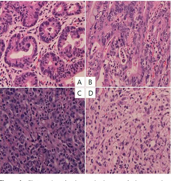
Figure 1. Photographs of microscopic images of tissue
sections according to histologic
differentiation status.
‥‥‥‥‥‥‥‥‥‥‥ 17
Figure 2. Methylation patterns in experimental group
for each cancer stage.
‥‥‥‥‥‥‥‥‥‥ 20
Figure 3. Methylation patterns in the experimental
group according to histologic differentiations
of cancer.
‥‥‥‥‥‥‥‥‥‥‥‥‥‥‥‥ 21
Figure 4. Comparison of the differences in methylation
patterns between PDCs and SRCCs in the
experimental group. ‥‥‥‥‥‥‥‥‥‥‥‥ 22
Figure 5. Methylation patterns upon 4 histologic
differentiation status in the validation group
‥‥‥‥‥‥‥‥‥‥‥‥‥‥‥‥‥‥‥‥‥‥ 24
Figure 6. Methylation patterns in the validation group
for intestinal and diffuse type.
‥‥‥‥‥‥ 25
Figure 7. Comparison of the differences in methylation
patterns between PDC and SRCC in the
validation group.
‥‥‥‥‥‥‥‥‥‥‥‥‥ 26
The List of Tables
Table 1. Primer sequences and annealing
temperatures for PCR and pyrosequencing. ‥ 10
Table 2. Primer sequences for qPCR. ‥‥‥‥‥‥‥‥ 13
Table 3. Clinicopathologic characteristics of Patients.
‥‥‥‥‥‥‥‥‥‥‥‥‥‥‥‥‥‥‥‥‥‥ 16
Table 4. The analysis result of qPCR. ‥‥‥‥‥‥‥‥ 29
Table 5.
H. pylori
infection and methylation.
‥‥‥‥ 31
ABSTRACT
Different Molecular Features of Methylation
Profile Between Intestinal and Diffuse Sporadic
Gastric Carcinogenesis
Gastric cancer (GC) is histologically classified into intestinal type and diffuse type, and diffuse type cancer can be further subdivided into poorly differentiated carcinoma (PDC) and signet ring cell carcinoma (SRCC). Recent evidence suggests early SRCC is an initial, differentiated form of diffuse GC that may evolve into PDC. This study aimed at identifying the molecular features of epigenetic methylation changes in histologic differentiation status of GC. Included in this study are 149 samples of paraffin embedded tissues and 115 fresh endoscopically biopsied tissues from Yonsei University Wonju Christian Hospital. Multiple paraffin tissues involving normal (N=22), dysplasias (GDs, N=39), differentiated cancers (DCs, N=35), PDCs (N=33) and SRCCs (N=20) were included as an experimental group. For the validation group, endoscopically biopsied tissues of DCs (N=50), PDCs (N=31), and SRCs (N=34) were analyzed. DNAs, isolated from each group were analyzed to determine the methylation status of 6 genes
(GDNF, RORA, MINT25, KLF7, CDH1, LINE1) using pyrosequencing. LINE1 hypomethylated in GCs compared to normal and GD. GDNF, RORA and MINT25 were more hypermethylated in intestinal type GCs than diffuse type GCs, whereas CDH1 showed opposite patterns of methylation status. Among diffuse type GCs, SRCCs showed lower level of methylation for GDNF, RORA, MINT25 and KLF7, and higher level for CDH1 compared to PDCs. In conclusion, the intestinal type of GCs shows different epigenetic methylation profiles compared to the diffuse one. Moreover, SRCCs have different methylation profiles compared with PDCs, suggesting a unique methylation pathway in the gastric carcinogenesis.
Key words: gastric cancer, gastric carcinogenesis, epigenetics, m ethylation, histologic differentiation
Different Molecular Features of Methylation
Profile Between Intestinal and Diffuse Sporadic
Gastric Carcinogenesis
Directed by Professor Hyun-Soo Kim
Department of Medicine
The Graduate School, Yonsei University
Misuk Yang
Introduction
Ⅰ
Gastric adenocarcinoma is one of the most common diseases in the world, and the main cause of cancer death in Asia.1 In particular, the incidence rate of GC (41.1~44.3/100,000) was barely changed from 1999 to 2008 in Korea. Total cancer deaths were 68,912, which account for 28% of all deaths during 2008. Among all cancers, the GC was the third leading cause of the cancer death (15%) after lung cancer (21%) and hepatocellular carcinoma (17%).2
Helicobacter pylori infection and smoking are known as risk factors for GC development.3-5 Furthermore, genetic and epigenetic factors also p lay a critical role in gastric carcinogenesis.6,7 For instance, genetic factors include single-nucleotide polymorphisms and DNA mutations,8-10 and epigenetic factors include post-translational modifications, aberrant DNA methylation and histone modification.7,8
Many researchers have attempted to classify gastric adenocarcinoma. Lauren classification is the most widely used method that distinguishes two histological types according to microscopic morphology.8,9,11,12 Intestinal type is the well-differentiated tumor with cohesive tumor cells with gland-like tubular structures. Diffuse type is undifferentiated tumors that infiltrate through the stomach wall, showing diffused thickness of the stomach wall.8,9,13 However, many gastric tumors are sorted as mixed type since not classified into either type of gastric tumors.8,14 While intestinal type of GCs frequently metastasizes to the liver, diffuse type GCs can be easily disseminate to the peritoneum.8,15 Gastric carcinogenesis progresses through serial steps involving atrophy, metaplasia, dysplasia and carcinoma. These steps are more commonly seen in the intestinal type.7,8
Molecular studies have provided evidence that GC result not only from the combined effects of environmental aspects and genetic modifications, but also from the cumulative genetic and epigenetic changes that play pivotal roles in tumorigenesis.8,10,16 Genetic and epigenetic mutations in various gene sets including oncogenes, tumor-suppressor, and others are closely associated with tumor
pathogenesis as well as progression.7,8,13,17 In GC, for instance, aberrant DNA methylations occur more frequently than mutations.11,18,19 DNA methylation changes even occuring in non-neoplastic gastric epithelial cells suggests that the epigenetic methylation is an early event in GC.7,17,20,21,22 Global DNA hypomethylation occurs in gastric or other tissues during the early stages of tumor growth.9,23 However, demethylation of individual gene probably occurs at developmental stages after global DNA hypomethylation.9,22 As hypermethylation in the gastric mucosa is related to the higher risk of GC, H. pylori infection is believed to accelerate gastric carcinogenesis by causing DNA hypermethylation in the gastric mucosa.21,22
We decided to see DNA methylation in the promoter of six genes for this study. Three genes (RORA, GDNF and MINT25) of the six were identified as sensitive molecular markers of early GC in the previous studies.18,24 These three genes showed different methylation patterns between normal and GC.18 Retinoic acid-related orphan receptor-alpha (RORA), a nuclear receptor, plays a critical role in the development of various organs.25 It can functionally associated with Wnt signaling pathway that is involved in oncogenic systems by attenuating the pathway.26 Glial cell derived neurotrophic factor (GDNF) is a small protein that strongly promotes the survival of various type of neurons.27 Interaction of up-regulated GDNF and RET ligand-receptor are possibly involved in the glucose-induced cancer progression.28 Specific methylation has been shown in several genes including MINT25 in GC.24 The Kruppel-like family (KLF) of transcription factors
have been shown to play pivotal roles in tumorigenesis of various tissues.29 The expression of CDH1, an important e-cadherin protein for cell adhesion, is frequently down-regulated in sporadic tumors and is associated with the poorly differentiated phenotype. Hypermethylation of this gene leads to gene silencing in GC.9,12,13,17,30 Global DNA hypomethylation of DNA repetitive elements within the human genome such as long interspersed nuclear element 1 (LINE1) is a common finding in cancer and is believed to cause chromosomal instability and proto-oncogene activation.11,17
GC proceeds through serial steps. Intestinal GC progresses through sequential steps after several genetic and epigenetic changes, while diffuse GC progresses directly to early cancer after similar genetic and epigenetic changes with intestinal GC.7,8,17 Judging from the fact that the pathway is different for each type of cancer, genes associated with the cancer seem to affect different aspects of the changes involved in each pathway.
Recent studies show that the pattern of lymph node metastasis is different between PDC and SRCC.31,32 Additionally, they also have different mechanisms of tumor invasion.33 These findings have reported that SRCC and PDC have different origins.
In this study, therefore, we investigate differences of DNA methylation patterns at the molecular level especially in diffuse type GC including SRCC and PDC.
Materials and Methods
Ⅱ
1. Sample Collection
Gastric tumor tissues of the experimental group were obtained from Korean patients who had surgical or endoscopic resection at Yonsei University Wonju Christian Hospital (Wonju, Korea) between January 2000 and December 2004. The 127 tumor tissue samples including 39 GDs, 35 DCs, 33 PDCs and 20 SRCCs from patients, were retrospectively and randomly collected and examined. In addition, normal gastric tissues were collected from 22 age-matched patients who experienced surgical resection due to the peptic ulcer disease during the same period. Tissues were sent to pathologists for histopatologic diagnosis and fixed with paraffin. These paraffin-embedded tissue blocks were sliced for serial sections, and each section was microdissected by an expert pathologist to distinguish the tumor regions. The extracted DNA for each sample was therefore more than 70% homogeneous. Additionally, photographs of representative tissue sections were taken.
For the validation group, we collected 115 tumor tissues obtained between October 2008 and February 2011 at the same hospital. Because a large number of samples had been collected for intestinal type cancer, randomly selected samples were used. Due to the small number of available diffuse GC samples, all them were used. Small
portions of these samples were used for histopathologic analysis and the rest of them were frozen immediately after biopsy.
Basic and clinicopathologic informations about each patient were used as raw data. This study was approved by the institutional review board of Yonsei University Wonju Christian Hospital. We also notified all patients and received consents.
2. DNA Extraction, Bisulfite Conversion and Polymerase
Chain Reaction
Genomic DNA (gDNA) was extracted using QIAamp DNA Mini Kit (Qiagen, Hilden, Germany). The methylation status of the gDNA was then characterized by first performing bisulfite conversion on the gDNA using EpiTect bisulfite kit (Qiagen). These two steps were carried out according to supplied protocols. Unmethylated cytosine is changed to uracil after bisulfite conversion, in which case, it is amplified as thymine during PCR. PCR was carried out twice except for LINE1, with the PCR product of the first reaction being used as template for the second reaction. In the first PCR reaction forward (F) and reverse (R) primers were used while in the second reaction F, reverse-universal (RU) and biotin-universal (BU) primers were used. The first PCR reaction conditions were 94℃ for 30 seconds, 55~58℃ for 30 seconds and 72℃ for 30 seconds, 45 cycles. The second PCR was performed under the same conditions except for the different annealing temperature (60℃ for all primers). The reagents for PCR were HotStarTaq Plus PCR mixture (Qiagen). Primer sequences and annealing temperatures are listed in Table 1.
Table 1. Primer sequences and annealing temperatures for PCR and pyrosequencing.
Gene name Type Primer Sequence 5'-3' Temp.
LINE1 F TTTTGAGTTAGGTGTGGGATATA 56℃ RU BU-AAAATCAAAAAA TTCCCTTTC S AGTTAGGTGTGGGATATAGT GDNF F AGGATTGAGAATTTTTGTTTTTGATT 55℃ R CCAAACCCTAAATTAAACATTAACTCCA RU BU-CCAAACCCTAAATTAAACATTAACTCCA S TTTTGTTTTTGATTTGTTG RORA F TTTGGTATTATAGAGTTGTTTTGAAAATAGAA 56℃ R ACCCAAACTAACTCCATATTTTTTCC RU BU-ACCCAAACTAACTCCATATTTTTTCC S TGAAAATAGAAGATAGAGGGA MINT25 F TGTTTGTAAAGGGTTGGAATTATT 55℃ R CCCRCCAAAACAACTTTA RU BU-CCCRCCAAAACAACTTTA S TAGTTTATTATTTTTAAGAG CDH1 F TTTTTTGATTTTAGGTTTTAGTGAGTTAT 58℃ R TACCRACCACAACCAATCAACAAC RU BU-TACCRACCACAACCAATCAACAAC S GATTTTAGGTTTTAGTGAGT KLF7 F AGGATTGAGAATTTTTGTTTTTGATT 56℃ R CCAAACCCTAAATTAAACATTAACTCCA RU BU-CCAAACCCTAAATTAAACATTAACTCCA S TTTTGTTTTTGATTTGTTG
3. Pyrosequencing
Pyrosequencing was then performed on the PCR products. Since biotin labeled universal reverse primer was used for PCR, the 5' ends of the reverse strands were labeled with biotin. For pyrosequencing, the PCR product is first captured using streptavidin-coated beads. After washing out unbound products, the PCR product is denatured leaving the reverse strand still bound to the beads, while the forward strand is released into the solution. A second washing step is carried out to remove the unbound forward strand, and the biotin labeled reverse strand acts as the sequencing template. As the sequencing proceeds, depending on whether the cytosine of CpG site is methylated or not, the laser detects cytosine or thymine and measures methylation percentage. For pyrosequencing, PyroMark Gold Q24 Reagents (Qiagen) and PyroMark Q24 machine (Qiagen) were used. The sequencing primers are listed with PCR primers in Table 1.
Through pyrosequencing, methylation levels were measured at 3~5 CpG sites of the promoter region in each gene and the results were shown as mean values. Because the methylation levels of each site were similar in one sample, their mean value can represent the methylation levels of whole promoter region.
4. Quantitative Real-Time PCR
We measured mRNA expression levels of two genes, CDH1 and RORA by qPCR. First, RNA was extracted using RNeasy Plus Mini kit (Qiagen) from tissue samples of several patients in the validation group, already used for pyrosequencing. After, we synthesized cDNA using QuantiTect Reverse Transcription Kit (Qiagen) and performed qPCR under the following conditions: denaturation step at 95℃ for 15 seconds, annealing and extension step at 60℃ for 60 seconds, 40 cycles. Pre-designed primer sets were used and their sequences are listed in Table 2.34,35,36 Paired samples were prepared from the same patient to compare the differences in expression levels between noncancer (adjacent to cancer) versus tumor tissues. Real time PCR was carried out using ABI 7900HT system (Applied Biosystems, Carlsbad, Cal). The SYBR green mixture used was made from KAPA SYBR FAST qPCR Kit (KAPA Biosystems, Woburn, MA). The data was analyzed using the comparative ΔCt method.
Table 2. Primer sequences for qPCR.
Gene name Type Primer Sequence 5'-3'
CDH1 F TGGGCAGCTATCCAGTGACTTGTTC R CTGTCTTTGGCTGCAGCACTTTAGG RORA F AAACCTGCCAATACTTGAGAGAA R CACAATTGCCACATCACCTC -actin β F GCGGGAAATCGTGCGTGACATT R GATGGAGTTGAAGGTAGTTTCGTG
5. Statistical analysis
In this study, all statistical analysis were carried out using SPSS for Windows, version 17 (SPSS, Inc, Chicago, IL) and PRISM software for Windows, version 5 (GraphPad Prism, Inc, San Diego, CA). Methylation level (in %) taken as a continuous variable, was analyzed and associated with stages or differentiation of the different GC types (95% confidence intervals), for each gene. The z score was utilized to normalize the data of the methylation levels. The z score was calculated according to the following formula: z score = (methylation level of each sample - mean value of methylation level)/standard deviation of methylation level.
Methylation levels of the experimental group were analyzed using one way ANOVA test according to cancer stage and type. Methylation levels of intestinal type and diffuse type including all types of each, were compared using one way ANOVA test and t-test. Subtypes of diffuse type, the poorly differentiated type and the signet ring cell type, were compared with t-test. Methylation levels in Helicobacter pylori positive and negative tissues were also compared using t-test. The results of qPCR were analyzed using paired sample t-test.
Correlation between methylation patterns and mRNA expression for CDH1 and RORA was analyzed using Spearman's correlation test.
Results
Ⅲ
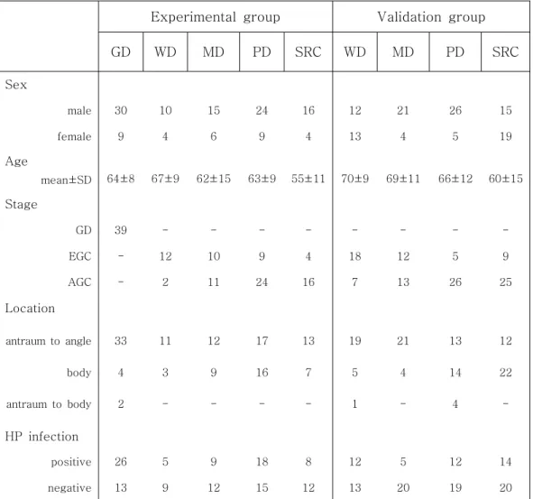
1. Clinicopathologic Characteristics of Patients
Clinical features of patients are summarized according to tumor types involving gastric dysplasia, early GC or advanced GC (Table 3). The number of male is more than female in this cohort. Since we collected the samples randomly, this result indicates that the cancer incidence is higher in men than in women. It is also observed that poorly differentiated tumors are associated with the younger patients. This means that undifferentiated cancers are more common in younger patients than in older patients. The number of diffuse type GCs is more than the number of intestinal type GC in advanced GC, and the opposite is true in early GC. Helicobacter pylori infection and differentiation of GC appears to be irrelevant.
Images of the different types of GC are shown according to differentiation status (Figure 1).
Table 3. Clinicopathologic characteristics of patients
GD. gastric dysplasia, WD. well differentiated gastric cancer, MD. moderately differentiated gastric cancer, PD. poorly differentiated gastric cancer, SRC. signet ring cell carcinoma, EGC. early gastric cancer, AGC. advanced gastric cancer
Experimental group Validation group
GD WD MD PD SRC WD MD PD SRC Sex male 30 10 15 24 16 12 21 26 15 female 9 4 6 9 4 13 4 5 19 Age mean±SD 64±8 67±9 62±15 63±9 55±11 70±9 69±11 66±12 60±15 Stage GD 39 - - - -EGC - 12 10 9 4 18 12 5 9 AGC - 2 11 24 16 7 13 26 25 Location antraum to angle 33 11 12 17 13 19 21 13 12 body 4 3 9 16 7 5 4 14 22 antraum to body 2 - - - - 1 - 4 -HP infection positive 26 5 9 18 8 12 5 12 14 negative 13 9 12 15 12 13 20 19 20
Figure 1. Photographs of microscopic images of tissue sections according to histologic differentiation status. A. well differentiated gastric cancer, B. moderately differentiated gastric cancer, C. poorly differentiated gastric cancer, D. signet ring cell carcinoma.
2. Different Methylation Profiles according to the Cancer
Differentiation Status in the Experimental Group.
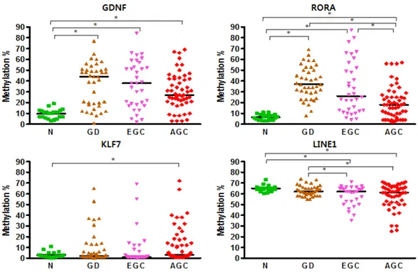
Methylation levels were quantitatively measured using pyrosequencing and analyzed according to tumor progression stages include normal, dysplasia, early GC and advanced GC and differentiation states include normal, GD, well differentiated cancer, moderately differentiated cancer, PDC and SRCC to determine the methylation levels of four selected genes in each type of GC.
Data obtained from the experimental group showed that the methylation levels of normal mucosa were highly conserved in all genes (Figure 2), whereas methylation patterns were changed as the disease developed from gastric dysplasia to advanced GC. GDNF and RORA showed high methylation levels in precancerous stage but the levels decreased as the cancer advanced. KLF had no significant changes but showed high methylation levels in advanced GC compared with normal. In contrast, methylation levels of LINE1 had no significant difference between normal and precancerous stage, but LINE1 became hypomethylated as the cancer advanced.
GDNF and RORA showed significantly low methylation levels in diffuse type GC compared with normal, dysplasia and intestinal type GC (Figure 3), while KLF7 showed increased methylation levels in diffuse type GC compared to normal tissues only. LINE1 was hypomethylated in both types of GC compared with normal and gastric
dysplasia.
We also compared the methylation levels of the diffuse GC subtypes in order to investigate any differences between PDC and SRCC (Figure 4). There were no significant differences except for LINE1, whose methylation levels were higher in SRCC.
Figure 2. Methylation patterns in experimental group for each cancer stage. N. Normal gastric mucosa, GD. Gastric dysplasia, EGC. Early gastric cancer, AGC. Advanced gastric cancer. (* P < 0.05).
Figure 3. Methylation patterns in the experimental group according to histologic differentiations of cancer. N. Normal gastric mucosa, GD. Gastric dysplasia, WD. Well differentiated gastric cancer, MD. Moderately differentiated gastric cancer, PD. Poorly differentiated gastric cancer, SRC. Signet ring cell carcinoma (* P < 0.05).
Figure 4. Comparison of the differences in methylation patterns between PDCs and SRCCs in the experimental group (* P < 0.05).
3. Validation of Different Methylation Profiles according to
the Cancer Differentiation Status.
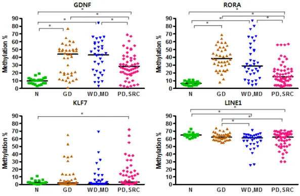
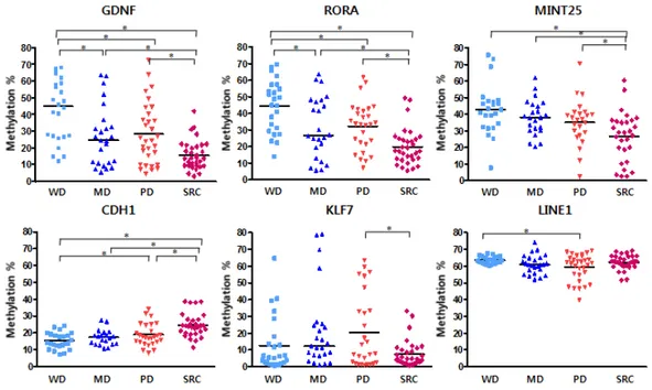
In the validation group, methylation levels were measured by pyrosequencing and the data were analyzed according to the differentiation status involving WD, MD, PD and SRC.
From the data, GDNF and RORA showed similar methylation patterns (Figure 5). Their methylation levels were remarkably low in moderately differentiated and signet ring cell GC. Specifically, the methylation levels of SRCCs were conspicuously low compared to other types. MINT25 had no statistical significance but showed a similar pattern and was significantly hypomethylated in SRCCs. CDH1 was hypermethylated in SRCCs. KLF7 had a significant difference between PDC and SRCC. Other types had no differences. LINE1 showed different methylation levels between well and poorly differentiated cancer.
GDNF, RORA and MINT25 showed significantly lower methylation levels in diffuse type GCs (Figure 6). On the other hand, the methylation levels of CDH1 were significantly higher. KLF7 and LINE1 showed inconsistent methylation pattern in the experimental group.
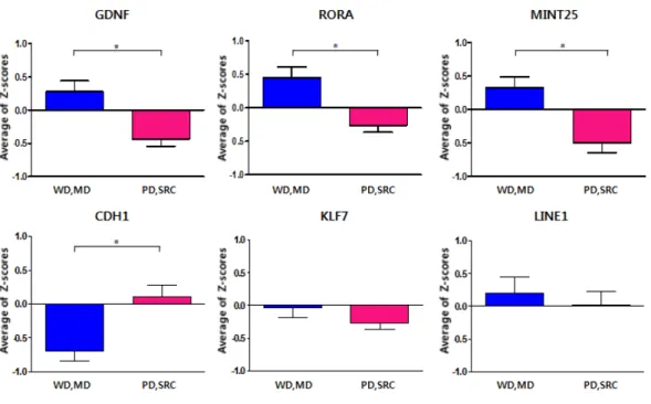
As a result of comparison between PDC and SRCC, CDH1 showed highly methylated pattern in SRCC (Figure 7). In the other four genes with the exception of LINE1, the methylation levels of SRCCs were significantly decreased compared with PDCs.
Figure 5. Methylation patterns upon 4 histologic differentiation status in the validation group (* P < 0.05).
Figure 6. Methylation patterns in validation group for intestinal and diffuse type (* P < 0.05).
Figure 7. Comparison of the differences in methylation pattrerns between PDC and SRCC in validation group (* P < 0.05).
4. mRNA Expression of CDH1 and RORA
We investigated mRNA expressions of CDH1 and RORA in the 19 pairs of samples that had been utilized for pyrosequencing. In the validation group, we picked the same number of samples for each type. Expression levels of mRNA were measured by quantitative real time PCR and analyzed using comparative Δct method, after isolating total RNA from tissues. The mRNA expressions in the cancer tissues were lower than the mRNA expressions in the noncancer tissues for both genes (Figure 8 and Table 4). High methylation levels of these genes corresponded to low mRNA expressions in the cancer tissues. Although there was no significant difference in CDH1 expression levels between noncancer and cancer tissues, correlation between methylation levels and mRNA expressions were significant (p=0.041). RORA showed opposite result (p=0.249).
Table 4. The analysis result of qPCR.
Paired sample t-test
N Paired Differences Sig.(2-tailed) Mean SD Std. Error Mean CDH1 18 3.943 8.941 2.107 0.079 RORA 16 3.658 6.535 1.634 0.041
5.
Helicobacter pylori
Infection and Methylation Pattern
Histopathologic examination was used to determine if patients had H. pylori infection or not. Though many studies have reported that H. pylori infection is associated with aberrant DNA methylation, we could not identify significant correlation between H. pylori infection and methylation pattern except for GDNF in the validation group (Table 5).
Table 5. H. pylori infection and methylation
A. Experimental Group
HP infection N Mean Std.Deviation p-value
GDNF negative 77 27.052 19.265 0.034 positive 71 33.915 19.694 RORA negative 77 24.117 18.194 0.273 positive 71 27.676 21.127 KLF7 negative 77 10.273 15.463 0.333 positive 71 7.887 14.338 LINE1 negative 71 59.451 10.004 0.342 positive 66 60.939 8.097 B. Validation Group
HP infection N Mean Std.Deviation p-value
LINE1 negative 75 60.824 5.859 0.064 positive 44 62.774 4.799 RORA negative 76 26.744 18.053 0.356 positive 44 30.133 21.355 GDNF negative 70 30.756 15.467 0.719 positive 44 31.937 19.294 MINT25 negative 64 35.270 14.900 0.928 positive 39 35.548 15.200 KLF7 negative 72 14.311 17.478 0.552 positive 44 16.453 20.714 CDH1 negative 65 19.332 7.030 0.511 positive 43 20.247 7.109
Discussion
Ⅳ
This study identified six molecular markers that can be used to distinguish histologic differentiation status of GC : intestinal type and diffuse type, poorly differentiated type and signet ring cell type. These markers can be utilized as diagnostic or prognostic tools. However, for them to be useful and effective markers, noninvasive detection methods from the gastric mucosa are necessary.
GC is one of the worldwide common diseases which may result in death and has been studied by many researchers.1 Recently, genome-wide epigenetic analysis have been performed to determine epigenetic mutations in diverse cancers.13,22 Currently, a lot of epigenetic research in gastric carcinogenesis is ongoing in the Asia. Papers detailing the step-by-step methylation changes of GC have been on the increase in recent years, but there are few large studies about carcinogenesis of SRCC, due to the difficulty in collecting GC tissues. Particularly, cancer specific biomarkers are considered to be helpful for prognosis since some cancers like the undifferentiated GC have poor prognosis and the markers would help in distinguishing them from other cancers. The undifferentiated GCs are common in younger female patients,8,30,32 which was not evident in this study. There were no significant differences in the methylation levels of SRCC between females under the age of 50 and rest of patients (data was not shown).
DNA methylation plays a role in X-chromosome inactivation, genome imprinting, and inactivation of DNA repetitive sequences.17,37 In cancer cells, two conflicting epigenetic events can coexist together. These two are global hypomethylation and regional hypermethylation. Global hypomethylation frequently occurs in DNA repetitive sequences and is believed to cause chromosomal instability and proto-oncogene activation.9,19,22,37 Regional hypermethylation is frequently observed in CpG islands of promoter regions and brings about inactivation of tumor suppressor genes.6-10,17,22 As one of the main epigenetic events in carcinogenesis, transcriptional inactivation of certain genes occurs through hypermethylation of promoter CpG islands that leads to continued gene silencing.9,16,21 In this study, LINE1 tends to be hypomethylated in advanced GC. Specifically, hypomethylation of LINE1 was frequently observed in PDC. This would explain the chromosomal instability that is commonly associated in poorly differentiated or undifferentiated cancers.
DNA methylation has been the theme of research in various tumor cells. The cancer cells show altered DNA methylation profiles compared to the normal cells.6,20 A previous study determined 6 genes that are prone to hypermethylation (MINT25, GDNF, RORA, PRDM5, ADAM23, MLF1) in gastric neoplasia.18 In particular, GDNF and MINT25 show high methylation patterns in gastric tumor samples regardless of tumor stage and can therefore be used as biomarkers for gastric tumors.19 Similarly, in this study, methylation levels of GDNF and RORA are dramatically high in precancerous stages and they tend
to decreased in the advanced cancer stages. Therefore, these markers can be utilized to detect early stage gastric neoplasia.
Furthermore, in GC, 11 genes including MINT25 are known to display specific methylation patterns as previously reported.24 In line with this, GDNF, RORA and MINT25 show significant differences between PDC and SRCC in this study. This suggests that GDNF, RORA and MINT25 can be adopted as sensitive biomarkers to distinguish poorly differentiated type from signet ring cell type as well as biomarkers for the detection of early stage of gastric tumor.
Interestingly, CDH1 also has significantly higher methylation levels in SRCCs compared to other cancers, which seems to correspond to results of a previous study that CDH1 plays a key role as a tumor suppressor gene in hereditary diffuse GCs.8,12,13,30,39 In other words, the suppression of CDH1 gene expression by methylation can promote carcinogenesis.9-11,17 This fact is more evident in undifferentiated GCs, especially SRCCs shown in this study, than in the other GCs. Therefore, these results show that the methylation of CDH1 is involved in the carcinogenesis of undifferentiated cancers. Accordingly, CDH1 has the potential to be a specific biomarker for undifferentiated cancers indicating poor prognosis. This strongly suggests that the methylation changes of different genes are involved in carcinogenesis of the two types of undifferentiated GCs.
Meanwhile, the current study suggests that SRCC has similar clinicopathologic characteristics with differentiated cancers but has differences with other undifferentiated cancers including PDCs.31-33
KLF7 methylation patterns supports this theory, since the patterns are similar between SRCCs and DCs but different from the PDC.
We examined correlation analysis between methylation levels and mRNA expressions for CDH1 and RORA. Only CDH1 showed a significant correlation. This means that hypermethylation of CDH1 leads to down regulation of its mRNA expression which corresponds to the inactivation of e-cadherin genes that commonly occurs due to promoter methylation.22
Many papers report that Helicobacter pylori infection leads to gastric carcinogenesis by causing aberrant DNA methylation.9,17,21,22,38 Our study, however, could not find any relationship between H. pylori
infection and DNA methylation .
From this study, the genes that show differences in epigenetic methylation profiles among different cancer phenotypes were identified. We believe expanding the study to investigate gene functions and expressions in tissues would improve the understanding of carcinogenesis in different phenotypes of GCs.
Conclusion
Ⅴ
Collectively, at the molecular level, the intestinal type of GCs have different epigenetic methylation profiles compared to the diffuse one. Furthermore, signet ring cell GCs have different methylation profiles compared with poorly differentiated GCs, suggesting a unique gastric carcinogenesis pathway.
References
1. Jemal, A., R. Siegel, et al. (2007). "Cancer statistics, 2007." CA Cancer J Clin 57(1): 43-66.
2. Jung, K. W., S. Park, et al. (2011). "Cancer statistics in Korea: incidence, mortality, survival, and prevalence in 2008." Cancer Res Treat 43(1): 1-11.
3. Nardone, G. and A. Morgner (2003). "Helicobacter pylori and gastric malignancies." Helicobacter 8 Suppl 1: 44-52.
4. Asaka, M. and B. A. Dragosics (2004). "Helicobacter pylori and gastric malignancies." Helicobacter 9 Suppl 1: 35-41.
5. Kang, H. Y., N. Kim, et al. (2006). "Progression of atrophic gastritis and intestinal metaplasia drives Helicobacter pylori out of the gastric mucosa." Dig Dis Sci 51(12): 2310-2315.
6. Choi, I. S. and T. T. Wu (2005). "Epigenetic alterations in gastric carcinogenesis." Cell Res 15(4): 247-254.
7. Yasui, W., K. Sentani, et al. (2006). "Molecular pathobiology of gastric cancer." Scand J Surg 95(4): 225-231.
8. Vogiatzi, P., C. Vindigni, et al. (2007). "Deciphering the underlying genetic and epigenetic events leading to gastric carcinogenesis." J Cell Physiol 211(2): 287-295.
9. Tamura, G. (2006). "Alterations of tumor suppressor and tumor-related genes in the development and progression of gastric cancer." World J Gastroenterol 12(2): 192-198.
10. Resende, C., A. Ristimaki, et al. (2010). "Genetic and epigenetic alteration in gastric carcinogenesis." Helicobacter 15 Suppl 1: 34-39.
11. Ushijima, T. and M. Sasako (2004). "Focus on gastric cancer." Cancer Cell 5(2): 121-125.
12. Humar, B., V. Blair, et al. (2009). "E-cadherin deficiency initiates gastric signet-ring cell carcinoma in mice and man." Cancer Res 69(5): 2050-2056.
13. Panani, A. D. (2008). "Cytogenetic and molecular aspects of gastric cancer: clinical implications." Cancer Lett 266(2): 99-115.
14. Kim, W. H, et al. (2005). "A standardized pathology report for gastric cancer" Korean J Pathol 39: 106-13
15. Marrelli, D., F. Roviello, et al. (2004). "Risk factors for liver metastases after curative surgical procedures for gastric cancer: a prospective study of 208 patients treated with surgical resection." J Am Coll Surg 198(1): 51-58.
16. Crew, K. D. and A. I. Neugut (2006). "Epidemiology of gastric cancer." World J Gastroenterol 12(3): 354-362.
17. Yamamoto, E., H. Suzuki, et al. (2011). "Role of DNA methylation in the development of diffuse-type gastric cancer." Digestion 83(4): 241-249.
18. Watanabe, Y., H. S. Kim, et al. (2009). "Sensitive and specific detection of early gastric cancer with DNA methylation analysis of gastric washes." Gastroenterology 136(7): 2149-2158.
methylation markers." Nat Rev Cancer 3(4): 253-266.
20. Kang, G. H., S. Lee, et al. (2003). "Profile of aberrant CpG island methylation along multistep gastric carcinogenesis." Lab Invest 83(4): 519-526.
21. Ksiaa, F., S. Ziadi, et al. (2009). "Biological significance of promoter hypermethylation of tumor-related genes in patients with gastric carcinoma." Clin Chim Acta 404(2): 128-133.
22. Compare, D., A. Rocco, et al. (2011). "Global DNA hypomethylation is an early event in Helicobacter pylori-related gastric carcinogenesis." J Clin Pathol.
23. Bariol, C., C. Suter, et al. (2003). "The relationship between hypomethylation and CpG island methylation in colorectal neoplasia." Am J Pathol 162(4): 1361-1371.
24. Hiraki, M., Y. Kitajima, et al. (2011). "Aberrant Gene Methylation Is a Biomarker for the Detection of Cancer Cells in Peritoneal Wash Samples from Advanced Gastric Cancer Patients." Ann Surg Oncol.
25. Jetten, A. M. (2009). "Retinoid-related orphan receptors (RORs): critical roles in development, immunity, circadian rhythm, and cellular metabolism." Nucl Recept Signal 7: e003.
26. Lee, J. M., I. S. Kim, et al. (2010). "RORalpha attenuates Wnt/beta-catenin signaling by PKCalpha-dependent phosphorylation in colon cancer." Mol Cell 37(2): 183-195.
27. Airaksinen, M. S. and M. Saarma (2002). "The GDNF family: signalling, biological functions and therapeutic value." Nat Rev
Neurosci 3(5): 383-394.
28. Liu, H., Q. Ma, et al. (2011). "High glucose promotes cell proliferation and enhances GDNF and RET expression in pancreatic cancer cells." Mol Cell Biochem 347(1-2): 95-101. 29. Moore, D. L., A. Apara, et al. (2011). "Kruppel-like transcription
factors in the nervous system: Novel players in neurite outgrowth and axon regeneration." Mol Cell Neurosci.
30. Humar, B. and P. Guilford (2009). "Hereditary diffuse gastric cancer: a manifestation of lost cell polarity." Cancer Sci 100(7): 1151-1157.
31. Kim, J. H., Y. C. Lee, et al. (2009). "Endoscopic resection for undifferentiated early gastric cancer." Gastrointest Endosc 69(4): e1-9.
32. Tong, J. H., Z. Sun, et al. (2011). "Early gastric cancer with signet-ring cell histologic type: risk factors of lymph node metastasis and indications of endoscopic surgery." Surgery 149(3): 356-363.
33. Kim, H. M., K. H. Pak, et al. (2011). "Early gastric cancer of signet ring cell carcinoma is more amenable to endoscopic treatment than is early gastric cancer of poorly differentiated tubular adenocarcinoma in select tumor conditions." Surg Endosc. 34. Lee, J. M., I. S. Kim, et al. (2010). "RORalpha attenuates
Wnt/beta-catenin signaling by PKCalpha-dependent phosphorylation in colon cancer." Mol Cell 37(2): 183-195.
cell lines representing four histopathological subtypes with gene expression profiling using quantitative real-time PCR." Cancer Cell Int 10: 2.
36. Odawara, H., T. Iwasaki, et al. (2009). "Activation of aromatase expression by retinoic acid receptor-related orphan receptor (ROR) alpha in breast cancer cells: identification of a novel ROR response element." J Biol Chem 284(26): 17711-17719.
37. Baylin, S. B. (2005). "DNA methylation and gene silencing in cancer." Nat Clin Pract Oncol 2 Suppl 1: S4-11.
38. Shin, C. M., N. Kim, et al. (2011). "Genome-Wide DNA Methylation Profiles in Noncancerous Gastric Mucosae with Regard to Helicobacter pylori Infection and the Presence of Gastric Cancer." Helicobacter 16(3): 179-188.
39. Barber, M., A. Murrell, et al. (2008). "Mechanisms and sequelae of E-cadherin silencing in hereditary diffuse gastric cancer." J Pathol 216(3): 295-306.
국문요약
장형과 미만형 위암화 과정에서
메칠화 패턴의 분자적 특성
지도교수 김현수
연세대학교 대학원 의학과
양 미 숙
위암은 병리학적으로 장형과 미만형으로 구분되며 미만형의 암은 저분화, 암과 반지세포 미분화암으로 세분된다 최근의 연구결과들은 조기의 반지세포. 미분화암이 저분화암으로 발전할 것으로 예상되는 미분화암 초기의 분화된, 형태라는 것을 시사한다 이 연구에서는 병리학적으로 서로 다른위암화과정에. 서 후성유전학적 메칠화 변화의 분자적 특성을 규명하고자 한다 본 연구에서. 는 연새대학교 원주기독병원에서 얻어진 149개의 파라핀 조직과 내시경 절제 한 115개의 신선 조직을 이용하였다 시험군으로 정상. 22 ,개 이형성증 39 ,개 분화암 35 ,개 저분화암 33 ,개 반지세포 미분화암 20개의 파라핀 조직을 이용 하였다 확인군으로는 분화암. 50 ,개 저분화암 31 ,개 반지세포 미분화암 34개 의 내시경 생검 조직을 이용하여 분석하였다 각 군에서 분리해낸. DNA는 6개의 유전자의 메칠화 상태를 pyrosequencing을 통해 측정하기 위해 이용되 었다. LINE1은 정상과 이형성증에 비해 저메칠화 되었다. GDNF, RORA, 는 장형의 위암에서 과메칠화된 반면 은 미만형의 위암에서 현 MINT25 , CDH1 저하게 저메칠화되었다 미만형의 위암 중. GDNF, RORA, MINT25, KLF7은 반지세포 미분화암에서 저분화암보다 메칠화 수준이 더 낮았다 결론적으로. , 장형의 위암은 미만형의 위암의 후성유전학적 메칠화 경향은 서로 다르다 게. 다가 반지세포 미분화암의 메칠화 경향 또한 저분화암과 달라 서로 다른 암화 과정을 거침을 시사한다. 핵심 되는 말 위암 위 암화과정 후성유전학 메칠화 조직학적 분화: , , , ,