저작자표시-비영리-변경금지 2.0 대한민국 이용자는 아래의 조건을 따르는 경우에 한하여 자유롭게 l 이 저작물을 복제, 배포, 전송, 전시, 공연 및 방송할 수 있습니다. 다음과 같은 조건을 따라야 합니다: l 귀하는, 이 저작물의 재이용이나 배포의 경우, 이 저작물에 적용된 이용허락조건 을 명확하게 나타내어야 합니다. l 저작권자로부터 별도의 허가를 받으면 이러한 조건들은 적용되지 않습니다. 저작권법에 따른 이용자의 권리는 위의 내용에 의하여 영향을 받지 않습니다. 이것은 이용허락규약(Legal Code)을 이해하기 쉽게 요약한 것입니다. Disclaimer 저작자표시. 귀하는 원저작자를 표시하여야 합니다. 비영리. 귀하는 이 저작물을 영리 목적으로 이용할 수 없습니다. 변경금지. 귀하는 이 저작물을 개작, 변형 또는 가공할 수 없습니다.
Bone Regeneration Using Block-type
Deproteinized Porcine Bone Mineral with
Collagen Membrane Using
3,4-dihydroxyphenylalanine as Bone Adhesive
Joo Hyun Kang
Department of Dentistry
The Graduate School, Yonsei University
[UCI]I804:11046-000000520470
[UCI]I804:11046-000000520470
Bone Regeneration Using Block-type
Deproteinized Porcine Bone Mineral with
Collagen Membrane Using
3,4-dihydroxyphenylalanine as Bone Adhesive
Directed by Professor Seong-Ho Choi
The Doctoral Dissertation
submitted to the Department of Dentistry
and the Graduate School of Yonsei University
in partial fulfillment of the requirements for the degree of
Ph.D. in Dental Science
Joo Hyun Kang
i
Table of Contents
List of Figures ··· ii
List of Tables ··· iii
Abstract (English) ··· iv
I. Introduction ··· 1
II. Material & Methods ··· 4
1. Materials ··· 4 2. In vitro experiments ··· 5 3. In vivo experiments ··· 6 4. Evaluation methods ··· 8 III. Results ··· 11 1. In vitro study ··· 11 2. In vivo study ··· 11 IV. Discussion ··· 15 References ··· 19 Figure Legends ··· 23 Tables ··· 25 Figures ··· 27 Abstract (Korean) ··· 34
ii
List of Figures
Figure 1.
Clinical photographs of the experimental sitesFigure 2.
Peel resistance testFigure 3.
Cell cytotoxicity testFigure 4.
Three-dimensional reconstructed micro-CT images of defects at 2 weeksFigure 5.
Three-dimensional reconstructed micro-CT images of defects at 8 weeksFigure 6.
Histologic view after 2 weeks of healingiii
List of Tables
Table 1.
Mean ± SD values of NBV measured by micro-CT grey valueiv
Abstract
Bone Regeneration Using Block-type Deproteinized
Porcine Bone Mineral with Collagen Membrane Using
3,4-dihydroxyphenylalanine as Bone Adhesive
Joo Hyun Kang, M.S.D.;
Department of Dentistry
The Graduate School, Yonsei University
(Directed by Professor Seong-Ho Choi, D.D.S., M.S.D., PhD.)
Purpose: (i) To assess 3,4-dihydroxyphenylalanine (DOPA) as bone adhesive by
testing adhesiveness and cell cytotoxicity (ii) To evaluate effectiveness of cross-linked collagen membrane with DOPA in guided bone regeneration.
Materials and methods: Peel resistance test and cell cytotoxicity test were
performed to compare DOPA and cyanoacrylate. Four circular defects in 9 rabbit calvaria were randomly allocated to i) blood clot only (control) ii) membrane only (membrane) iii)
v
deproteinized porcine bone mineral (DPBM) covered with collagen membrane with DOPA (DOPA) iv) DPBM covered with collagen membrane with cyanoacrylate (cyanoacrylate). Animals were sacrificed at 2 (n=4) and 8 weeks (n=5) for micro-computed tomography and histomorphometric analysis.
Results: DOPA showed little resistance to peeling load but high cell viability. In
micro-CT, both cyanoacrylate and DOPA groups showed significantly higher new bone volume (NBV), 38.00±3.50mm3 and 35.41±2.62mm3 respectively, compared to control and membrane groups at 2 weeks (5.73±4.01mm3 and 9.54±8.61mm3, respectively, p<0.029). At 8 weeks, the highest NBV was in DOPA group (28.91±5.12mm3) with no significant difference from cyanoacrylate group (27.63±8.52mm3). A significantly higher area of new bone was observed for DOPA group at 8 weeks (4.29±1.64mm2, p<0.029). Moreover, bone formation was significantly increased from 2 to 8 weeks in DOPA group only (p<0.016).
Conclusions: In vitro effect of DOPA via application onto collagen membrane
showed high cell viability with little adhesiveness, In vivo study revealed predictable performance as barrier membrane in bone regeneration.
1
Bone Regeneration Using Block-type Deproteinized
Porcine Bone Mineral with Collagen Membrane Using
3,4-dihydroxyphenylalanine as Bone Adhesive
Joo Hyun Kang, M.S.D.;
Department of Dentistry
The Graduate School, Yonsei University
(Directed by Professor Seong-Ho Choi, D.D.S., M.S.D., PhD.)
I. Introduction
For the past decades, the area of research in guided bone regeneration (GBR) has been on high demand due to worldwide prevalence of chronic periodontitis and its consequent periodontal destruction1. The investigation and subsequent development of procedure have proven its efficiency and predictability via various protocols and materials2. The challenges presented by lack of periodontal tissues have since been lowered, and the possibilities for using implants for means of restoring missing dentition have become high in return.
2
Four types of graft materials for replacing hard tissue are used in GBR procedures; autogenous or autografts, allografts, xenografts and alloplasts. Among these, autograft is considered a gold standard being a source of the most osteogenic organic material. It is however not without limits, such as donor site morbidity and limited dimensions of graft obtainable3. To overcome these advantages, other types of natural transplants and synthetic materials have been developed for mass-production. Xenografts are non-human source materials obtained from another species and are widely used in clinical applications. Next to the bovine-derived bone mineral, the use of porcine bone graft tissue has been introduced by several researchers based on its structural and physiological compatibility between human and swine bone tissue4,5. Deproteinized porcine bone mineral (DPBM) has shown comparable bone regeneration and volume stability when used in maxillary sinus graft and ridge preservation after tooth extraction6,7.
Since the discovery of a principle of tissue healing by Nyman and Karring, the role of barrier membranes in GBR has been established; to exclude undesired cells from a given bony defect and also to form a stable space into which desired cells are allowed to migrate8,9. In clinical situations, bone substitute particles are often not well-retained at the defect site, and their displacement may hinder osteoconductivity before adequate bone formation is achieved. There have been several attempts to overcome this challenge, and by fixating membrane, displacement of bone substitutes outside desired area can be prevented and space occupied by material can be thus maintained. Various means of fixating membrane onto defect area have been tested, such as pin, screw, tag and
bio-3
adhesive10. Using pins or screws is a conventional method to achieve rigid fixation of membrane or grafting materials. Despite obvious advantages, there are some potential drawbacks including the need for removal after a healing period, risk of pin or screw fracture during removal, stress shielding and patient discomfort11.
Recently, the efficiency of adhesive materials in soft and hard tissue fixation has been studied12. In the midst of developing various bio-adhesives, mussel adhesive proteins (MAPs) have been a subject of active research in tissue and biomedical engineering13. Their biocompatibility and surface-independent adhesiveness have presented opportunities for developing novel type of bio-adhesive14,15. It is the current understanding that 3,4-dihydroxyphenylalanine (DOPA), which is the hydroxylated form of tyrosine is accountable for the adhesiveness of MAPs16. Several reports have focused on DOPA-based adhesives and shown its ability to form strong hydrogen bonds and show affinity toward hydroxyapatite which is abundant in bone17-19.
By utilizing DOPA molecule onto a barrier membrane, and consequently making the membrane adhere to bone materials, more stability can be achieved which in turn maximize the formation of new bone. Thus, the aim of our study has been to evaluate the in vitro adhesiveness and cell cytotoxicity of DOPA, and in vivo potential of guided bone regeneration when DPBM is used with DOPA as functional binder material in rabbit model.
4
II. Material and methods
1. Materials
Xenograft bone substitute. A disc type (diameter=8.0mm and height=3.0mm)
comprised of deproteinized porcine bone mineral (DPBM) and highly purified organic bovine-derived collagen (type I) was purchased (THE Graft Collagen, Purgo Biologics, Seoul, Korea). The bone material was prepared by mixing DPBM particles with collagen suspension by the ratio of 12:88(weight percentage) using freeze drying methods.
Barrier membrane. A commercially available collagen membrane cross-linked using
1-ethyl-3-(3-dimethylaminopropyl) carbodiimide (EDC) was used (Rapigide, Dalim Tissen, Seoul, Korea). The membrane consists of two layers, of which one facing the connective tissue is a compact non-porous film layer, and the other facing the bone defect is a porous sponge layer.
3,4-Dihydroxy-DL-phenylalanine (DOPA). 3,4-Dihydroxy-DL-phenylalanine (DOPA)
was purchased (DOPA, Sigma-Aldrich Co., MO, USA) to be used as adhesive for fixating collagen membrane in this study.
Cyanoacrylate. Cyanoacrylate (Histoacryl, B. Braun Surgical SA, Barcelona, Spain)
5
2. In vitro experiments
Peel resistance test. In order to test the peel resistance membrane immersed with two solutions containing cyanoacrylate and DOPA, a simplified version of Standard Test Method for Peel Resistance of Adhesives (T-Peel Test) by the American Section of the International Association for Testing Materials (ASTM D 1876) was derived. A disc type of hydroxyapatite (diameter=10mm) was manufactured and was clamped in the test grips of a tension testing machine, Universal Testing Machine (Instron 3366). One side of collagen membrane specimen was applied with either cyanoacrylate or DOPA solution and the wet surface of the membrane was attached to the upper surface of disc. A load of a constant head speed (254mm/min) was applied and an autographic recording of the load versus the head movement was made. The test was repeated three times for each solution.
Cell cytotoxicity test. Direct contact test was performed using murine preosteoblast
cells (MC3T3-E1, ATCC, USA). The cells were grown in α-modified minimum essential medium (Welgene, Korea) supplemented with 10% fetal bovine serum (Gibco, USA) and 1% penicillin-streptomycin (Gibco, USA) within a humidified 5% CO2 balanced air incubator at 37°C. Cells were removed from the flasks and seeded at 1x10⁵ cells/mL/well in a 12-well plate in the presence of following specimens of same size (15mm*10mm)
Blank control group (BC) : no specimen
6
Positive control group (PC) : latex glove Control group (C) : collagen membrane only
Cyanoacrylate group (T1): collagen membrane immersed in cyanoacrylate DOPA group (T2) : collagen membrane immersed in DOPA
After incubation period of 24 hours, EZ-Cytox cell viability water-soluble tetrazolium salt assay kits (Daeil Laboratory Service, Seoul, Korea) were used to measure cell proliferation. After another 3 hours, optical density was measured at an absorbance of 450nm on a microplate spectrophotometer (Epoch, BioTek, USA).
3. In vivo experiments
Animals
Nine male New Zealand White rabbits weighing 2.8-3.2 kg and aged 16–20 weeks were used in this study. Animal selection and management, surgical protocol, and preparation followed routines approved by the Institutional Animal Care and Use Committee (Yonsei Medical Center, Seoul, Korea; approval number 2017-0115). All protocols followed the ARRIVE (Animal Research: Reporting of In Vivo Experiments) guidelines for the study design20.
Study design
Four defects of 8mm in diameter were created in calvarium of each animal. The depth of the defects comprised full thickness of the calvarial bone, with slight variation in
7
thickness according to individual specimen and location within the calvarium. Thus the defects were randomly assigned to the following 4 groups (Figure 1).
Control group : blood clot only
Membrane group : collagen membrane only
Cyanoacrylate group : bone graft plus collagen membrane and cyanoacrylate DOPA group : bone graft plus collagen membrane and DOPA
The animals were euthanized at either 2 weeks (n=4) or 8 weeks (n=5) postoperatively and specimens were harvested.
Surgical protocol
General anesthesia was induced in all animals using alfaxan (5mg/kg, subcutaneous injection) and isoflurane (2-2.5%, inhalation). The head of the rabbit was shaved and disinfected using povidone iodine prior to local anesthetic injections at the surgical site using 2.2ml lidocaine hydrochloride 2% with adrenaline 1:80,000. An incision was made along the midline of the cranium from the frontal bone to the occipital bone in order to expose the entire calvarium. A full-thickness flap was elevated. Under copious saline irrigation, four standardized round defects, each 8mm in diameter, were created using a trephine bur. The resected bone windows were removed carefully to avoid injury to the underlying brain tissue. The four treatment groups described above were randomly applied to the defects created. For the groups containing bone graft, the amount of graft particles were standardized to completely fill each defect by application of gentle
8
pressure using a surgical instrument. For the groups containing the collagen membrane, the membrane was cut to the size of 10×10 mm to cover the entire perimeter of each defect. The flaps were repositioned and sutured with absorbable suture material (Vicryl, Ethicon, Somerville, NJ, USA). The animals were sacrificed at either 2 weeks (n=4) or 8 weeks (n=5) postoperatively. The skin flaps were then reflected and the entire calvarium was harvested from each animal.
4. Evaluation methods
Clinical observations
Animals were carefully observed for inflammation, allergic reactions, and other complications around the surgical site throughout the 2 and 8 weeks healing periods. The specimens were also inspected at the time of sacrifice once the calvarial bone including the experimental sites were harvested from the animal.
Micro-CT analysis
The harvested specimens were scanned with a micro-computed tomography (Sky-Scan 1173, SkyScan, Kontich, Belgium) at a resolution of 13.85μm (130kV, 60μA). The scanned data sets were processed in DICOM (Digital Imaging and Communications in Medicine) format, and the region of interest was reconstructed with 3-dimensional (3D) reconstruction software (Nrecon reconstruction program, SkyScan, Kontich, Belgium). The region of interests (ROI) for the total augmented volume was defined by the outline
9
of the round defect margin laterally, soft connective tissue border superiorly, and the dura mater inferiorly. Radiopaque areas were distinguished from the total augmented area with 8-bit threshold gray-scale values. The gray-scale values were set from 68 to 255 for newly formed bone in the defects. Areas with gray-scale values lower than 68 were considered as fibrovascular connective tissue. Within the ROI, new bone volume (NBV; mm3) was defined as volumetric measurement of the newly formed bone within the defects.
Histological and histomorphometric analysis
The fixed specimens were decalcified in 5% formic acid for 14 days and then embedded in paraffin. Serial 5μm thick sections were cut through the central portion of each experimental site. Only sections located at the middle of the defects were selected, and stained with hematoxylin-eosin and Masson Trichrome for histologic observation and histomorphometric analysis. The specimens were examined under a microscope (DM LB, Leica Microsystems, Wetzlar, Germany) equipped with a camera (DC300F, Leica Microsystems, Wetzlar, Germany) by one blinded examiner. Images of the slides were acquired and saved as digital files. After the conventional microscopic examination, computer-assisted histometric measurements in the calvarial defect were performed using an automated image analysis system (CaseViewer 2.1; 3DHISTECH ltd., Budapest, Hungary). The following parameters were measured from each histologic section of the defect areas.
10
Total augmented area (TAA; mm2): Total area contained within the membrane or periosteum superiorly, lateral boundaries of the defect and the dura matter inferiorly. This consists of the sum of the area of new bone, residual particles, connective tissue, adipose tissue and blood vessels within the defect.
New bone area (NBA; mm2): Area of newly formed bone within the defect. Residual material area (RMA; mm2): Area of the remaining bone graft
material within the defect.
Statistical analysis
The statistical analysis was performed using a commercially available software program (SPSS 23, SPSS, Chicago, IL). Micro CT values and histomorphometric records from the calvarial defect samples were used to calculate the mean and standard deviation (SD) values of the four groups. Kruskal Wallis test and Mann-Whitney U test (nonparametric analysis of variance) were used to analyze the difference between the groups at each time periods, and also to compare the same experimental group between the two healing periods. Statistical significance was considered when p<0.05.
11
III. Results
1. In vitro study
Peel resistance test. The collagen membrane applied with cyanoacrylate showed
maximum load of 2.64±0.41N whereas the one with DOPA showed minimal load of 0.06±0.04N (Figure 2).
Cell cytotoxicity test. The viabilities of the cells are expressed as relative percentages to
BC group (Figure 3). The MC3T3-E1 cell cultures growing in the presence of DOPA showed the highest viability of 99.69±6.82%, followed by C group (97.55±4.29%) and NC group (94.05±3.10%). The cultures containing cyanoacrylate showed the lowest viability of 10.20±0.09%, next to PC group with 13.73±4.81% of viability.
2. In vivo study
Clinical observations
None of the animal presented any severe postoperative complication such as excessive bleeding or swelling. Would healing was uneventful without any infection or flap exposure. There was no specific inflammatory process observed at surgical sites throughout the healing period.
12
Table 1 presents the summary of micro CT analysis. At 2 weeks, cyanoacrylate group showed the highest NBV (38.00±3.50mm3) followed by DOPA group (35.41±2.62mm3) (Figure 4). Both groups showed significant differences from control and membrane groups (5.73±4.01mm3 and 9.54±8.61mm3, respectively, p<0.029). At 8 weeks, the highest NBV was shown in DOPA group (28.91±5.12mm3) with significant differences between control group (14.36±3.25mm3, p<0.008) and membrane group (12.15±5.54mm3, p<0.008), but without any significant difference from cyanoacrylate group (27.63±8.52mm3) (Figure 5).
Histologic observations Control group
At 2 weeks, the defect spaces appeared to be mostly empty with small amount of new bone being formed only on the margins (Figure 6A). The spaces were mostly occupied with loose connective tissue. At 8 weeks, however, markedly increased new bone formation could be observed spreading towards the center of the defect (Figure 7A). On certain histological specimen, almost full closure of defect by bony bridge could be observed; however the height and volume of newly formed bone was far less than that of native adjacent bone. The remaining spaces were filled with soft tissues.
Membrane group
13
adaptation to the native bone at the perimeter of the defects (Figure 6B). There was a limited amount of newly formed bone on the defect margins. Remaining areas were occupied by non-mineralized connective tissue underneath. At 8 weeks, some specimens showed partial resorption of the membrane, whereas in others they remained intact (Figure 7B). The new bone formation appeared to be inconsistent among specimens, some showing a complete closure of the defect margin by bony bridges, while others revealed only small amount of bone formed on the border of defects.
Cyanoacrylate group
At 2 weeks, the spaces were well-maintained by bone graft materials with overlying intact collagen membrane (Figure 6C). The bone graft particles appeared to occupy more space than that of the original bone, leading to greater augmented areas. This is due to the packing of the bone graft materials onto the underlying dura mater with pressure during the surgical procedure. New bone was observed to be formed from the margin of the defect towards the center, with loose fibrous tissue in-between bone graft particles. At 8 weeks, newly formed bone had matured and some parts of membrane appeared to be degraded compared to 2 weeks, with loose connective tissue still filling up the spaces (Figure 7C). The remnants of collagen suspension of block-type DPBM are still present throughout the area occupied by the bone graft at 2 week, whereas at 8 weeks almost all the collagen fibers disappeared.
14
DOPA group
At 2 weeks, the collagen membranes were well-adapted to the defect with supporting bone graft particles underneath (Figure 6D). Similar to cyanoacrylate group, the augmented areas were often greater than the original bone area. New bone formation was mainly observed on the periphery of the defects. At 8 weeks, the new bone was well formed inside the bone graft scaffold (Figure 7D). The appearance of collagen remnants from the DPBM block at 2 weeks corresponds to the findings in cyanoacrylate group.
Histomorphometric analysis
Table 2 shows the summary of histomorphometric analysis. The area of total augmentation was highest in cyanoacrylate group (14.55±3.35mm2), followed by DOPA group (11.59±4.61mm2), control group (9.57±5.18 mm2) and membrane only group (9.04±2.26mm2) at 2 weeks. At 8 weeks, the highest in group was DOPA group (17.68±5.42mm2), followed by cyanoacrylate group (16.96±6.01mm2), control group (11.85±4.60mm2) and membrane only group (11.01±9.77mm2). At both time periods, the differences between the treatment modalities showed no significance. Within the augmented area, there was no significant difference between each group at 2 week. However, a significantly higher bone formation was observed for DOPA group (4.29±1.64mm2) compared to membrane group (1.94±0.37mm2, p<0.008) and cyanoacrylate group (2.40±0.59mm2, p<0.032) at 8 weeks. Moreover, bone formation increased significantly from 2 to 8 weeks in DOPA group only (p<0.016).
15
IV. Discussion
The aim of the present study was to perform in vitro investigation of DOPA for adhesiveness and cell cytotoxicity, and in vivo animal experiment using DPBM and collagen membrane for guided bone regeneration in order to evaluate DOPA as functional binder material. The results of this study showed that DOPA had high cell viability, close to the control group, whereas it lacked the desired effect in adhesiveness when compared to cyanoacrylate group. Histomorphometric analysis revealed the use of DOPA significantly increased the new bone formation at 8 weeks compared to membrane and cyanoacrylate groups in the same period. When using DOPA, the increase in new bone from 2 weeks to 8 weeks was significantly higher as well.
Ever since much attention has been given to excellent adhesive ability of mussels to various types of inorganic and organic surfaces, DOPA has been identified as the main component of protein composites secreted by mussels. The research on mussel-inspired adhesive has been mainly focused on tissue-specific medical application, such as wound treatment for diabetes, sutures for corneal tissue, and surgical repair of nerves21-23. A method of multifunctional polymer coatings using simple immersion of substrates in a solution of dopamine has also been explored, which has proven to be facile approach to surface modification17. From these recent inventions, the idea of coating collagen membrane with DOPA in order to attach the membrane onto bone graft material has previously been explored in another study24. By using a composite of bone substitute and barrier membrane attached by DOPA, it was shown that not only the operation time was
16
shortened, but also similar performance on new bone formation was achieved compared to applying bone material and membrane separately. This result is in accordance with other findings which suggests improved bone regeneration by increasing the retention of bone graft particles25,26. Although the adhesiveness of DOPA tested in this study has proven to be far less than that of cyanoacrylate, it should be considered that, being highly vulnerable to spontaneous oxidation, controlling the DOPA redox remains a crucial challenge when using it in adhesion applications13. Further engineering of DOPA in order to enhance its adhesiveness is needed.
We used cyanoacrylate to compare the performance of DOPA in both in vivo and in vitro settings. Cyanoacrylate adhesives are extensively used for many medical purposes, such as repair of peripheral nerves, skin lacerations, incisions of skin and mucosa27-29. However, contradictory results have been reported, among which show increased inflammation and fibrosis caused by cyanoacrylate30. In an experiment using rabbits, n-2-butyl cyanoacrylate was used to fixate bone block grafts on rabbit mandibles. The level of graft necrosis was found to be significantly higher when using cyanoacrylate than using screw31. It should be noted that, in the present study, a significantly lower new bone formation with cyanoacrylate group was shown compared to DOPA group at 8 weeks. Due to a low biodegradability of cyanoacrylate, the presence of unresorbed adhesive between bone graft and membrane could prevent new bone infiltration between graft materials. This could also point to a possibility of adhesive interfering the healing processes and consequently with the integration of the graft to the new bone forming
17
from the parent bone. This may explain our findings of membrane only group with higher formation of new bone compared to cyanoacrylate group at 2 weeks, and both control and cyanoacrylate groups showing similar values in new bone formation at 8 weeks. On the other hand, the presence of DOPA did not seem to affect the generation of new bone. Previous researches have shown the osteoinductiveness of DOPA molecule in vitro and in vivo32,33. An animal study has confirmed both the binding of DOPA-containing MAP to DBBM particles and its effects on new bone formation in rat calvaria34. The researchers suggested that MAP has osteoinductive activity based on recruitment of cells and active molecules through its unique adhesion ability. In accordance with this theory, in vivo bone regeneration may have been accelerated by DOPA, which stimulated the subsequent bone remodeling process. It can be concluded that DOPA could be successfully utilized as a functional biomaterial for bone tissue engineering when applied to barrier membrane.
The defect size used in this study was 8mm, smaller than critical size defect of 15mm in many other studies conducted for investigation of bone regeneration35,36. Other researchers, however, have selected a smaller size of defect for investigating the early events of regeneration or comparing various implant materials37,38. According to a study where defects of different diameters were compared for spontaneous healing capacity, it was concluded that four 8 mm defects in rabbit calvaria could be used to investigate the early phase healing response and to simultaneously compare several materials whilst avoiding individual variation39. Although the block-type DPBM used in this study was specifically fabricated with a diameter of 8mm, for technical reasons, it was difficult to
18
achieve a consistent fitting of the material onto the defect area with no gap between block bone margin and the periphery of defect on each animal. Nonetheless, no soft tissue ingrowth was observed between material and recipient bone, and the innate healing capacity which originates from the defect margin appeared to be constant throughout every defects.
The present study investigated the in vitro qualities DOPA and in vivo performances of block-type DPBM with collagen membrane applied with DOPA. Within the limitations of this study, the obtained results suggest that DOPA, with its low cell cytotoxicity and potential osteoinductiveness, may provide its role in GBR procedure when applied to collagen membrane. Furthermore, future improvement on its adhesiveness may enhance its role as a bio-adhesive.
19
References
1. Albandar JM. Epidemiology and risk factors of periodontal diseases. Dent Clin
North Am. 2005;49:517-532, v-vi.
2. Hammerle CH, Jung RE. Bone augmentation by means of barrier membranes.
Periodontol 2000. 2003;33:36-53.
3. McAllister BS, Haghighat K. Bone augmentation techniques. J Periodontol. 2007;78:377-396.
4. Nannmark U, Sennerby L. The bone tissue responses to prehydrated and collagenated cortico-cancellous porcine bone grafts: a study in rabbit maxillary defects. Clin Implant Dent Relat Res. 2008;10:264-270.
5. Kim S-H, Shin J-W, Park S-A, et al. Chemical, structural properties, and osteoconductive effectiveness of bone block derived from porcine cancellous bone.
Journal of Biomedical Materials Research Part B: Applied Biomaterials.
2004;68B:69-74.
6. Lee J-S, Shin H-K, Yun J-H, et al. Randomized Clinical Trial of Maxillary Sinus Grafting using Deproteinized Porcine and Bovine Bone Mineral. Clinical Implant
Dentistry and Related Research. 2017;19:140-150.
7. Festa VM, Addabbo F, Laino L, et al. Porcine-derived xenograft combined with a soft cortical membrane versus extraction alone for implant site development: a clinical study in humans. Clin Implant Dent Relat Res. 2013;15:707-713.
8. Sture N, Thorkild K, Jan L, et al. Healing following implantation of periodontitis‐affected roots into gingival connective tissue. Journal of Clinical
Periodontology. 1980;7:394-401.
9. Thorkild K, Flemmino I, Sture N, et al. New attachment formation on teeth with a reduced but healthy periodontal ligament. Journal of Clinical Periodontology. 1985;12:51-60.
10. Wang HL, Boyapati L. "PASS" principles for predictable bone regeneration.
20
11. Suuronen R, Kallela I, Lindqvist C. Bioabsorbable plates and screws: Current state of the art in facial fracture repair. J Craniomaxillofac Trauma. 2000;6:19-27; discussion 28-30.
12. Maurer P, Bekes K, Gernhardt CR, et al. Tensile bond strength of different adhesive systems between bone and composite compared: an in vitro study. Journal
of cranio-maxillo-facial surgery : official publication of the European Association for Cranio-Maxillo-Facial Surgery. 2004;32:85-89.
13. Kaushik NK, Kaushik N, Pardeshi S, et al. Biomedical and Clinical Importance of Mussel-Inspired Polymers and Materials. Mar Drugs. 2015;13:6792-6817.
14. Silverman HG, Roberto FF. Understanding marine mussel adhesion. Mar
Biotechnol (NY). 2007;9:661-681.
15. Cha HJ, Hwang DS, Lim S. Development of bioadhesives from marine mussels. Biotechnol J. 2008;3:631-638.
16. Sever MJ, Weisser JT, Monahan J, et al. Metal-mediated cross-linking in the generation of a marine-mussel adhesive. Angew Chem Int Ed Engl. 2004;43:448-450. 17. Lee H, Dellatore SM, Miller WM, et al. Mussel-Inspired Surface Chemistry for Multifunctional Coatings. Science. 2007;318:426-430.
18. Yufei A, Jun N, Gang W, et al. The DOPA-functionalized bioadhesive with properties of photocrosslinked and thermoresponsive. Journal of Applied Polymer
Science. 2014;131.
19. Sedo J, Saiz-Poseu J, Busque F, et al. Catechol-based biomimetic functional materials. Adv Mater. 2013;25:653-701.
20. Kilkenny C, Browne WJ, Cuthill IC, et al. Improving Bioscience Research Reporting: The ARRIVE Guidelines for Reporting Animal Research. PLOS Biology. 2010;8:e1000412.
21. Lee H, Scherer NF, Messersmith PB. Single-molecule mechanics of mussel adhesion. Proc Natl Acad Sci U S A. 2006;103:12999-13003.
22. Ho CC, Ding SJ. Structure, properties and applications of mussel-inspired polydopamine. J Biomed Nanotechnol. 2014;10:3063-3084.
23. Brodie M, Vollenweider L, Murphy JL, et al. Biomechanical properties of Achilles tendon repair augmented with a bioadhesive-coated scaffold. Biomed Mater.
21
2011;6:015014.
24. Cha JK, Joo MJ, Yoon S, et al. Sequential healing of onlay bone grafts using combining biomaterials with cross-linked collagen in dogs. Clin Oral Implants Res. 2017;28:76-85.
25. Baldini A, Zaffe D, Nicolini G. Bone-defects healing by high-molecular hyaluronic acid: preliminary results. Annali di Stomatologia. 2010;1:2-7.
26. Schwartz Z, Goldstein M, Raviv E, et al. Clinical evaluation of demineralized bone allograft in a hyaluronic acid carrier for sinus lift augmentation in humans: a computed tomography and histomorphometric study. Clin Oral Implants Res. 2007;18:204-211.
27. Pineros-Fernandez A, Rodeheaver PF, Rodeheaver GT. Octyl 2-cyanoacrylate for repair of peripheral nerve. Ann Plast Surg. 2005;55:188-195.
28. Bruns TB, Robinson BS, Smith RJ, et al. A new tissue adhesive for laceration repair in children. The Journal of Pediatrics. 1998;132:1067-1070.
29. Gulalp B, Seyhan T, Gursoy S, et al. Emergency wounds treated with cyanoacrylate and long-term results in pediatrics: a series of cases; what are the advantages and boards? BMC Research Notes. 2009;2:132.
30. Sarıkaya S, Bişkin S, Damar M, et al. Histopathological Effects of Fibrin Glue and Cyanoacrylate on the Maxillary Sinus. Turkish Archives of Otorhinolaryngology. 2016;54:63-68.
31. Bas B, Ozden B, Bekcioglu B, et al. Screw fixation is superior to N-butyl-2-cyanoacrylate in onlay grafting procedure: a histomorphologic study. Int J Oral
Maxillofac Surg. 2012;41:537-543.
32. Jones GT, Jian XC, Laskin DM. The effect of L-dopa on the healing of a rat mandibular defect. J Oral Maxillofac Surg. 1996;54:470-473.
33. Liu YT, Lee TM, Lui TS. Enhanced osteoblastic cell response on zirconia by bio-inspired surface modification. Colloids Surf B Biointerfaces. 2013;106:37-45. 34. Choi B-H, Cheong H, Ahn J-S, et al. Engineered mussel bioglue as a functional osteoinductive binder for grafting of bone substitute particles to accelerate in vivo bone regeneration. Journal of Materials Chemistry B. 2015;3:546-555.
22
bone healing of autogenous bone grafts in critical-size defects. J Clin Periodontol. 2009;36:775-783.
36. Li Y, Chen S-K, Li L, et al. Bone defect animal models for testing efficacy of bone substitute biomaterials. Journal of Orthopaedic Translation. 2015;3:95-104. 37. Messora MR, Nagata MJ, Dornelles RC, et al. Bone healing in critical-size defects treated with platelet-rich plasma activated by two different methods. A histologic and histometric study in rat calvaria. J Periodontal Res. 2008;43:723-729. 38. Wang D, Tabassum A, Wu G, et al. Bone regeneration in critical-sized bone defect enhanced by introducing osteoinductivity to biphasic calcium phosphate granules. Clin Oral Implants Res. 2017;28:251-260.
39. Sohn J-Y, Park J-C, Um Y-J, et al. Spontaneous healing capacity of rabbit cranial defects of various sizes. Journal of Periodontal & Implant Science. 2010;40:180-187.
23
Figure legends
Figure 1.
Clinical photographs of the experimental sites. (A) Four circumferential defects with 8mm diameter in rabbit calvarium. (B) Random assignment of defects, from top left in clockwise; membrane group, cyanoacrylate group, DOPA group and control group.Figure 2.
Peel resistance test. Cyanoacrylate, when applied to collagen membrane, shows higher resistance to peeling load compared to DOPA.Figure 3.
Cell cytotoxicity test. Cyanoacrylate (T1) shows the lowest cell viability whereas DOPA (T2) shows the highest value. (BC: blank control, NC: negative control, PC: positive control, C: control, T1: cyanoacrylate, T2: DOPA)Figure 4.
Three-dimensional reconstructed micro-CT images of defects at 2 weeks. (A) control group (B) membrane group (C) cyanoacrylate group (D) DOPA group.Figure 5.
Three-dimensional reconstructed micro-CT images of defects at 8 weeks. (A) control group (B) membrane group (C) cyanoacrylate group (D) DOPA group.24
Figure 6.
Histologic view after 2 weeks of healing showing (A) control group; defect space is mostly empty (B) membrane group; collagen membrane is intact with good adaptation (C) cyanoacrylate group; space is well-maintained by bone graft materials with overlying intact collagen membrane (D) DOPA group; collagen membrane is overlaid on defect with supporting bone graft particles. (Masson Trichrome, bar=1mm)Figure 7.
Histologic view after 8 weeks of healing showing (A) control group; almost full closure of defect by bony bridge is observed (B) membrane group; only a small amount of bony islands is formed with partially resorbed collagen membrane (C) cyanoacrylate group; newly formed bone from the margin of defect is present (D) DOPA group; more mature bone is present inside the bone graft scaffold compared to 2 weeks. (Masson Trichrome, bar=1mm)25
Tables
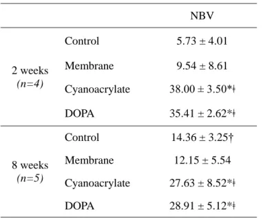
Table 1.
Mean ± SD values of NBV measured by micro-CT grey valueNBV 2 weeks (n=4) Control 5.73 ± 4.01 Membrane 9.54 ± 8.61 Cyanoacrylate 38.00 ± 3.50*ǂ DOPA 35.41 ± 2.62*ǂ 8 weeks (n=5) Control 14.36 ± 3.25† Membrane 12.15 ± 5.54 Cyanoacrylate 27.63 ± 8.52*ǂ DOPA 28.91 ± 5.12*ǂ Values are presented as mean ± standard deviation (mm3).
* Statistically significant difference compared to the control group. ǂ Statistically significant difference compared to the membrane group
† Statistically significant difference compared to the corresponding groups at 2 weeks. NBV=new bone volume;
26
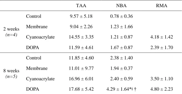
Table 2.
Histomorphometric analysisTAA NBA RMA
2 weeks (n=4) Control 9.57 ± 5.18 0.78 ± 0.36 Membrane 9.04 ± 2.26 1.23 ± 1.66 Cyanoacrylate 14.55 ± 3.35 1.21 ± 0.87 4.18 ± 1.42 DOPA 11.59 ± 4.61 1.67 ± 0.87 2.39 ± 1.70 8 weeks (n=5) Control 11.85 ± 4.60 2.38 ± 1.40 Membrane 11.01 ± 9.77 1.94 ± 0.37 Cyanoacrylate 16.96 ± 6.01 2.40 ± 0.59 3.50 ± 1.10 DOPA 17.68 ± 5.42 4.29 ± 1.64*ǂ † 4.80 ± 2.23 Values are presented as mean ± standard deviation (mm2).
* Statistically significant difference compared to the membrane group. ǂ Statistically significant difference compared to the cyanoacrylate group
† Statistically significant difference compared to the corresponding groups at 2 weeks. TAA=total augmented area; NBA=new bone area; RMA=residual material area.
27
Figures
28
29
30
31
32
33
34
국문요약
3,4-디하이드록시페닐알라닌을 골 접착제로 사용하여 블록타입
탈단백 돼지뼈와 콜라겐 차폐막을 이용한 골재생술
<지도교수 최 성 호> 연세대학교 대학원 치의학과 강 주 현 본 연구의 목적은 접착성 및 세포독성에 대해 3,4-디하이드록시페닐알라닌 (DOPA)을 평가하고 골유도재생술식에 있어서 DOPA와 콜라겐 차폐막을 평가 하는 것을 목적으로 한다. 박리 저항성 및 세포독성 시험을 수행 하였으며, 9개의 토끼 두개골에 네 개의 골결손부를 형성하고 다음과 같은 재료를 무작위로 적용하였다: (i) 대조 군 (ii) 콜라겐 차폐막 (iii) DOPA를 적용한 콜라겐 차폐막으로 덮힌 탈단백질 돼지뼈 (iv) 시아노아크릴레이트를 적용한 콜라겐 차폐막으로 덮인 탈단백 돼 지뼈. 미세 전산화 단층 촬영과 조직 형태 측정 분석을 위해 2주(n=4)와 8주 (n=5)에 동물을 희생시켰다.35 DOPA는 낮은 박리 저항성을 나타내지만 높은 세포 생존력을 보였다. 시아 노아크릴레이트와 DOPA 그룹은 2주 (P<0.029)에서 대조군과 콜라겐 차폐막 그룹에 비해 새로운 골량이 유의하게 높았다. 8주째 DOPA 군에서 가장 높은 새로운 골량을 보였다. DOPA 군에서 8주째에 새로운 골부위가 유의하게 높 았다 (P<0.029). DOPA 군에서 2주에서 8주로 갈 수록 골형성이 증가 하였다 (P<0.016). DOPA는 높은 세포 생존력을 나타내었고 생체 내 연구 결과 뼈 재생에서 예측 가능한 성능을 보였다. 핵심되는 말 : 골유도재생술, 골 접착제, 3,4-디하이드록시페닐알라닌