Protein-Protein Interaction between Poly(A) Polymerase and Cyclophilin A Bull. Korean Chem. Soc. 2014, Vol. 35, No. 1 83 http://dx.doi.org/10.5012/bkcs.2014.35.1.83
Protein-Protein Interaction between Poly(A) Polymerase and Cyclophilin A in
Chemotactic Cells
Hyun-Sook Choi,†,‡ Hana Kim,† Changgook Lee,† Youngmi Kim,† and Younghoon Lee†,* †Department of Chemistry, KAIST, Daejeon 305-701, Korea. *E-mail: Younghoon.Lee@kaist.ac.kr
‡National Science Museum, Daejeon 305-705, Korea Received October 1, 2013, Accepted October 9, 2013
Poly(A) polymerase (PAP) play an essential role for maturation of mRNA by adding the adenylate residues at the 3' end. PAP functions are regulated through protein-protein interaction at its C-terminal region. In this study, cyclophilin A (CypA), a member of the peptidyl-prolyl cis-trans isomerase family, was identified as a partner protein interacting with the C-terminal region PAP. The interaction between PAP and CypA was inhibited by the immunosuppressive drug cyclosporine A. Deletion analysis revealed that the N-terminal 56 residues of CypA are sufficient for the interaction with PAP. Interestingly, we observed that PAP and CypA colocalize in the nucleus during SDF-1-induced chemotaxis, implying that CypA could be involved in the regulation of polyadenylation by PAP in the chemotactic cells.
Key Words : Poly(A) polymerase (PAP), Cyclophilin A (CypA), Protein-protein interaction, Stromal cell-de-rived factor-1 (SDF-1), Chemotactic cells
Introduction
The poly(A) tail of eukaryotic mRNA is synthesized by the polyadenylation machinery. The polyadenylation mach-inery comprises multiple trans-acting factors, including cleav-age and polyadenylation specificity factor (CPSF), cleavcleav-age- cleavage-stimulation factor (CstF), two cleavage factors (CFI and CFII), poly(A) polymerase (PAP) and poly(A)-binding pro-tein II (PAB II).1-5 The poly(A) tail is known to enhance the translation efficiency and the mRNA stability,6,7 so poly-adenylation can play important regulatory roles in diverse cellular processes.8-10
PAP is a key enzyme for the poly(A) tail synthesis. The N-terminal region of PAP has the catalytic domain,11,12 whereas the C-terminal region carries an RNA binding domain, two nuclear localization signals and a serine/threonine-rich regulatory domain.13 The activity and cellular localization of PAP could be regulated through interaction with various regulatory proteins in its C-terminal region. The U1A pro-tein, the component of U1 snRNP, is known to repress the PAP activity by interaction with C-terminal region of PAP.8 14-3-3, one of the members of the 14-3-3 protein family, binds to the C-terminal region of PAP. 14-3-3 inhibits the polyadenylation activity of PAP and redistributes PAP within the cell by increasing its cytoplasmic localization.14 The C-terminal region of PAP also interacts with splicing factor U2AF65 and cleavage factor I 25 kDa subunit which is component of polyadenylation complex.15,16 These evi-dences suggest that the C-terminal region of PAP could serve as a platform for binding partner proteins and the activity of PAP could be regulated in various cellular pro-cesses. However, there might be other regulatory proteins binding to the C-terminal region that would be involved in the regulation of PAP function.
In this study, we performed a yeast two hybrid screening by using the C-terminal region of PAP as a bait to search for another interaction partner of PAP. We identified cyclophilin A (CypA) as a binding partner for PAP. CypA is involved in pathogenesis of several diseases, including viral infection, cardiovascular disease, and cancer.17 CypA may act as a chaperone-like protein18 and play a role in protein-folding processes19 because it has a peptidyl-prolyl cis-trans isomer-ase activity.20 CypA is diffusely distributed in the cytoplasm of culture cells.21 However, CypA can be transported to the nucleus by transportin 1 through its binding to CXCR4, the chemokine receptor of SDF-1, during the stromal cell-derived factor 1 (SDF-1)-induced chemotaxis.22 In addition, CypA binds with high affinity to the immunosuppressive drug cyclosporine A23,24 and the CypA-cyclosporine A complex inhibits calcineurin, a serine-threonine phosphatase required for cytokine induction in response to stimulation of T cells.25,26 Here, we show that CypA interacts with the C-terminal region of PAP and that the N-C-terminal 56 residues of CypA are responsible for this interaction. We also observed the PAP and CypA colocalize in the nucleus during SDF-1 induced chemotaxis.
Experimental Section
Yeast Two-hybrid Screen. We used the Matchmaker LexA two-hybrid system (Clontech), with the C-terminal 268 residues of mouse PAP as bait in a pEQ202 LexA fusion-plasmid vector.14 A human HeLa cDNA library was pre-pared with the B42 transactivating domain fusion-plasmid vector and screened from 2 × 10⁶ primary transformants of yeast strain EGY48. Yeast cells were grown in SD media lacking histidine, tryptophan, and uracil. Clones expressing -galactosidase activity were screened on the plates by using
84 Bull. Korean Chem. Soc. 2014, Vol. 35, No. 1 Hyun-Sook Choi et al. 5-bromo-4-chloro-3-indolyl--D-galactopyranoside (X-gal)
as a substrate. One of the selected clones was identified as a cDNA coding for cyclophilin A. The coding sequence of mouse cyclophilin A cDNA (GenBank accession No. Y00052) was obtained by PCR-amplification from a mouse kidney cDNA library.
Preparation of Recombinant cDNA Constructs. Escherichia
coli expression plasmid pGST-CypA was constructed by
cloning the coding sequence of mouse CypA into the pGEX4T-1 vector (Amersham Pharmacia Biotech). For expression of GST-CypA and its derivatives in mammalian cells, full-length or defined regions of CypA were cloned into pEBG.16 A plasmid expressing FLAG-PAP-CTD was constructed by cloning the DNA sequence for the C-terminal 269 residues of PAP into p3XFLAG-CMV-7.1 (Sigma). A plasmid ex-pressing GFP-PAP was generated by cloning the PAP coding region into pEGFP-C1 (Clontech).
Purification of GST–CypA and its Derivatives. The E.
coli JM109 strain and the GST purification system
(Amer-sham–Pharmacia Biotech) were used for expression and purification of GST-fusion proteins.
Preparation of [35S]-labeled PAP. [35 S]-methionine-labe-led full-length PAP was obtained by a TNT Quick CoupS]-methionine-labe-led Transcription/Translation kit (Promega) according to the manufacturer’s instruction.
Antibodies. Antibodies specific for GST (Santa Cruz), FLAG (Sigma), PAP (Bethyl Lab), and CypA (Abcam) were purchased. The Cy3-conjugated secondary antibody was from Jackson Immunoresearch.
In Vitro Binding Assay. A GST pull-down experiment was performed for an in vitro binding assay. Purified GST or GST-fusion proteins were bound to glutathione beads (Amersham Pharmacia Biotech). The [35S]-labeled trans-lation mixture and glutathione beads were incubated in a 500 L binding buffer (20 mM Tris-HCl, pH 7.4, 50 mM NaCl, 2 mM MgCl2, 1 mM DTT, and 0.1% Nonidet-P 40) on ice for 2 h. The beads were recovered by centrifugation and were washed four times with the same fresh buffer. Then the beads were boiled in a SDS sample buffer, and analyzed by SDS-PAGE and autoradiography. To examine the effects of cyclosporine A or salt concentrations, cyclosporine A or the salt at different concentrations was included in the binding buffer.
In Vivo Binding Assay. HeLa and NIH3T3 cells were maintained in Dulbecco’s modified Eagle’s medium supple-mented with 10% fetal calf serum or 10% bovine serum. Cells (~1 × 10⁸) were transfected with DNA constructs (1 g) using Lipofectamine reagent (Invitrogen). Coimmuno-precipitations were performed as described previously.16 Similar, coimmunoprecipitation assays using lysates from untransfected HeLa cells were also performed with anti-PAP and anti-CypA antibodies.
Confocal Laser Scanning Microscopy. HeLa cells were cotransfected with cDNA (3 g) expressing GFP-PAP and FLAG-CypA. The transfected cells were treated with SDF-1 (Sigma) at a final concentration of 12.5 nM and taken out for analysis at different time intervals (0, 30, 60, 120, 200 min).
The cells were fixed with 3.7% formaldehyde and treated with anti-FLAG antibody, followed by Cy3-conjugated secondary antibody, as described previously.14 The GFP-PAP was directly visualized, while nuclei were stained with DAPI. Imaging was conducted on a Zeiss LSM510 meta confocal microscope.
Results
Identification of CypA as a New Interacting Partner of PAP. We searched for PAP-interaction proteins through a LexA-based yeast two-hybrid screen using the C-terminal 268 residues of mouse PAP as a bait from a human HeLa cDNA library (Fig. 1). Approximately two-million colonies were screened to find clones coding for proteins that would interact with the C-terminal region of PAP. One of the interacting clones showed was the cDNA encoding human CypA.
Interaction of PAP and CypA In Vitro. The coding sequence of mouse CypA cDNA was obtained by PCR-amplication from a mouse kidney cDNA library and this mouse CypA sequence was used throughout the study. To determine whether interactions between mouse PAP and CypA occur in vitro, we performed in vitro protein-protein interaction assays. GST-CypA fusion protein purified from
E. coli and [35S] methionine-labeled PAP translated in vitro were used for coimmunoprecipitation. The interaction between PAP and CypA was observed (Fig. 2, lanes 3-5). A significant fraction of the input PAP was bound to GST-CypA, but not to the GST control protein (Fig. 2, lane 7). This interaction was stable at the concentrations of 50 to 150 mM NaCl, but disrupted at a high concentration of 1000 mM NaCl. In addition, when cyclosporine A at the final concentration of 10 M was added in the binding buffer, the interaction between PAP and CypA was almost disrupted (Fig. 2, lane 2).
Interaction of PAP and CypA In Vivo. We performed a coimmunoprecipitation experiment using HeLa cells cotrans-fected with GST-CypA and FLAG-PAP-CTD constructs. Total lysates from the transfected cells were immunopreci-pitated with an anti-FLAG antibody, and the coimmuno-precipitated materials were immunoblotted with an
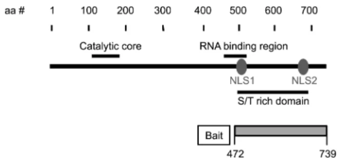
anti-Figure 1. Schematic diagram of the significant features of PAP.
The catalytic domain, an RNA-binding region, the S/T-rich region, and two nuclear localization signal sequences (NSL) are shown. The C-terminal 268 residues of mouse PAP (residues 472 to 739) were used for a bait for yeast two-hybrid screening.
Protein-Protein Interaction between Poly(A) Polymerase and Cyclophilin A Bull. Korean Chem. Soc. 2014, Vol. 35, No. 1 85
GST antibody. We found that CypA was present in PAP-CTD immunoprecipitates (Fig. 3(a)), confirming that that the C-terminal domain of PAP interacts with CypA in mam-malian cells.
Next, we performed coimmunoprecipitation assays to determine whether endogenous PAP and CypA interact in cells. Lysates of HeLa cells were immunoprecipitated with anti-PAP antibody, and blotted with an anti-CypA antibody. The immune blot indicated that the two proteins interact in the cells (Fig. 3(b)).
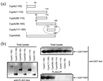
Regions of CypA Required for Binding to PAP. To delineate regions of CypA required for interaction with PAP, we generated several deletion plasmid constructs that could express truncated derivatives of CypA (Fig. 4(a)). Mutant proteins were tested for their ability to bind PAP by coimmuno-precipitation analyses. Strong interactions were observed with various fragments over the 111-165 fragment (the
terminal 56 residues) (Fig. 4(b)), suggesting that the C-terminal 56 residues of CypA are sufficient for the inter-action with PAP.
Colocalization of PAP and CypA. To examine whether PAP and CypA are colocalized in cells, we performed con-focal fluorescence microscopic analyses (Fig. 5). In normal cells, CypA was distributed in the cytoplasm diffusely and it was difficult to find the colocalization of PAP and CypA
Figure 2. In vitro interaction of PAP with CypA. The in vitro
translated PAP labeled with [35S]methionine and the GST–CypA
fusion protein were mixed with glutathione beads in the binding buffer. NaCl concentrations in the buffer are indicated above each lane. The pull-down materials were loaded on a SDS–PAGE gel and detected by autoradiography. Lane 1, [35S]methionine-labeled PAP alone; lanes 2 to 7, pull-down products; and lane 7, GST used as a negative control. To investigate the effect of cyclosporine A on the interaction between PAP and CypA, Cyclospoine A (10 M) was included in the buffer (lane 2).
Figure 3. In vivo interaction of PAP with CypA. (a)
Coimmuno-precipitation of GST-CypA with FLAG-PAP-CTD. Cell lysates from HeLa cells co-transfected with the GST-CypA and FLAG-PAP-CTD constructs were immunoprecipitated with anti-FLAG antibody (lanes 2 and 4). The GST construct was used as a negative control (lanes 1 and 3). The immunoprecipitates were subjected to immunoblotting using anti-GST antibody (lanes 3 and 4). For the ensuring the expression of GST-CypA or GST, 1% of total lysates were used (lanes 1 and 2). (b) Interactions between endogenous PAP and CypA. Coimmunoprecipitation was perform-ed using HeLa cell lysates. T, total lysate (1% input); C, control antibody; IP, immunoprecipitates with anti-PAP. The immuno-precipitates were subjected to immunoblotting using anti-CypA antibody.
Figure 4. Regions of CypA required for the interaction with PAP.
(a) Schematic diagram of FLAG-CypA derivatives. (b) Coimmuno-precipitation of GST-PAP with FLAG-CypA derivatives. Lysates of NIH3T3 cells transfected with clones encoding GST–PAP and FLAG-CypA derivatives were immunoprecipitated with anti-FLAG antibody and the resulting immunoprecipitates were blotted with anti-GST antibody. The total lysates (2% input) were also blotted with anti-FLAG antibody and anti-GST antibody for ensuring the expression of FLAG-CypA derivatives and GST-PAP, respectively.
Figure 5. Co-localization of PAP and CypA in cells. HeLa cells
were cotransfected with GFP-PAP and FLAG-CypA. The trans-fected cells were treated with SDF-1 (12.5 nM) and analyzed at different time intervals (0, 30, 60, 120, 200 min). The treated cells were fixed in 3.7% formaldehyde and incubated with a mouse anti-FLAG monoclonal antibody, followed by a Cy3-conjugated anti-mouse antibody. The GFP-PAP was directly visualized, while nuclei were stained with DAPI. FLAG-CypA, GFP-PAP, nuclei were visualized with a confocal microscope for green, red, and blue fluorescence. Images of FLAG-CypA alone (top), merged images of FLAG-CypA and nuclei (middle), and merged images of FLAG-CypA and GFP-PAP (bottom).
86 Bull. Korean Chem. Soc. 2014, Vol. 35, No. 1 Hyun-Sook Choi et al. (Fig. 5, 0 min). This is probably because PAP and CypA
mainly exist in the nucleus and the cytoplasm, respectively. Therefore, we supposed that the colocalization of PAP and CypA would be observed when the two proteins are present in the same organelle. It was known that CypA is imported into the nucleus during SDF-1-induced chemotaxis,22 so we determined the colocalization of the two proteins in the 1 treated cell. When HeLa cells were treated with SDF-1 at SDF-10 M, we observed that Cyp was imported to the nucleus, but appeared to be exported from the nucleus starting 30 min after the SDF-1 treatment (Fig. 5). PAP and the imported CypA were colocalized in the nucleus, suggesting that the binding of CypA to PAP may play more important roles probably in polyadenylation in chemotactic cells.
Discussion
In the polyadenylation machinery, PAP is a key enzyme responsible for the synthesis of the poly(A) tail. Therefore, the control of PAP activity could be important to regulate the level of gene expression. It is known that the C-terminal region of the PAP functions as a platform for protein-protein interactions.8,15 In this study, we performed a LexA-based yeast two-hybrid screening to identify PAP-interaction partners and identified the CypA as a partner protein. We show that CypA binds strongly to the C-terminal region of PAP and the C-terminal 56 residues of CypA is required for this interaction. CypA has the peptidyl-prolyl cis-trans isomerase activity20 and is involved in various diseases, such as viral infection, cardiovascular diseases and cancers.17 We found that cyclosporine A inhibits the PAP-CypA interaction, possibly due to the overlapping of the binding regions because the C-terminal 56 residues of CypA is essential for binding of cyclosporine A to CypA.27 We also observed that CypA was colocalized with PAP in the nucleus during SDF-1-induced chemotaxis.
Although the effect of the interaction between PAP and CypA on polyadenylation of mRNA is unrevealed, it might be involved in the regulation of the polyadenylation machi-nery. There is the growing possibility that CypA could affect the activity of the polyadenylation machinery. For example, another peptidyl-prolyl cis-trans isomerase Pin1 interacts with cytoplasmic polyadenylation element binding protein (CPEB), leading to the degradation of CPEB by the
Pin-induced conformational change.28 In this respect, our data imply that the interaction between PAP and CypA could play important roles in regulation of polyadenylation, especially in the chemotactic cells. However, the real function of this interaction remains to be demonstrated.
Acknowledgments. This work was supported by the National Research Foundation of Korea (NRF) Grant by the Korea government (MSIP) [2010-0029167, 2011-0020322].
References
1. Keller, W.; Bienroth, S.; Lang, K. M.; Christofori, G. EMBO J.
1991, 10, 4241.
2. Barabino, S. M.; Keller, W. Cell 1999, 99, 9.
3. Zhao, J.; Hyman, L.; Moore, C. Microbiol. Mol. Biol. Rev. 1999,
63, 405.
4. Shatkin, A. J.; Manley, J. L. Nat. Struct. Biol. 2000, 7, 838. 5. Takagaki, Y.; Manley, J. L. Mol. Cell. Biol. 2000, 20, 1515. 6. Beelman, C. A.; Parker, R. Cell 1995, 81, 179.
7. Tarun, S. Z., Jr.; Sachs, A. B. EMBO J. 1996, 15, 7168.
8. Gunderson, S. I.; Vagner, S.; Polycarpou-Schwarz, M.; Mattaj, I. W. Genes Dev. 1997, 11, 761.
9. Wells, D. G.; Dong, X.; Quinlan, E. M.; Huang, Y. S.; Bear, M. F.; Richter, J. D.; Fallon, J. R. J. Neurosci. 2001, 21, 9541.
10. Richter, J. D. Microbiol. Mol. Biol. Rev. 1999, 63, 446. 11. Martin, G.; Keller, W. EMBO J. 1996, 15, 2593.
12. Martin, G.; Keller, W.; Doublie, S. EMBO J. 2000, 19, 4193. 13. Raabe, T.; Murthy, K. G.; Manley, J. L. Mol. Cell. Biol. 1994, 14,
2946.
14. Kim, H.; Lee, J. H.; Lee, Y. EMBO J. 2003, 22, 5208. 15. Vagner, S.; Vagner, C.; Mattaj, I. W. Genes Dev. 2000, 14, 403. 16. Kim, H.; Lee, Y. Biochem. Biophys. Re.s Commun. 2001, 289, 513. 17. Liu, X.; Zhao, Z.; Liu, W. Viruses 2013, 5, 182.
18. Baker, E. K.; Colley, N. J.; Zuker, C. S. EMBO J. 1994, 13, 4886. 19. Gothel, S. F.; Marahiel, M. A. Cell. Mol. Life Sci. 1999, 55, 423. 20. Galat, A. Eur. J. Biochem. 1993, 216, 689.
21. Sarris, A. H.; Harding, M. W.; Jiang, T. R.; Aftab, D.; Handschumacher, R. E. Transplantation 1992, 54, 904.
22. Pan, H.; Luo, C.; Li, R.; Qiao, A.; Zhang, L.; Mines, M.; Nyanda, A. M.; Zhang, J.; Fan, G. H. J. Biol. Chem. 2008, 283, 623. 23. Handschumacher, R. E.; Harding, M. W.; Rice, J.; Drugge, R. J.;
Speicher, D. W. Science 1984, 226, 544.
24. Kim, I. K.; Park, S. J.; Park, J. H.; Lee, S. H.; Hong, S. E.; Reed, J. C. BMB Rep. 2012, 45, 482.
25. Rao, A.; Luo, C.; Hogan, P. G. Annu. Rev. Immunol. 1997, 15, 707. 26. Schreiber, S. L.; Crabtree, G. R. Immunol. Today 1992, 13, 136. 27. Kallen, J.; Spitzfaden, C. S.; Zurini, M. G. M.; Wider, G.; Widmer,
H.; Wuthrich, K.; Walkinshaw, M. D. Nature 1991, 353, 276. 28. Nechama, M.; Lin, C. L.; Richter, J. D. Mol. Cell. Biol. 2013, 33,