저작자표시 2.0 대한민국 이용자는 아래의 조건을 따르는 경우에 한하여 자유롭게 l 이 저작물을 복제, 배포, 전송, 전시, 공연 및 방송할 수 있습니다. l 이차적 저작물을 작성할 수 있습니다. l 이 저작물을 영리 목적으로 이용할 수 있습니다. 다음과 같은 조건을 따라야 합니다: l 귀하는, 이 저작물의 재이용이나 배포의 경우, 이 저작물에 적용된 이용허락조건 을 명확하게 나타내어야 합니다. l 저작권자로부터 별도의 허가를 받으면 이러한 조건들은 적용되지 않습니다. 저작권법에 따른 이용자의 권리는 위의 내용에 의하여 영향을 받지 않습니다. 이것은 이용허락규약(Legal Code)을 이해하기 쉽게 요약한 것입니다. Disclaimer 저작자표시. 귀하는 원저작자를 표시하여야 합니다.
A THESIS
FOR THE DEGREE OF DOCTOR OF PHILOSOPHY
In vivo and In vitroEffects of
Chestnut (Castanea crenata) Inner Shell on
Antioxidant and Cardiovascular-Related
Parameters
Min-Sook Kang
Department of Food Science and Nutrition
GRADUATE SCHOOL
JEJU NATIONAL UNIVERSITY
In vivo and In vitroEffects of
Chestnut (Castanea crenata) Inner Shell on
Antioxidant and Cardiovascular-Related Parameters
Min-Sook Kang
(Supervised by professor Jung-Sook Kang)
A thesis submitted in partial fulfillment of the requirement for the degree of DOCTOR OF PHILOSOPHY
2013. 02.
This thesis has been examined and approved by
Thesis director, You-Jin Jeon, Professor of Marine Life Science
Nam-Ho Lee, Professor of Chemistry
In-Sook Kwun, Professor of Food and Nutrition
So-Mi Kim, Professor of Faculty of Biotechnology
Jung-Sook Kang, Professor of Food Science and Nutrition
Date
Department of Food Science and Nutrition
GRADUATE SCHOOL
JEJU NATIONAL UNIVERSITY
CONTENTS
LIST OF TABLES ... iv LIST OF SCHEME ... iv LIST OF FIGURES... v INTRODUCTION ... 1 LITERATURE REVIEW ... 4 1... R isk factors of cardiovascular disease ... 41) ... A therosclerosis ... 4
2) ... O xidative stress and inflammation in atherosclerosis... 6
3) ... H ypertension ... 9
2... P revention properties of polyphenols on cardiovascular disease ... 11
3... P otential possibility of utilization of chestnut inner shell as bio-resource... 13
Part I. In vivo study for the effects of chestnut (Castanea crenata)
inner shell on antioxidant and cardiovascular-related parameters
ABSTRACT... 17MATERIALS AND METHODS ... 19
1... M aterials ... 19 1) ... A
nimals and diets... 19 2) ... P
reparation of chestnut inner shell extract ... 19 2... C
ollection of samples ... 21 3... S
ample analysis ... 21 1) ... W
hole blood platelet aggregation ... 21 2) ... P
lasma and liver lipid assay ... 22 3) ... P
latelet rich plasma (PRP) and liver TBARS productions ... 22 4) ... E
rythrocyte Na efflux channels ... 23 5) ... P
lasma GOT and GPT levels ... 27 6) ... L
ung angiotensin converting enzyme (ACE) activity ... 28 4... S
tatistical analysis ... 28 RESULTS ... 30
1. ...B ody weights, plasma and liver lipids... 30
2. ...H ematocrit and whole blood platelet aggregation ... 33
3. ...P lasma GOT and GPT levels ... 35
4. ...E rythrocyte Na efflux and ACEinhibitory effect ... 37
5. ...E rythrocyte Na-leak ... 39
6. ...P latelet rich plasma (PRP) and liver TBARS productions ... 41 DISCUSSION ... 43
Part II. In vitro study for the effects of chestnut (Castanea crenata)
inner shell on antioxidant and cardiovascular-related parameters
ABSTRACT... 50 MATERIALS AND METHODS ... 52 1... M
aterials ... 52 1) ... C
hemical materials ... 52 2) ... S
olvent fractionation of chestnut inner shell extract... 52 3) ... B
lood and lung samples preparation... 53 4) ... L
ymphocyte isolation... 54 2... M
ethods ... 54
1) ...F ree radical scavenging activity... 54
2) ...D etermination of total polyphenolic content ... 56 3) ... C
omet assay for determination of DNA damage ... 57 4) ... A
ngiotensin converting enzyme (ACE) inhibition activity... 58 5) ... W
6) ... A ssay for pro-inflammatory mediators... 60 3... S
tatistical analysis ... 63 RESULTS... 64 1... F
ree radical scavenging activities of Ech and the major polyphenols in Ech 64 2... D
PPH radical scavenging activities and total polyphenolic contents of Ech and its solvent fractions ... 68 3... P
rotective effect against H2O2-induced DNA damage... 70
4... A ngiotensin converting enzyme (ACE) inhibition activity... 72 5... W
hole blood platelet aggregation... 74 6... E
ffects on NO production in LPS-stimulated RAW 264.7 cells ... 76 7... E
ffects on PGE2 production in LPS-stimulated RAW 264.7 cells ... 78
8... E ffects on protein levels of iNOS and COX-2 in LPS-stimulated RAW 264.7 cells ... 80 9... E
ffects on the production of pro-inflammatory cytokines in LPS-stimulated RAW 264.7 cells... 82 DISCUSSION ... 86
LIST OF TABLES
Table 1-1. Diet composition... 20
Table 1-2. Effects of chestnut inner shell powder (Pch) and extract (Ech), statin and coenzyme Q10 on body weight and plasma and liver lipid in rats fed with cholesterol-based diet ... 32
Table 2-1. SC50 of Ech and the major polyphenols in Ech against DPPH and
hydroxyl radicals... 69
Table 2-2. SC50 of Ech and its solvent fractions against DPPH radical ... 69
Table 2-3. Total polyphenolic contents of Ech and its solvent fractions ... 69
LIST OF SCHEME
Scheme 2-1. Systematic purification using solvent partitioning from chestnut (Castaneacrenata) inner shell powder ... 53
LIST OF FIGURES
Figure 1. Response to injury hypothesis of atherosclerosis... 5
Figure 2. Hydrolysable tannins ... 12
Figure 3. Precursor of condensed tannins ... 12
Figure 1-1. Model of erythrocyte Na efflux channels ... 27
Figure 1-2. Effects of chestnut inner shell powder (Pch) and extract (Ech), statin and coenzyme Q10 on hematocrit and platelet aggregation in rats fed with cholesterol-based diet... 34
Figure 1-3. Effects of chestnut inner shell powder (Pch) and extract (Ech), statin and coenzyme Q10 on plasma GOT and GPT levels in rats fed with cholesterol-based diet... 36
Figure 1-4.Effects of chestnut inner shell powder (Pch) and statin on erythrocyte Na efflux in rats fed with cholesterol-based diet ... 38
Figure 1-5. Effects of chestnut inner shell powder (Pch) and statin on angiotensin converting enzyme activity in rats fed with cholesterol-based diet... 38
Figure 1-6. Effects of statin, coenzyme Q10, and chestnut inner shell extract (Ech) on intact and AAPH treated erythrocyte Na leak in rats fed with cholesterol-based diet... 40
Figure 1-7. Effects of chestnut inner shell powder (Pch) and extract (Ech), statin and coenzyme Q10 on platelet rich plasma (PRP) and liver thiobarbituric acid reactive substance (TBARS) in rats fed with cholesterol-based diet... 42
Figure 2-1. Absorbance changes by difference drying conditions ... 59
Figure 2-2. DPPH radical scavenging activities of Ech and the major polyphe-nols in Ech ... 66
Figure 2-3. Hydroxyl radical scavenging activities of Ech and the major poly-phenols in Ech ... 67
Figure 2-4. Protective effects of Ech, its solvent fractions, and the major poly-phenols in Ech against H2O2-induced DNA damage in lymphocyte 71
Figure 2-5. Effects of Ech, its solvent fractions, and the major polyphenols in Ech on angiotensin converting enzyme (ACE) activity... 73
Figure 2-6. Effects of Ech, its solvent fractions, and the major polyphenols in Ech on platelet aggregation... 75
Figure 2-7. Effects of Ech, its solvent fractions, and the major polyphenols in Ech on nitric oxide (NO) production in LPS-stimulated RAW 264.7 cells ... 77
Ech on prostaglandin E2 (PGE2) production in LPS-stimulated RAW
264.7 cells... 79
Figure 2-9. Effects of Ech, its solvent fractions, and the major polyphenols in Ech on protein levels of iNOS and COX-2 in LPS-stimulated RAW 264.7 cells... 81
Figure 2-10. Effects of Ech, its solvent fractions, and the major polyphenols in Ech on TNF-α production in LPS-stimulated RAW 264.7 cells ... 83
Figure 2-11. Effects of Ech, its solvent fractions, and the major polyphenols in Ech on IL-1βproduction in LPS-stimulated RAW 264.7 cells ... 84
Figure 2-12. Effects of Ech, its solvent fractions, and the major polyphenols in Ech on IL-6 production in LPS-stimulated RAW 264.7 cells ... 85
INTRODUCTION
Cardiovascular disease (CVD) is one of the leading causes of death and disability in the world.1)An estimated 17.3 million people died from CVE in 2008, representing that 30% of all global deaths and almost 23.6 million people will die from CVD, mainly from heart disease and stroke by 2030.2)According to 2010 statistics in Korea, CVD was the second cause of death following cancer, corresponding 23.0% of all death.3)Incidence of CVD is associated with multiple risk factors such as raised total cholesterol and LDL-cholesterol, hypertension, increased platelet reactivity, alterations in glucose metabolism, and smoking.4)Recent findings have suggested that
oxidative stress, vascular inflammation, and endothelial dysfunction may play roles in developing atherosclerosis which is the major incidence of CVD.5)
Statins, 3-hydroxy-3-methylglutaryl coenzyme A (HMG-CoA) reductase inhibitors, are commonly used drugs to treat hyperlipidemia, consequently preventing coronary artery disease and stroke. Statin is known to deplete coenzyme Q10 (CoQ10) by halting production of mevalonate, a CoQ10 precursor in cholesterol biosynthesis. In fact, the use of statins can decrease the synthesis of CoQ10 up to 40%.6)CoQ10 acts as electron carrier in mitochondrial electron transport system (ETS), and depletion of CoQ10 directly impacts on active muscles. Myopathic complications such as myalgia and rhabdomyolysis are most serious side effects after statin therapy and constipation, diarrhea, dizziness, headaches, rashes, and upset stomach are also reported as minor symptoms. Besides function in ETS, the reduced form of CoQ10 (CoQH2), ubiquinol plays role in protecting cell membrane from oxidative damage by scavenging free radical generated in physiological system. Dhanasekaran et
al.7)proposed that a lipophilic antioxidant CoQH2 participates regeneration of α– tocopherol and ascorbate in plasma membrane and prevents lipid peroxidation.
Recently, the emphasis in medical care has changed from “treatment” to “prevention” in which functional food and food therapy play an important role. This approach reflects the oriental medicine paradigm of “prevent illness before it began” and “food and medicine have the same root”. With this concepts, there has been growing interest in complementary and alternative medicine (CAM) for treatment of diseases, illness prevention and maintenance of health. Many Americans are now pursuing CAM as an alternative or supplement to conventional medicine, and a 2007 National Health Interview Survey (NHIS) showed that approximately 38% of adults in America use CAM therapy.8)
Over the past few years, various epidemiological studies have shown an inverse relation between the consumption of polyphenols or polyphenol-rich foods and the risk of CVD.9)10)Much attention has been paid to natural polyphenols of silymarin, curcumin, green tea, and grape seed extracts,11) which provide strong antioxidants
against free radicals, thereby reducing cell damage and risk of certain diseases such as cancer,12) inflammation,13)and CVD.9)10)
Chestnut is one of the favorite nuts cultivated for a long time and widely used including Korean traditional ceremonies. In the last few years, the consumption of fresh or previously transformed chestnuts has gradually increased due to their nutritional qualities and palatability as foods. In the process of getting edible nut, considerable amounts of the inedible waste part, the pericarp (outer shell) and integument (inner shell) are generated, and much efforts has been made to regenerate the chestnut bark (inner and outer shells) to valuable coproduct with economic view
point in European countries.14)
Ellagitannin and gallotannin in chestnut bark are hydrolyzed to strong antioxidant polyphenols, ellagic acid and gallic acid. Despite valuable phenolic compounds of chestnut bark, there are not many studies have been reported on its protective effects against CVD.
Accordingly, the first part of this study attempted to investigate the effects of chestnut inner shell of powder and extract in vivo on cardiovascular-related parameters, such as plasma and liver lipids, platelet aggregation, erythrocyte Na efflux, ACE activity, and plasma GOT and GPT levels using animal model of guinea pigs which were fed with statin-added cholesterol diets. In the second part, study focused on in vitro or ex vivo effects of ethanol extract of chestnut inner shell and the major polyphenol compounds such as ellagic acid, gallic acid, and catechin using lung tissue, platelet, lymphocytes, and RAW 264.7 cells. This study also included the comparison of anti-inflammatory activity in fractions partitionated by polarity (n-hexane, ethyl acetate, butanol, and water) of ethanol extract of the chestnut inner shell, providing valuable data to elucidate the association of the chestnut inner shell with CVD development.
1. Risk factors of CVD
1) Atherosclerosis
Atherosclerosis, the primary cause of CVD and stroke, is a progressive disease characterized by the build-up of lipids and fibrous elements in the large arteries. The prevalent view of spontaneous atherosclerosis is the “response-to-injury” hypothesis based on early proposals made by Virchow15) and subsequently modified by Ross16)more than a century later.
In this hypothesis,17)atherosclerosis is initiated as a response to various forms of injury to arterial endothelium. Endothelial cell injury from hyperlipidemia, hypertension, diabetes mellitus, or smoking etc, disrupts normal endothelial cell function. Endothelial dysfunction leads to increased permeability so that lipids and circulating cells including monocytes and T-lymphocytes, enter the sub-endothelial space and form the initial characteristic lesion of atherosclerosis, the fatty steak. Accumulation of cells and lipids may increased endothelial disruption leading to thrombogenic surfaces to which platelets adhere. Platelets, macrophages, endothelial cells and probably smooth muscle cells (SMCs) themselves can release growth modulatory factors, leading to proliferation of SMCs and fibroblasts and this process leads to the formation of the fibrous plaque and ultimately to the complex advanced lesion of atherosclerosis (Figure 1). Intimal vascular SMCs synthesize extracellular matrix proteins such as collagen, elastin, and proteoglycans that lead to the development of a fibrous cap.18)19)Vulnerable plaque are characterized by lipid
accumulation that expands the intima, degradation of the extracellular matrix, a decrease in connective tissue proteins, and a necrotic core. It is thought that lesions with large amounts of such lipids are particularly unstable and liable to rupture, leading to thrombosis and vessel occlusion. Plaque rupture and thrombosis induce acute cardiovascular events that result in myocardial infarction and stroke.20)
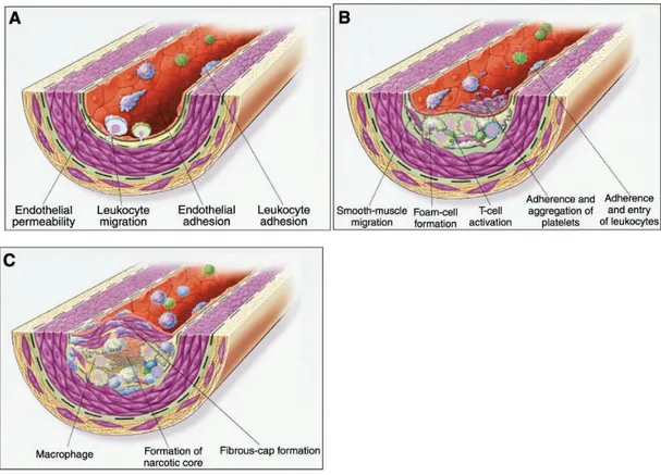
Figure 1. Response-to-injury hypothesis of atherosclerosis
In this hypothesis, atherosclerosis begins with endothelial injury or dysfunction (A) that is characterized by enhanced endothelial permeability and low-density lipoprotein (LDL) deposition in the subendothelial space. This is followed by leukocyte adhesion and transmigration across the endothelium. In intermediate stages (B), atherosclerosis is characterized by foam cell formation and an inflammatory response including T-cell activation, the adherence and aggregation of platelets, and further entry of leukocytes into the arterial wall along with migration of smooth muscle cells into the intima. Finally, advanced atherosclerosis (C) is characterized by continued macrophage accumulation, fibrous cap formation, and necrosis in the core of the lesion. [From Stocker
et al,21]copyright 2004 Physiol Rev.]
It is now widely accepted that oxidative stress and inflammation play a pivotal role in the initiation and progression of atherosclerosis and CVD.22)Oxygen is essential for metabolic processes in the body. However, oxygen derived free radicals, namely reactive oxygen species (ROS) are reported to exert detrimental effects, such as membrane lipid peroxidation, alteration of lipid-protein interactions, enzyme inactivations, and DNA breakage.23)
The production of free oxidative radical is believed to induce endothelial dysfunction through several mechanisms. Firstly, ROS, especially hydroxyl radicals, directly injure cell membranes and nuclei. Lipid peroxidation by ROS causes particularly destructive effects on cell membranes, furthermore severe lipid peroxidation causes myocardial necrosis.24)H2O2 has been demonstrated to induce
vascular smooth muscle cell (VSMC) death,25)which may occur by apoptosis.26) Secondly, ROS was proposed to modulate vasomotion and the atherogenic process by interacting with endogenous vasoactive mediators formed in endothelial cells. Endogenous nitric oxide (NO) known as endothelium-derived relaxing factor (EDRF) not only acts as potent endogenous vasodilator, but also inhibits vascular smooth muscle cell (VSMC) migration and proliferation, platelet adhesion and aggregation, LDL oxidation, and vascular inflammation.27)28)Endogenous NO synthesized from L-arginine by endothelial nitric oxide synthase (eNOS) get dampened by ROS. ROS inactivate NO via three different mechanisms: 1) direct inactivation of NO by superoxide (O2-), resulting in the formation of peroxynitrite
(ONOO-),29)2) reducing NOS activity with increased levels of asymmetric dimethylarginine (ADMA), an endogenous NOS inhibitor,30)and 3) uncoupling
eNOS with increased oxidation of cofactor tetrahydrobiopterin (BH4).31)Decreased
NO bioavailability with decreased NO production and/or increased NO inactivation causes endothelial dysfunction by an imbalance of NO and ROS, that cause atherosclerosis.32)
Thirdly, ROS expedites atherosclerosis by formation of the key mediator, oxidized lipoprotein (LDL) via peroxidizing lipid components. Hydroxyl radicals may initiate the peroxidation of long-chain polyunsaturated fatty acids within LDL molecule, giving rise to conjugated dienes and lipid hydroperoxy radicals (LOO·). This process is self-propagation, such that LOO· can attack adjacent fatty acids until completion of chain fragmentation. Unlimited amount of oxidatively modified LDL (oxLDL) is taken up by sub-endothelial macrophages via the “scavenger” receptor pathway to form a “foam cell” in the arterial intima.33)In addition, oxidized LDL exerts several biological effects; induction of inflammation, inhibition of eNOS and vasoconstriction, stimulation of cytokines such as interleukin-1 (IL-1), and activation of platelets.34)
Inflammatory processes play an important role in all stages of atherosclerosis.35)Unlike circulating monocytes in the blood, resident monocytes in healthy arteries patrol healthy tissue for sites of inflammation. In response to oxLDL, local tissue produces directional chemical signals, called chemokines that direct monocytes to areas of inflammation where they differentiate into macrophage. In genetically altered mice, inhibition of chemokine receptors results in almost complete prevention of macrophage accumulation and atherosclerosis.36)Once differentiated into macrophages, they secrete pro-inflammatory mediators, resulting in upregulation of vascular cell adhesion molecule-1 and intercellular adhesion
molecule-1 on activated endothelial cell surfaces. This leads to further recruitment of T-cells and macrophages to the arterial wall. Smooth muscle cells also participate synthesis of pro-inflammatory cytokines including interleukin-1 (IL-1) and tumor necrosis factor-alpha (THF-α). In atheroma, T-cells undergo activation after interacting with macrophages and dendritic cells, both of which process and present antigen such as oxLDL.37)
Inflammation not only promotes development of atherosclerosis but also influence plaque vulnerability. The immediate site of plaque rupture or erosion is marked by an active inflammatory process where activated monocytes, macrophages and T-cells may play roles in destabilizing the fibrous cap and rupturing plaque. Atherosclerotic plaques from unstable symptomatic patients exhibit significant infiltration by leukocytes, which secrete matrix-degrading enzymes and thrombogenic substances, resulting in plaque disruption and local thrombosis.38)
Inflammatory processes, which play a key role in the development and progression of atherosclerosis, are characterized by increased circulating levels of pro-inflammatory cytokines [interleukin-6 (IL-6), tumor necrosis factor-alpha (TNF-α), interleukin-1beta (IL-1β), and inducible nitric oxide synthase (iNOS)], soluble adhesion molecules, and cytokine-responsive acute phase protein like C-reactive protein (CRP).39)40)
Prostaglandins (PGs) and nitric oxide (NO) exert numerous vascular and inflammatory effects. Production of PGs or NO by the constitutive isoenzymes, cyclooxygenase-1 (COX-1), or eNOS, is implicated in regulation of vascular tone and homeostatic functions. In contrast, COX-2 and iNOS are not generally expressed in resting cells, but are induced following appropriate stimulation with
pro-inflammatory agents such as cytokines and lipopolysaccharide (LPS).41)Activation of these inducible enzymes results in overproduction of PGs and NO, which play a key role in the pathophysiology of arthritis and other inflammatory conditions.42)NO is also able to enhance the production of TNF-α and IL-1β, which participate in the macrophage dependent inflammation.43)
3) Hypertension
Hypertension is a known risk factor for the development of atherosclerosis and in hypertensive patients with elevated plasma rennin-angiotensin activity, a five folds increased incidence of myocardial infarction was demonstrated44)Rennin-angiotensin system (RAS) plays a part in blood pressure regulation, sodium homeostasis, and blood volume maintenance in the human body.45)The angiotensin-converting enzyme (ACE) is a highly glycosylated zinc dipeptidyl-carboxypeptidase that plays an important role in the RAS, where the latter regulated the arterial blood pressure and the electrolyte balance in mammals.46)ACE catalyzes the degradation of angiotensin I to angiotensin II, a potent vasoconstrictor, by removing the carboxyl terminal dipeptide His-Leu.47)Accordingly, ACE hydrolyzes the angiotensin I analogue, hippuryl-L-histidyl-L-leucine (HHL) forming hippuric acid (His-Leu), levels of which determine ACE activity.Clinical studies have demonstrated that ACE inhibitorssignificantly reduced the morbidity and mortalityin myocardial infarction patients and theincidence of recurrent myocardial infarction and ischemicevents in patients with coronary artery disease,even in the absence of blood pressure lowering.48)~50)The mechanism by which ACE inhibitors affectatherosclerosis is not
well understood, but it has beenpostulated that these agents may have multiple effects,including blood pressure lowering, anti-proliferative effecton vascular cells, inhibitory effect on platelet aggregationand inhibition of lipid peroxidation.51)52)Currently, several synthetic drugs that act as ACE inhibitors
havebeen synthesized and are used to treat arterial hypertension inhumans, such as Captopril and Enalapril. Nevertheless,the identificationof natural sources that act as ACE inhibitors had also beenreported. Epidemiological studies have associated the consumptionof flavonoid-rich foods with reduced risk of cardiovasculardiseases and decreased blood pressure in humans.53)54)
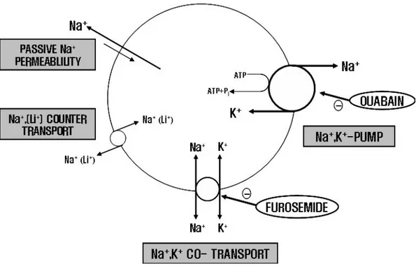
Electrolytic Na+ and K+ are important in controlling extracellular fluid volume
including blood. It has long been recognized that alternation of Na+ distribution within the cells can be related to the trigger mechanism of hypertension. If the amount of Na+ is increased within the cells, there can be change in cell volume followed by increased cell membrane Ca2+ and electric pressure. These changes cause an increase in blood vessel resistance and blood pressure. There are four Na+
transport pathways; Na-K ATPase, Na-K cotransport, Na-(Li+) counter transport and Na-passive transport. Na-K ATPase is known to be sensitive and inhibited by a cardiac glycoside, ouabain and Na-K cotransport is known to be sensitive and inhibited by furosemide. Both ouabain and furosemide have natriuretic and diuretic effects, thereby being used for hypotensive drugs. Use of red blood cells for measurement of Na+ efflux has great advantage, because it can be gotten easily.Na-K ATPase isused for the indicator of the study as the pathological adjunctive factors of diseases such as hypertension, arthritis andcardiac disorder.55)~57)Na-K ATPase in blood vessel and heartventricle was more suppressed in the hypertensive patients
than thenormotensive entity.58)59)
2. Prevention properties of polyphenols on cardiovascular disease
Epidemiologic studies suggest that consumption of fruits and vegetables has been linked with lower prevalence of coronary heart disease.60)Drinking tea has also been linked with reduced mortality arising from cardiovascular disease,61)although some epidemiological data are inconclusive.62)Fruits, vegetables and teas contain a wide range of antioxidant compounds, including phenolic compounds and vitamins. Phenolic compounds, such as anthocyanins, flavan-3-ols, flavonols, and tannins, are widespread in fruits and vegetables, with especially high quantities being found in berries and teas. Among the most well-known polyphenols are the flavonoids, and they have long been recognized to possess inflammatory, antioxidant, anti-allergic, hepato-protective, anti-thrombotic, anti-viral, and anti-carcinogenic activities.63)
Tannins are a unique group of phenolic metabolites that are widely distributed in almost all plant foods and beverages.64)They are classified into two main groups, hydrolysable tannins and condensed tannins. The hydrolysable tannins65)are readily hydrolyzed by acids or enzymes into a polyalcohol and a phenolic carboxylic acid. Depending on the nature of the phenolic carboxylic acid, the hydrolysable tannins are usually subdivided into gallotannins and ellagitannins. Hydrolysis of gallotannins yields gallic acid while that of ellagitannins, hexahydroxydiphenic acid, which is isolated normally as its stable dilactone, ellagic acid (Figure 2). The condensed tannins or proanthocyanidins64)are polyflavonoids in nature, consisting of chains of
flavan-3-ol units. The most common class of proanthocyanidins are the procyanidins which consist of chains of catechin and/or epicatechin linked 4→6 or 4→8 (Figure 3).
Tannins exhibit a variety of bioactivities such as inflammatory and anti-allergic properties,66)anti-diabetic effect,67)anticancer activity,68)and antimicrobial and
anti-viral properties.69)70)Several studies have reported on the oxidant, anti-mutagenic, and anti-inflammatory activities of ellagic acid.71)72)
Figure 2. Hydrolysable tannins
The hydrolysable tannins are readily hydrolyzed by acids (or enzymes) into a sugar (1) or a related polyhydric alcohol and a phenolic carboxylic acid. Depending on the nature of the phenolic carboxylic acid, the hydrolysable tannins are usually subdivided into gallotannins (2) and ellagitannins (4). Hydrolysis of gallotannins yields gallic acid (3) while that of ellagitannins, hexahydroxydiphenic acid (5), which is isolated normally as its stable dilactone, ellagic acid (6) [From Garro Galvez et al,73]copyright 1997Holzforschung]
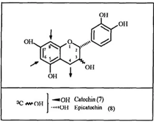
Figure 3. Precursor of condensed tannins
The condendsed tannins or proanthocyanidins are polyflavonoids in nature, consisting of chains of flavan-3-ol units. The most common class of proanthocyanidins are the procyanidins which consist of chains of catechin (7) and/or epicatechin (8) linked 4→6 or 4→8. [From Garro Galvez et
Gallic acid is one of the constituents of hydrolysable tannins and possesses anti-carcinogenic, anti-oxidative, anti-allergic, and anti-inflammatory activities.74)Tannin with gallate group has various physiological functions, such as bacterial, anti-allergic, scavenging free radical, lowering blood pressure and serum and hepatic cholesterol concentrations and increasing fecal sterol excretion in rats with hypercholesterolemia.75)Hydrolyzable gallotannins from Rhus coriaria (Sumac) show anti-ischemic activity and endothelium-dependent vasorelaxant effect in isolated rabbit heart and thoracic aorta.76)
Condensed tannins are known to have antioxidant properties.77)Yokozawa et
al.78)fed rats with condensed tannins and found that lipid peroxidation in plasma and
tissues decreased significantly in the presence of the supplemented polymeric tannins, and the antioxidant effect of condensed tannins was as effective as that of vitamin E. The lower incidence of coronary heart disease in France has been linked to the high consumption of proanthocyanidin-rich red wine, since proanthocyanins possess long lasting antihypertensive and vasorelaxing properties linked to endothelium-related factors in normotensive and hypertensive rats, in which nitric oxide is involved.79)A diet rich in flavanols and procyanidins can improve oxidant defense and reduce tissue markers for oxidative stress, although these effects can be tissue specific.80)As a result, tannins have recently attracted attention for their potential cardiac protective properties.81)Chestnut inner shell also contains relatively high amount of polyphenols,
with gallic acid, ellagic acid, and catechin predominant among hydrolysable and condensed tannins.82)
The chestnut belongs to the genus Castanea in the Fagaceae family and 13
Castanea species in total are recognized. They are native to warm temperate areas of
the Northern Hemisphere, but many countries cultivate different varieties of chestnut mainly for fruit as Castanea crenata Sieb.et Zucc.(Japan and Korea), Castanea
mollissima Blume (China), Castanea sativa Mill.(Europe), and Castanea dentate Borkhausen (America). All Castanea species and their hybrids are edible and some
are commercialized as nut products around the world.
Chestnut trees have been cultivated long time ago in Korea and chestnut fruit are used in traditional Korean ceremonies. Chestnuts are still consumed as a favorite food because of their nutritional values. Chestnuts are not only excellent energy source due to its high starch content, but also good food source containing unsaturated fatty acids and polyphenolic compounds. Several studies have investigated the presence of bioactive antioxidant molecules, not only in edible kernels of chestnut,82)~84)but also in chestnut waste.82)86)There is strong evidence that polyphenols provide protection against harmful effects of free radicals and are known to reduce the risk of several diseases including certain types of cancer, coronary heart disease (CHD), type-2 diabetes, and inflammation. The action of polyphenols on health is also to protect against environmental stresses.87)The fruit of chestnut is typically consumed whole (raw, boiled, or roasted, without skin) or used as ingredient in a variety of processed foods, especially in bakery and confectionery products. From the processing of chestnut productions, large amount of inedible byproduct is generated, representing about 8~14% of outer shell and 6~10% of inner shell.
relatively high amount of tannins, which are well-known polyphenolic antioxidants. Barreira et al.82)analyzed the phenolic contents in chestnut leaves, flowers, skins and fruits, and then reported that contents of polyphenols and flavonoids showed in the following order:outer skins > inner skins > flowers > leaves > fruits.The other study on the chestnut shell, also showed high content of total phenolics as well as a good antioxidant activity in comparison with the results from the same analysis performed with eucalyptus bark samples.89)Despite these reports, the byproducts of chestnut, including inner shell, are usually discarded without recycling. The other hand, extraction yields of chestnut shell with boiling water were higher in inner shell (21.6%) than outer shell (4.98%), therefore the resource recycling with chestnut inner shell is more effectively.
According to trade trends of annual agricultural products in Korea rural economic institute, Korea’s chestnut harvests were about 70,000 tons, and 15~25% are exported, the remaining is consumed in the nation. A large amount of inner shell is discarded from chestnut production processing. Hence, if bioactive materials which obtained from chestnut inner shell by extraction or fraction can re-product as medicinal materials or health supplements, the results can enhance public health with the economic benefit.
PARTⅠ
In vivo study for effects of chestnut
(Castanea crenata) inner shell on antioxidant
and cardiovascular-related parameters
ABSTRACT
The first part of this study focused to compare in vivo effects of simvastatin versus chestnut inner shell on cardiovascular-related parameters and CoQ10 versus chestnut inner shell on antioxidant-related parameters using guinea pigs.
Fifty guinea pigs were divided into five groups and fed one of the following diets; 0.25% cholesterol-based control diet, control diet plus 10% chestnut inner shell powder (Pch), control diet plus 0.03% simvastatin (STN), control diet plus 0.03% simvastatin and 0.3% CoQ10 (STN + CoQ10), and control diet plus 0.03% simvastatin and 3% ethanol extract of Pch (STN + Ech) for four weeks.The final weights significantly decreased in all groups fed statin-containing diet, compared to cholesterol-fed control group, which is paralleled with the pattern of significant decreasing in average daily feed intake (ADFI) (P< 0.01 for both final body weight and daily feed intake). Plasma total cholesterol were significantly lower in the statin plus CoQ10 group than those of other groups (P< 0.01). Liver total cholesterol decreased significantly in groups of statin and statin plus CoQ10 compared with that of the control (P< 0.05). Hematocrit was significantly higher in the statin plus CoQ10 group than that of Pch group (P< 0.05). GOT increased and GPT decreased significantly in all groups fed statin diets compared with those of the control (P< 0.01, P< 0.05, respectively). Erythrocyte Na-K ATPase and intracellular Na and K in groups of Pch and statin decreased significantly compared to those of the control (all,
P< 0.01) with no difference in ACE of lung tissue between groups. Na-K cotransport
and Na passive transport decreased significantly in group of Pch compared with the control or statin group (P< 0.05,P< 0.01, respectively). Na leak in
2,2-azobis-(2-amidinopropane) dihydrochloride (AAPH) treated erythrocyte decreased significantly in groups of statin plus CoQ10 and statin plus Ech compared with that of statin alone (P< 0.01). TBARS productions in liver and platelet rich plasma increased in statin group and decreased in group of statin plus CoQ10, and the difference between these two groups was significant (P< 0.01). Present study showed thatstatin or chestnut inner shell which contains high amount of tannins did not affect plasma cholesterol. Statin with CoQ10 decreased cholesterol and triglyceride more efficiently than statin alone, assuming synergic effects of CoQ10 with statin. Ethanol extract of chestnut inner shell and CoQ10 seemed to enhance thepositive effects of statin by reducing oxidative stress and cell damage, thereby protecting liver and cardiovascular systems from CVD such as atherosclerosis.
MATERIALS AND METHODS
1. Materials
1) Animals and diets
Fifty of six weeks old guinea pig (Orient Bio Co Ltd, Gapyung, Korea) were divided into five groups and fed the following diets : 0.25% cholesterol-based control diet; control diet plus 10% chestnut (Castanea crenata)inner shell powder; control diet plus 0.03% simvastatin (20 mg/kg BW); statin plus 0.3% CoQ10 (200 mg/kg BW); statin plus 3% ethanol extract of chestnut inner shell powder. Doses in detail for chestnut inner shell (herbal medicine shop in Jeju), CoQ10 (Yunjin Pharm Co, Korea) and simvastatin (Choongwae Pham Co, Korea) are in diet composition (Table 1-1). Diet was pelletized by the assistance of Korea Food Research Institute. Guinea pig had free access to water and were housed individual cages in a room maintained at 20 ~ 25°C with a 12-hour dark-light cycle.
(1) Preparation of chestnut inner shell extract
Chestnut inner shell powder was purchased from herbal medicine shop in Jeju. Chestnut inner shell powder (4 kg) were extracted with 12ℓ of 80% ethanol (EtOH) three times for 24 hours each at room temperature and filtered. The filtrate was then concentrated in vacuo at 40°C. Residual ethanol was removed by freeze dryer. The yields of the chestnut inner shell powder extrat (Ech) was 526.5 g (13.16%). The prepared extracts were stored at -20°Cuntil further examined.
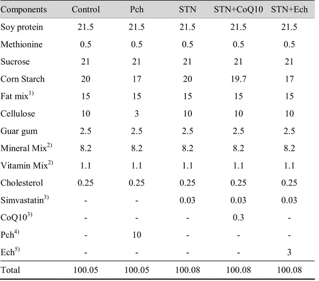
Table 1-1.Diet composition (%)
Components Control Pch STN STN+CoQ10 STN+Ech
Soy protein 21.5 21.5 21.5 21.5 21.5 Methionine 0.5 0.5 0.5 0.5 0.5 Sucrose 21 21 21 21 21 Corn Starch 20 17 20 19.7 17 Fat mix1) 15 15 15 15 15 Cellulose 10 3 10 10 10 Guar gum 2.5 2.5 2.5 2.5 2.5 Mineral Mix2) 8.2 8.2 8.2 8.2 8.2 Vitamin Mix2) 1.1 1.1 1.1 1.1 1.1 Cholesterol 0.25 0.25 0.25 0.25 0.25 Simvastatin3) - - 0.03 0.03 0.03 CoQ103) - - - 0.3 - Pch4) - 10 - - - Ech5) - - - - 3 Total 100.05 100.05 100.08 100.08 100.08
1 Fat mix contains olive oil, palm oil, and safflower oil (1:2:1.8), high in lauric and myristic acids
2 Mineral and vitamin mix adjusted to meet NRC requirements for guinea pigs. Detailed composition of the
vitamin and mineral mix has been reported elsewhere.90)
3 Simvastatin, Choongwage Pharma Co. Korea; 0.03% statin equivalent to 20mg/kg BW/day.
0.3% CoQ10 (Yungjin Pharm Co) equivalent to 200mg/kg BW/day when calculated from the daily foodconsumption of 40g/day
4 Pch : Chestnut inner shell powder
2. Collection of samples
After 4 weeks ad libitum feeding, guinea pigs were anesthetized with ether after fasted for 12 hours, and blood samples were obtained by cardiac puncture into heparinized vacuum tubes. Whole blood platelet aggregation, hematocrit, and erythrocyte Na efflux were performed with fresh blood.
The Platelet rich plasma (PRP) was obtained by centrifugation at 300 × g for 10 minutes and stored at -70°C for TBARS analysis. The plasma samples were obtained by centrifugation at 1,000 × g for 10 minutes and stored at -70°C for lipid profiles, glutamic oxaloacetic transaminase (GOT), and glutamic pyruvic transaminase (GPT) analysis.
The liver and lung samples were quickly removed and measured weight, then stored at -70°Cfor later assays.
3. Sample analysis
1) Whole blood platelet aggregation
Platelet aggregation was measured using a Chronolog Whole Blood Aggregometor (model 500-Ca, Havertown, Pennsylvania, USA). Fresh whole blood was diluted with isotonic saline (1: 4) to give platelet concentration of approximately 200,000 platelets/μℓ. Adenosine diphosphate (ADP, 2μM; Chronolog, Havertown, Pennsylvania, USA) was added to initiate aggregation, and three readings of impedance changes were averaged for each guinea pig to determine the maximum
aggregation and the initial slope. The instrumental principle is based on the increase in impedance (Ω) across two platinum electrodes as platelet aggregation proceeds.
2) Plasma and liver lipid assay
Plasma total cholesterol, HDL-cholesterol and triglyceride were assayed using enzymatic kits (Asan Pharmaceuticals Co., Ltd, Seoul, Korea). Ten μℓ of plasma was used for the assays of total cholesterol and triglyceride For HDL cholesterol, 200 μℓ plasma was incubated with dextran sulfate to precipitate apo B containing lipoprotein, and 50 μℓof the supernatant was used.
Solvents for liver extract were supplied by Merck (Merck KcaA, Darmstadt, Germany). Liver lipids were extracted by a modified Folch method.91)One gram of liver tissue was homogenized for 5 min in 6mℓ of Folch solution [chloroform (2): methanol (1)] and 2 mℓ H2O. After centrifugation for 10 minutes, the lower phase
that contains liver lipids was separated. Lower phase of lipid fractions was assayed after treating with tritonX-100 : chloroform (25 μℓ : 475 μℓ) for total cholesterol or with methanol for triglyceride, using enzymatic kits (Asan Pharmaceuticals Co., Ltd, Seoul, Korea).
3) Platelet rich plasma (PRP) and liver TBARS productions
Platelet rich plasma (PRP) were obtained after centrifuging whole blood at 300 × g for 10 minutes. PRP TBARS (Thiobarbaituric acid reactive substance) production was measured with a modified Buege and Aust method.92)An half mℓ PRP in 1.5
mℓPBS (Phosphate buffered saline) was incubated at 100°Coil bath for 20 minutes after adding 2 mℓTBA solution (15g TBA, 0.139g TCA, 1.81mℓ 12N HCl in 85mℓ D-H2O). After cooled under tap water, the incubated mixture was centrifuged at
1,000 × g for 10 minutes. Finally the TBARS in supernatant was measured with the absorbance at 532 nm on spectrophotometer using deionized H2O as blank.
Buege and Aust method92)was also used for the liver TBARS. One gram of minced liver sample in 2 mℓ PBS was incubated at 100°C oil bath for 20 minutes after adding 2 mℓ TBA or non-TBA solution (0.139 g TCA, 1.81 mℓ 12N HCl in 85mℓ D-H2O). The incubated mixture was cooled down and centrifuged at 1,000 × g for 10
minutes. The TBARS in supernatant was measured of spectrophotometer at 532 nm using deionized H2O as blank. All samples were performed in duplicate and the
values ofTBARS was the outcome subtracting the concentration of thenon-TBA treated from the TBA treated.
The liver protein was measured with Lowry et al.93)method using bovine serum albumin (BSA) as standard protein. The absorbance for protein concentration was read at 750 nm with spectrophotometer using D-H2O as blank.
4) Erythrocyte Na efflux channels
(1) Red cell preparation
Chemicals for mediums including ouabain, furosemide and MOPS were purchased from Sigma chemical company (MO, USA). Blood was centrifuged at 1,000 × g for 10 minutes, and the plasma and buffy coat were removed. Red blood cells were washed 5 times with a cold isotonic washing solution [150 mM choline chloride, 10
mM Tris-4 morpholinopropane sulfonic acid (MOPS), pH 7.4 at 4 °C], and centrifuged at 1,000 × g for 5 minutes after each wash. The RBC pellet was resuspended in the choline chloride washing to give 40~50% hematocrit, then hematocrit was measured.The RBC suspension was kept in ice for measurement intracellular Na and K concentration, K ATPase, K cotransport, and Na-passive transport.
(2) Na-K ATPase activity
Ouabain sensitive Na-K ATPase can be blocked by ouabain, and Na+ efflux via Na-K ATPase is calculated from the difference between the efflux into MgCl2
medium with and without ouabain.
Fourmℓeach of erythrocyte was added to 40 mℓ medium 1 [70mM MgCl2, 10mM
KCl, 85mM sucrose, 10mM glucose, 10mM Tris-MOPS, pH 7.4 at 37°C] and medium 2 [medium 1 plus 10mM ouabain] then mixed gently and aliquot 10 tubes. The tubes were transferred in duplicates to an ice bath after incubation at 37°C in a shaking water bath for 0, 2, 4, 6, 8 minutes. The tubes were subsequently centrifuged at 1,000 × g for 10 minutes, the supernatant was removed.
(3) Na-K cotransport
Furosemide sensitive Na-K cotransport can be blocked by furosemide, and Na efflux via Na-K cotransport is calculated from the difference between the efflux into choline chloride medium with and without furosemide.
Twomℓ each of erythrocyte was added to 30 mℓ medium 3 [150mM choline chloride, 1.0mM ouabain, 10mM glucose, 10mM Tris-MOPS, pH 7.4 at 37℃] and
medium 4 [medium 3 plus 0.3308g/ℓ furosemide] then mixed gently and aliquot 10 tubes. The tubes were transferred in duplicates to an ice bath after incubation at 37°Cin a shaking water bath for 0, 10, 20, 30, 40 minutes. The tubes were subsequently centrifuged at 1,000 × g for 10 minutes, the supernatant was removed for determining Na concentration.
(4) Na-passive transport
Na-passive transport is the efflux into choline chloride medium containing ouabain and furosemide under condition of both Na-K ATPase and Na-K cotransport blocked.
For Na-passive transport measurement, erythrocyte was added medium 4 [150mM choline chloride, 1.0mM ouabain, 10mM glucose, 0.3308g/ℓ furosemide, 10mM Tris-MOPS, pH 7.4 at 37°C].
(5) Intracellular Na and K concentration
For intracellular Na+ concentration measurement, 50μℓ of the RBC suspension was
added to 5 mℓ of 0.02% acationox (a metal free detergent, Scientific products, McGraw Park, Illinois, USA) and Na concentration was measured using atomic absorption spectrophotometer (AAS, AA6701F Shimazu Co., Japan). For intracellular Kmeasurement, RBC suspension in acationox was diluted with distilled water in 1 to 10 and K concentration was measured using AAS.
(6) Erythrocyte Na-leak
Sodium leak is defined as sodium efflux through passive sodium channels occurring under inhibition of ouabain-sensitive Na-pumps and furosemide-sensitive
Na-K cotransport. Sodium leak can be increased through damage of erythrocyte membranes after exposure to a free radical generating system such as 2,2-azobis-(2-amidinopropane)dihydrochloride (AAPH).
All mediums and tubes were kept and handled in ice, and Na concentrations from Na efflux channels (Na-K ATPase, Na-K cotransport, Na-passive transport) were measured using AAS (AA6701F Shimazu Co., Japan).
[Calculations] - Na efflux ;
[Na μg/mℓ] [60min] [μmole] [44mℓ-(4 mℓ×Hct)]
[min] × [hr] × [23 μg] × [4mℓ×Hct)] = Na mmole/ℓRBC·hr -1 - Intracellular Na ; [Na μg] [μmole] [101] [mℓ] × [23 μg] × [Hct] = Na mmole/ℓRBC - Intracellular K ; [K μg] [μmole] [101] [mℓ] × [39 μg] × [Hct] = Na mmole/ℓRBC
Figure 1-1. Model of erythrocyte Na efflux channels
5) Plasma GOT and GPT levels
The plasma glutamic oxaloacetic transaminase (GOT) and glutamic pyruvictransaminase (GPT) was measured with a spectrophotometric diagnostic kit purchased from the Asan pharmaceutical Company (Seoul, Korea). Briefly, 1 mℓ GOT or GPT substrate solution was incubated at 37°Cshaking water bath for 5 minutes and 0.2 mℓ plasma sample was added. After GOT or GPT incubated at 37°Cfor 60 minutes and 30 minutes respectively, 1 mℓ of color reagent was added to each of samples and incubated for 20 minutes at room temperature. After incubation, 10 mℓ 0.4 N NaOH was added mixed well, incubated for 10 minutes at room
temperature. The absorbance at 505 nm was read, using spectrophotometer and results are expressed as IU/ℓ.
6) Lung angiotensin converting enzyme (ACE) activity
ACE activity was determined modified methods of Cushman and Cheung.94)Tissue ACE activity is determined by measuring hippuric acid (HA) released from hippuryl-L-his-tidyl-L-leucine (HHL) in incubation medium. Two hundreds mg of lung tissue was homogenized with 2mℓ 20 mM phosphate buffer (1M K2HPO4, plus 1M
KH2PO4, pH 8.3) containing 0.01% tritonX-100 and centrifuged at 600 × g for 10
minutes. Supernatant containing membrane bound ACE was transferred to a clean tube. Total 250μℓ medium including 10μℓ supernatant, 100 μℓ 5 mM HHL, 190μℓ100 mM K-phosphate buffer with 30 mM NaCl in effendorf tube was incubated at 37°Cfor 30 minutes, then reaction was stopped by 500 μℓ1 N HCl and medium was centrifuged at 1,000 × g for 10 minutes. Supernatant of 500 μℓwas transferred to a glass tube containing 1.5 mℓ ethyl acetate, vortexed for 15 seconds and centrifuged at 1,000 × g for 5 minutes. One mℓ of upper ethyl acetate layer was transferred to a new tubeand dried for 30 minutes on 120°C heating block. Dried HA was dissolved in deionized waterand read the absorbance at 228 nm on UV spectrophotometer using each blank prepared with the same procedure without HHL in incubation medium. Standard HA was prepared in the same procedure as tissue samples and ACE activity is expressed as HA in μmole/mg lung tissue.
Values were analyzed using the SPSS version 18.0 (SPSS, Inc Chicago, IL, USA). Analysis of variance were conducted in a completely randomized block design. Duncan's multiple test was applied to compared individual means when F-value was significant. A value of P< 0.05 was considered significant.
RESULTS
1. Body weight, plasma and liver lipids
Final body weight, average daily feed intake (ADFI), and liver weight/body weight ratio (L.W/B.W) are shown in Table 1-2. Body weight increased in the Pch group and the control group with no weight gain in all groups fed statin containing diet for 4 weeks, the final body weight was significantly different between the control and any of statin groups. Similarly, average daily feed intake (ADFI) increased in Pch group and decreased in the other groups. There was no difference in liver weight / body weight (L.W/B.W) ratio among groups. Considering the proper weight gain in animals, chestnut inner shell is safe as diet material and can be used for complementary medicine.
As listed in Table 1-2, Pch or statin alone rather increased plasma cholesterol compared to the control showing no cholesterol lowering effect. Statin plus CoQ10 effectively decreased plasma total cholesterol and the difference between group with statin plus CoQ10 and any of other groups was statistically significant (P < 0.01).No difference was observed in plasma HDL cholesterol among groups, and plasma triglyceride levels somewhat decreased in groups of the Pch and statin plus CoQ10, but the difference was not significant.
Liver cholesterol levels of the statin and statin plus CoQ10 groups were significantly lower than that of control (P < 0.05) (Table 1-2). Groups of Pch and statin plus Ech tended to be lower in liver cholesterol levels than the control group. All groups fed diets containing statin decreased in liver triglyceride with the lowest
in statin plus CoQ10 group, but not statistically different. In present study with guinea pigs, statin at dose of 20 mg/kg BW decreased liver cholesterol and triglyceride without affecting plasma total cholesterol and HDL-cholesterol. Statin with CoQ10 was more effective in cholesterol lowering than statin alone.
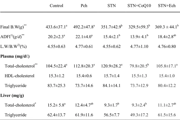
Table 1-2. Effects of chestnut inner shell powder (Pch) and extract (Ech), statin and coenzyme Q10 on body weight and plasma and liver lipid in rats fed with cholesterol-based diet
Control Pch STN STN+CoQ10 STN+Ech
Final B.W(g)** 433.6±37.1a 492.2±47.8a 351.7±42.9b 329.5±59.3b 369.3 ± 44.1b ADFI1)(g/d)** 20.2±2.3a 22.1±4.0a 15.4±2.1b 13.9± 4.1b 18.4±2.8ab L.W/B.W2)(%) 4.55±0.63 4.77±0.61 4.55±0.62 4.77±1.10 4.76±0.80 Plasma (mg/dℓ) Total-cholesterol** 104.5±22.4a 112.8±20.3a 120.9±28.2a 79.8±20.5b 105.8±17.1a HDL-cholesterol 15.3±1.2 15.4±0.6 15.7±1.4 15.5±1.3 15.4±1.0 Triglyceride 83.7±25.3 73.7±14.6 84.1±14.1 73.7±12.9 80.4±12.2 Liver (mg/g) Total-cholesterol* 15.2± 5.8a 12.4±4.7ab 9.3±1.7b 9.3±2.4b 11.1±2.7ab Triglyceride 62.4±13.7 61.9±11.6 56.5±7.7 49.3±17.2 61.5±15.6
1)ADFI : Average daily feed intake
2) L.W/B.W : Liver weight / Body weight ratio
Values are means ± SD of 9 guinea pigs.
* Values in the same row not sharing the same superscript differ (P< 0.05). ** Values in the same row not sharing the same superscript differ (P< 0.01).
2. Hematocrit and whole blood platelet aggregation
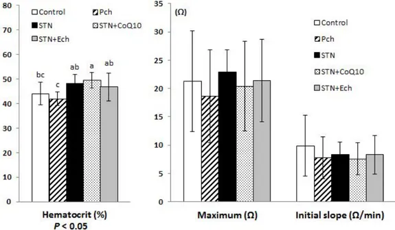
Hematocrit and platelet aggregation are shown in Figure 1-2. Hematocrit was increased in groups with statin and decreased in Pch group, showing statistical difference between Pch and statin plus CoQ10(P< 0.05).
Maximum of platelet aggregation tended to decrease in groups of Pch and statin plus CoQ10 and tended to increase in group of statin. Initial slope of platelet aggregation decreased in all experimental groups compared with the control, but not statistically different. Both of the maximum and initial slope of platelet aggregation were the lowest in Pch group.
Figure 1-2. Effects of chestnut inner shell powder (Pch) and extract (Ech), statin and coenzyme Q10 on the hematocrit and platelet aggregation in rats fed with cholesterol-based diet
Maximum aggregation in ohm at the point where platelet aggregate is dissociated. Initial slope is the ohm change for the first one minute of aggregation.
3. Plasma GOT and GPT levels
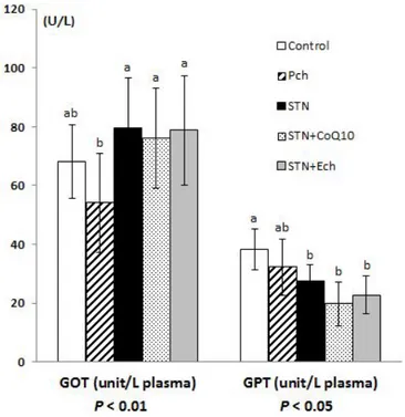
Plasma glutamic oxaloacetic transaminase (GOT) and glutamic pyruvic transaminase (GPT) are shown in Figure 1-3. Plasma GOT increased in groups of statin, statin plus CoQ10, and statin plus Ech, and the differences between these groups and Pch group were significant (P< 0.01). Unlikely, plasma GPT decreased in all groups fed statin containing diets, and the difference between these groups and the control were significant (P< 0.05).
GOT is present in most organs including liver, heart, skeletal muscle, kidney, brain and lung, while GPT located predominantly in liver with significant quantity. They are released into the blood when cells are damaged. Even though they changed in range of normal levels, high GOT with low GPT in present study can hardly be explained.
Figure 1-3. Effects of chestnut inner shell powder (Pch) and extract (Ech), statin and coenzyme Q10 on plasma GOT and GPT levels in rats fed with cholesterol-based diet
GOT; plasma glutamic oxaloacetic transaminase GPT; plasma glutamic pyruvic transaminase Values are mean ± SD of 9 guinea pigs.
4.Erythrocyte Na efflux and ACE inhibitory effect
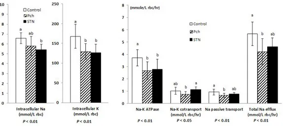
Intracellular Na and K concentrations and erythrocyte Na efflux channels are shown in Figure 1-4. Intracellular Na and K showed the similar tendency with decrease in groups of Pch and statin compared to the control. Both of intracellular Na and K levels were significantly decreased in the statin group compared with those in the control (P< 0.01). The major efflux channel, Na-K ATPase decreased in groups of Pch and statin compared with the control, which were the same trend with intracellular Na and K concentrations. Group of Pch consistently decreased in both Na-K cotransport and Na passive transport and the differences between Pch and the control were significant (P< 0.05, P< 0.01 respectively). Total Na efflux in group of Pch was significantly low compared to the control group (P< 0.01). Group of statin significantly decreased in Na-K ATPase (P< 0.01) and Na passive transport compared to the control, resulting an overall decrease in total Na efflux compared to the control group. Total Na efflux was mostly contributed by Na-K ATPase channel, consequently depending on Na-K ATPase activity. Angiotensin converting enzyme (ACE) activity which is involved in Na transport channels was measured to examine the correlations between ACE and Na channels (Figure 1-5). ACE activity of lung tissue in group of Pch increased compared with the control and group of statin, but not statistically different.
Figure 1-4. Effects of chestnut inner shell powder (Pch) and statin on erythrocyte sodium efflux in rats fed with cholesterol-based diet
Intracelluar Na and K; upper values are for intact red blood cells. Na-K ATPase is ouabain sensitive Na efflux.
Na-K cotransport is furosemide sensitive Na efflux.
Na passive transport is Na efflux under the blockage of Na-K ATPase and Na-K cotransport. Values are mean ± SD of 9 guinea pigs.
Figure 1-5. Effects of chestnut inner shell powder (Pch) and statin on angiotensin converting enzyme activity in rats fed with choles-terol-based diet
The angiotensin converting enzyme (ACE) activity expressed as hippuric acid (HA) concentrated released from HHL (2mM/mg lung tissue) using 0.2mg lung tissue. Values are mean ± SD of 9 guinea pigs.
5. Erythrocyte Na-leak
Depletion of reduced form ubiquinol by statin therapy leads to a loss of antioxidant capacity and erythrocyte membrane stability. Accordingly, Na passive leak through erythrocyte membrane after exposure to free radical generating system such as AAPH [2,2'-azobis(2-amidinopropane)dihydrochloride] was measured as parameter.
Assessment of Na leak lowering effects by erythrocyte membrane protection was performed among the groups with statin. In present study, Na leak through the erythrocyte are observed effects of statin plus coenzyme Q10 and chestnut inner shell ethanol extract compared with the statin only.
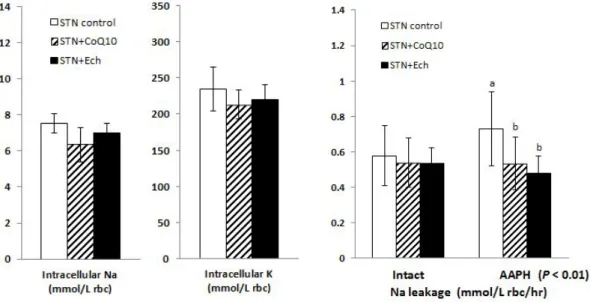
Intracellular Na and K, and Na passive leak in intact cells are shown in Figure 1-6. Intracellular Na and K in intact cells were not different. Na passive leak in intact cells was not also different among the groups.
Na leak in AAPH treated cells was significantly increased in statin control group but decreased in statin plus CoQ10 and statin plus chestnut inner shell extract groups, and there was significant difference (P< 0.01).
Figure 1-6. Effects of statin, coenzyme Q10, and chestnut inner shell extract (Ech) on intact and AAPH treated erythrocyte Na-leak in rats fed with cholesterol-based diet
Intact Na-leak is erythrocyte Na efflux through passive pathway in intact red blood cells.
AAPH; Na leakage is erythrocyte Na efflux through passive pathway in AAPH [2,2'-azobis(2-amidino-propane) dihydrochloride] treated red blood cell.
6. Platelet rich plasma (PRP) and liver TBARS productions
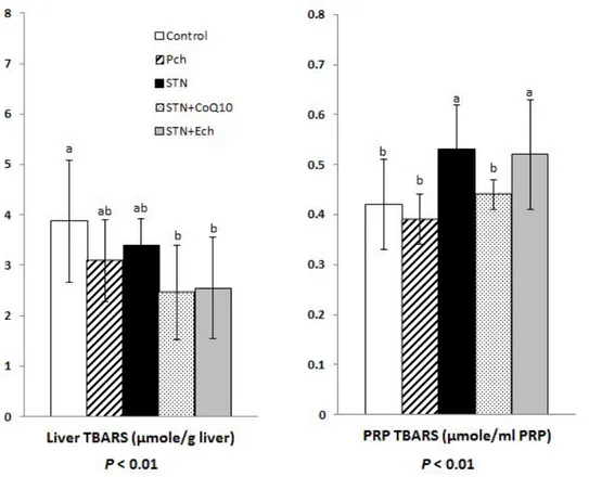
Thiobarbituric acid reactive substance (TBARS) productions in liver and platelet rich plasma (PRP) are shown in Figure 1-7. TBARS are formed as a byproduct of lipid peroxidation and can be measured as productions of the damage produced by oxidative stress, because reactive oxygen species (ROS) have extremely short half-lives.
Liver TBARS production decreased in all experimental groups compared to the control group, showing statistical difference between the control and statin plus CoQ10 or the control and the statin plus Ech (both P< 0.01).
However, PRP TBARS productions tended to increase in groups of statin and statin plus Ech, showing statistical difference between the control and statin or the control and statin plus Ech (both P < 0.01). There was no difference between the control and Pch or statin plus CoQ10 in PRP TBAR production.
Figure 1-7. Effects of chestnut inner shell powder (Pch) and extract (Ech), statin and coenzyme Q10 on platelet rich plasma (PRP) and liver thiobarbituric acid reactive substance (TBARS) in rats fed with cholesterol-based diet
Platelet rich plasma (PRP) was obtained after centrifuging whole blood at 300 X g for 10 minutes. Values are mean ± SD of 9 guinea pigs.
DISCUSSION
In present study, guinea pigs fed chestnut inner shell containing diet showed normal growth rate and feed intake, suggesting that chestnut inner shell in its dietary composition is safe and can be a useful source for complementary alternative medicine. Unlike normal growth in rat study,95)guinea pigs fed 0.03% simvastatin containing diet had low growth rate with low feed intake in present study. Similarly, West et al.96)reported that final weights of guinea pigs fed 0.05% simvastatin diet containing bile absorption inhibitor (SC-435) were significantly lower than those of the other groups. WT (wild type) mice and apo E knockout mice fed 0.5 mg/kg BW rosuvastatin did not have normal weight gain during 18 weeks experimental period.97)Statin with CoQ10 seemed to further suppress growth rate with decreased feed intake in present study. Although NOAEL dose (No Observed Adverse Effect Level) of CoQ10 is reported to be 600 mg/kg BW in male rats98)and low dose of CoQ10 (15 mg/kg BW) induced normal growth rate in rats.95)In present study, high
dose of CoQ10 (200 mg/kg BW) seemed to negatively influence on weight gain in guinea pigs when it was fed with statin. Although many studies have reported that the supplementation of CoQ10 is necessary for statin user in antioxidant view point, further studies are needed to confirm the beneficial effects of the combined therapy of statin and CoQ10. High doses of statin and CoQ10 may have metabolic impact affecting appetite, feed intake and growth rate. In present study with guinea pigs, statin of decreased liver cholesterol and triglyceride without affecting plasma total cholesterol and HDL-cholesterol. In rat studies, high simvastatin of 30 mg/kg BW did not decrease liver and serum cholesterol95)and low simvastatin of 2 mg/kg BW
even increased in serum cholesterol.99)Grothusen et al.100)reported that atorvastatin of 1 mg/kg BW in ApoE (-/-) mice tended to increase serum total cholesterol and LDL-cholesterol levels. Unlike these results, Aggawal et al.101)reported that high atorvastatin (0.05 % or 30 mg/kg BW) in guinea pigs significantly decreased serum cholesterol and triglyceride. In present study, statin at dose of 20 mg/kg BW did not decrease plasma cholesterol, but decreased liver cholesterol. Statin plus CoQ10 (200 mg/kg BW) decreased plasma and liver cholesterol as well as liver triglyceride more efficiently.
One of the most serious side effects from statin use is the exhaustion of CoQ10 which shares the cholesterol biosynthetic pathway. The alternative plan that is minimized these side effect would be to prescribe statin with CoQ10. In present study showed that supplemented CoQ10 seemed to potentiate hypocholesterolemic and hypolipidemic effects of statin, particularly lowering effect of plasma and liver triglyceride levels. Statin, a HMG-CoA reductase inhibitor and CoQ10 may have synergistic actions in lowering cholesterol. CoQ10, an intermediate product of cholesterol synthetic pathway may have negative feedback inhibition on HMG-CoA reductase. Further systemic researchesare necessary to clarify metabolic action of statin and CoQ10 in cholesterol metabolism. Although Pch did not affect cholesterol, but tended to decrease plasma triglyceride.Insoluble tannins and soluble fibers are known to inhibit the absorption of cholesterol and lipid.102)103)It is assumed that the
tannins and soluble fibers of chestnut inner shell also seemed to inhibit intestinal absorption of triglyceride. Considering lipid lowering effect of Pch for 4 weeks, Pch can be a good dietary source for complementary alternative medicine for statin therapy. Functions of statin are known to inhibition of HMG-CoA reductase activity
and cholesterol synthesis because of similar structure to HMG-CoA reductase. However, hypocholesterolemic effects of statin were different among mouse, rat, and guinea pigs within rodents, which should be further studied.
In present study, hematocrit increased in all groups with statin, and especially increase above normal in group of statin plus CoQ10 suggested that there might be some compensations for depleting status of oxygen in metabolizing statin and/or CoQ10.
Kim et al.95)observed simvastatin of 30 mg/kg BW did not affect the maximum platelet aggregation in rat, but statin plus CoQ10 (15 mg/kg BW) tended to decrease platelet aggregation. In present study with guinea pig, statin plus high dose CoQ10 of 200 mg/kg BW caused the lowest initial slope. Dajani et al.104)reported pravastatin treatment in primary hypercholesterolemic patients inhibited platelet aggregation and TXB2synthesis. It is known that hypercholesterolemia itself increased
hyper-sensitivity of platelet. Dajanietal.104)explained that inhibition effect of pravastatin on platelet aggregation coincided with cholesterol lowering effect of it. In present study, there was no cholesterol lowering effect in statin group with an increased platelet aggregation, inferring that increased cholesterol correlated with increased platelet aggregation. Considering no cholesterol lowering effects in guinea pigs with high statin (20 mg/kg BW) above applicable dose to human, there might be species difference between animal and human in metabolism and cardiovascular system. Plant antioxidants like herbal extracts decreased platelet aggregation,105)but 5% green tea powder diet increased somewhat in Kim's study.95)Pch in present study reduced platelet aggregation of both the initial and the maximum compared with the control, whereas statin plus Ech did not affect. Kawaguchi et al.106) reported that the platelet