저작자표시-비영리-동일조건변경허락 2.0 대한민국 이용자는 아래의 조건을 따르는 경우에 한하여 자유롭게 l 이 저작물을 복제, 배포, 전송, 전시, 공연 및 방송할 수 있습니다. l 이차적 저작물을 작성할 수 있습니다. 다음과 같은 조건을 따라야 합니다: l 귀하는, 이 저작물의 재이용이나 배포의 경우, 이 저작물에 적용된 이용허락조건 을 명확하게 나타내어야 합니다. l 저작권자로부터 별도의 허가를 받으면 이러한 조건들은 적용되지 않습니다. 저작권법에 따른 이용자의 권리는 위의 내용에 의하여 영향을 받지 않습니다. 이것은 이용허락규약(Legal Code)을 이해하기 쉽게 요약한 것입니다. Disclaimer 저작자표시. 귀하는 원저작자를 표시하여야 합니다. 비영리. 귀하는 이 저작물을 영리 목적으로 이용할 수 없습니다. 동일조건변경허락. 귀하가 이 저작물을 개작, 변형 또는 가공했을 경우 에는, 이 저작물과 동일한 이용허락조건하에서만 배포할 수 있습니다.
Prediction of Compensated Liver Cirrhosis by
Ultrasonography and Routine Blood Tests in
Patients with Chronic Viral Hepatitis
by
Hong Sub Lee
Major in Medicine
Department of Medical Sciences
The Graduate School, Ajou University
Prediction of Compensated Liver Cirrhosis by
Ultrasonography and Routine Blood Tests in
Patients with Chronic Viral Hepatitis
by
Hong Sub Lee
A Dissertation Submitted to The Graduate School of Ajou University
in Partial Fulfillment of the Requirements for the Degree of Master of
Medical Sciences
Supervised by
Sung Won Cho, M.D., Ph.D.
Major in Medicine
Department of Medical Sciences
The Graduate School, Ajou University
This certifies that the dissertation
of Hong Sub Lee is approved
.
SUPERVISORY COMMITTEE
Sung-Won Cho
Kwang Jae Lee
Jae Youn Cheong
The Graduate School, Ajou University
December, 23rd, 2010
- ABSTRACT -
Prediction of Compensated Liver Cirrhosis by Ultrasonography and
Routine Blood Tests in Patients with Chronic Viral Hepatitis
Background: A liver biopsy is the standard method for the diagnosis of liver cirrhosis in patients with chronic hepatitis. Because the liver biopsy is an invasive method, non-invasive methods have been used to diagnose compensated liver cirrhosis in patients with chronic hepatitis. The current study was designed to evaluate ultrasonography and routine blood tests for diagnosing compensated liver cirrhosis in patients with chronic viral hepatitis.
Materials and methods: Two hundred three patients with chronic viral hepatitis who underwent liver biopsy were included in this study and ultrasonography and routine blood tests were analyzed retrospectively. Ultrasonographic findings, such as surface nodularity, parenchyma echogenecity, and spleen size, were evaluated. The diagnostic accuracy of ultrasonography and routine blood tests were examined.
Results: Discriminant analysis with forward stepwise selection of the variables showed that liver surface nodularity, platelet count, and albumin level were independently associated with compensated liver cirrhosis (p<0.05). Cross-tabulation revealed that the following 4 variables had > 95% specificity: platelet count < 100,000 /uL; albumin level < 3.5 g/dL; INR > 1.3; and surface nodularity. If at least one of the 4 variables exists in a patient with chronic viral hepatitis, we can predict liver cirrhosis with 90% specificity and 61% sensitivity.
ii
Conclusions: These results suggest that 4 variables (platelet count < 100,000 /uL, albumin level < 3.5 g/dL, INR > 1.3, and surface nodularity) can be used to identify liver cirrhosis in patients with chronic viral hepatitis with high specificity.
Key words : liver cirrhosis, ultrasonography, diagnosis, blood test, liver biopsy
TABLE OF CONTENTS
ABSTRCT··· ⅰ TABLE OF CONTETS ··· ⅲ TABLE OF TABLES ···iv
. INTRODUCTION Ⅰ ··· 1. PATIENTS AND METHODS Ⅱ ··· 3 A. Patients··· 3 B. abdominal ultrasonography ··· 5 C. liver biopsy ··· 6 D. data analysis ··· 6 . RESULTS Ⅲ ··· 7
A. The interobserver agreement of ultrasonographic findings··· 7
B. Blood tests and ultrasonographic variables in the diagnosis of compensated liver cirrhosis ··· 7
C. The usefulness of each variable in the diagnosis of compensated liver cirrhosis 11 . DISCUSSION Ⅳ ··· 14 . CONCL Ⅴ USION ··· 18 REFERENCES ··· 19 국문요약 ··· 23
iv
LIST OF TABLES
Table 1. Baseline characteristics of patients··· 4
Table 2. Area under curve of each variable for compensated liver cirrhosis ··· 9
Table 3. Results of logistic regression: each variable versus pathologic diagnosis for compensated liver cirrhosis··· 10
Table 4. Diagnostic accuracy, sensitivity, and specificity of compensated liver cirrhosis by different variables··· 12
Table 5. Diagnostic accuracy, sensitivity, and specificity of compensated liver cirrhosis by combination variables ··· 13
I. INTRODUCTION
Chronic hepatitis B (CHB) and C (CHC) are major causes of chronic hepatitis (Anthony et al., 1977). Chronic carriers of the hepatitis B virus (HBV) are currently estimated to number > 350 million humans worldwide (Maynard, 1990). Chronic carriers of hepatitis C virus (HCV) are estimated to number approximately 170 million people (Alter, 1997). Chronic hepatitis can progress to compensated liver cirrhosis. Compensated liver cirrhosis may then progress to liver failure and hepatocellular carcinoma with attendant complications, such as hepatic encephalopathy and varix bleeding. Therefore, early detection of liver cirrhosis in patients with chronic hepatitis has become an important clinical issue for physicians(Sorrell et al., 2009; Paik, 2008). Because the diagnosis of liver cirrhosis requires histologic demonstration of abnormal regenerative nodules surrounded by fibrosis, liver biopsy is still considered to be the gold standard for assessing fibrosis (Bravo et al., 2001). Liver biopsy is limited, however, by the invasiveness of the procedure, cost, risk of complications (pain, bleeding, pneumothorax, bile peritonitis, and perforation), poor acceptance by patients, availability of expert practitioners, and intra- and inter-observer variability. An overall mortality one of 1 person per 10,000 has been reported in patients undergoing liver biopsy. False negative probability due to sampling error is reported to be 20%-30%6-9(Bravo and Sheth, 2001; Piccinino et al., 1986; Abdi et al., 1979; Bedossa et al., 2003).
Therefore, it is necessary to establish non-invasive diagnostic methods to diagnose liver cirrhosis more easily. Non-invasive and more reproducible alternative methods, such as a fibroscan and blood indices, have been proposed and tested as a means for detection of liver cirrhosis; however, the clinical use is limited because those methods have not been confirmed
- 2 -
and are difficult to use(Laharie et al., 2006; Forns et al., 2002; Chen et al., 2008). A liver biopsy is rarely performed in the clinical diagnosis of compensated cirrhosis, and compensated cirrhosis is usually diagnosed by using blood tests and abdominal ultrasonography. However, the diagnostic criteria for liver cirrhosis and accuracy of the tests may vary according to the hospital. The present study was designed to determine the accuracy of abdominal ultrasonography and blood tests in the diagnosis of compensated cirrhosis in patients with CHB and CHC and to determine the optimal point with high specificity in the diagnosis of liver cirrhosis. We analyzed patients with CHB and CHC who underwent abdominal ultrasonography, blood tests, and a liver biopsy at Ajou University Hospital.
II. PATIENTS AND METHODS
A. Patients
This retrospective study included 203 consecutive patients with CHB or CHC who underwent percutaneous liver biopsies, abdominal ultrasonography, and blood tests at Ajou University Hospital of Ajou University College of Medicine in Suwon, Korea between December 2003 and October 2009. Two hundred eighty patients underwent liver biopsies during the enrollment period. Seventy-seven patients were excluded because of poor quality ultrasonographic findings. Liver biopsies were performed to assess the degree of liver fibrosis and inflammation before antiviral treatment. Sonographic findings and blood tests were compared with histologic findings.
The patients included 167 males and 36 females (156 patients with CHB and 47 patients with CHC) with a mean age of 37.37 ± 1.32 years (range, 18–72 years; Table 1). Histologic examination revealed 167 F0-3 and 36 F4. In this study, CHB was defined as positivity for serum hepatitis B surface antigen, and a serum alanine aminotransferase and/or aspartate aminotransferase (AST) level at least a 2-fold above the upper limit of normal for > 6 months. CHC was defined as positivity for serum anti-HCV antibody and HCV RNA. All of the patients were compensated for liver function. Exclusion criteria included autoimmune liver disease, evidence of alcoholic or fatty liver disease, and combined positivity for other hepatitis virus markers and other spleen and gallbladder-related diseases. Blood tests were performed within 1 week of abdominal ultrasonography and included a complete blood count, routine chemistries, such as albumin and bilirubin, and blood clotting factors, such as the international normalized ratio (INR).
- 4 - Table 1. Baseline characteristics of patients
Patients (N=203) Age, years (SD) 37.37 (±1.3) Male / female, n (%) 167 / 36 (82.3 / 37.7) Hepatitis B / hepatitis C, n (%) 156 / 47 (76.8 / 23.2) ALT, U/L (SD) 140.39 (±326.8) AST, U/L (SD) 96.50 (±239.9) Total bilirubin, mg/dL (SD) 1.30 (±1.542) Hemoglobin, g/dL (SD) 14.89 (±1.0)
White blood cell count, /uL (SD) 5948 (±1575.1) Histology F1-3 / F4, n (%) 167 / 36 (82.3 / 37.7)
*Values are expressed as the mean ± SD for continuous variables and n (%) for categorical variables. *Liver histology was evaluated semi-quantitatively according to the Metavir system
B. Abdominal ultrasonography
Ultrasonography (US) examinations were performed within 1 week of the liver biopsy by two physicians (S. W. C. and J. Y. C.) using Logiq 7 (General Electric Company) [Mitaka-shi, Tokyo, Japan] or Prosound SSD-5000SV (Aloka Company) [Miwaukee, Wisconsin, USA] equipped with a 3.5–5.0 MHz convex probe. Ultrasonographic findings were reviewed by two gastroenterologists (S. W. C. and J. Y. C.) and a radiologist (J. K. K.). For 30 patients, the inter-observer variability of the US findings was tested to improve the objectivity of ultrasonographic findings. The reviewers were unaware of the pathologic diagnoses of the patients.
The US score was graded using three US variables which have been reported to be associated with the presence of cirrhosis and are currently utilized in clinical practice. Liver boundaries were carefully examined on the inferior surface of both lobes, especially in relation to the gallbladder and right kidney, and distinguished as normal (=0) and nodular (=1). Liver echogenecity was classified as normal (=0), coarse, and irregular (=1) in relation to the echogenecity and distribution of parenchyma echoes. Spleen size was assessed by the longitudinal size as this variable has been demonstrated to correlate with the actual spleen volume and is considered normal up to 12 cm. Each variable was classified as normal (=0) or frankly pathologic (=1). Therefore, a similar weight was given to the single variables, as there is no adverse evidence suggesting different relevance of one or more of these variables in the diagnosis of cirrhosis.
To analyze agreement between each examiner, kappa values were calculated. If there was inconsistency of the findings, representative values were defined when at least two inspectors came to the same conclusion.
- 6 -
C. Liver biopsy
Liver biopsies were performed using a 1.2-mm Menghini needle under consent. Tissue was more than at least 1 cm in length. Liver histology was evaluated semi-quantitatively according to the Metavir system. Fibrosis was classified into a 0–4 scale, as follows: F0, no fibrosis; F1, portal fibrosis; F2, periportal fibrosis; F3, septal fibrosis; and F4, cirrhosis based on the recommended criteria(Park et al., 1999; Poynard et al., 1997).
D. Data Analysis
SPSS 16.0 was used for the statistical data analysis. To assess the diagnostic accuracy of each non-invasive index, receiver-operating characteristic (ROC) curves were constructed and the areas under the ROC curves (AUROC) were calculated. Then, to evaluate the usefulness of the new predictive method, the sensitivity, specificity, positive predictive value (PPV), and negative predictive value (NPV) were calculated. P values < 0.05 were considered statistically significant.
III. RESULTS
A. The interobserver agreement of ultrasonographic findings
Among 3 reviewers, the kappa values for agreement of surface nodularity between 2 reviewers were 0.279 (p <0.001), 0.407 (p <0.001), and 0.335 (p <0.001). The kappa coefficients were between 0.2 and 0.4, which indicated fair agreement.
The kappa values for the agreement of liver parenchyma echo between 2 of 3 reviewers were 0.509 (p <0.001), 0.518 (p <0.001), and 0.435 (p <0.001). The kappa coefficients were between 0.4 and 0.6, which indicated moderate agreement.
B. Blood tests and ultrasonographic variables in the diagnosis of
compensated liver cirrhosis
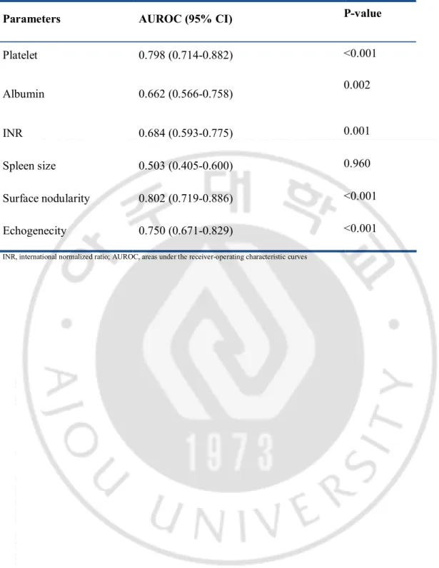
To validate the diagnostic values of non-invasive tests for cirrhosis, we calculated the AUROC of each variable (Table 2). The AUROC value of the spleen size for the diagnosis of cirrhosis was 0.503 and the p value of the test was 0.960. Thus, cut-off values could not be obtained. Five variables were identified as predictors of liver cirrhosis. These variables were as follows (in increasing order): albumin (P=0.002); INR (P=0.001); platelet count (P<0.001); surface nodularity (P<0.001); and parenchyma echogenicity (P<0.001). In particular, the AUROC value of surface nodularity, platelet count, and parenchyma echogenicity was high (>0.8).
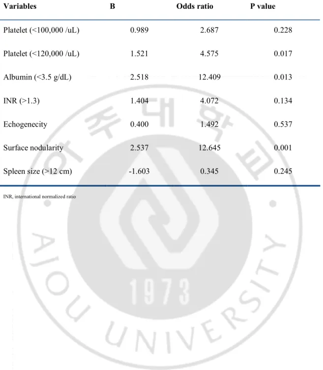
Logistic regression analysis showed that surface nodularity was significantly associated with liver cirrhosis and the odds ratio was 12.645. A platelet count < 120,000 / uL and an albumin level < 3.5 g / dL had significant odds ratio of 4.575 and 12.409, respectively. The other factors
- 8 -
Table 2. Area under curve of each variable for compensated liver cirrhosis Parameters AUROC (95% CI) P-value
Platelet 0.798 (0.714-0.882) <0.001 Albumin 0.662 (0.566-0.758) 0.002 INR 0.684 (0.593-0.775) 0.001 Spleen size 0.503 (0.405-0.600) 0.960 Surface nodularity 0.802 (0.719-0.886) <0.001 Echogenecity 0.750 (0.671-0.829) <0.001
- 10 -
Table 3. Results of logistic regression: each variable versus pathologic diagnosis for compensated liver cirrhosis
Variables B Odds ratio P value
Platelet (<100,000 /uL) 0.989 2.687 0.228 Platelet (<120,000 /uL) 1.521 4.575 0.017 Albumin (<3.5 g/dL) 2.518 12.409 0.013 INR (>1.3) 1.404 4.072 0.134 Echogenecity 0.400 1.492 0.537 Surface nodularity 2.537 12.645 0.001 Spleen size (>12 cm) -1.603 0.345 0.245
C. The usefulness of each variable in the diagnosis of compensated liver
cirrhosis
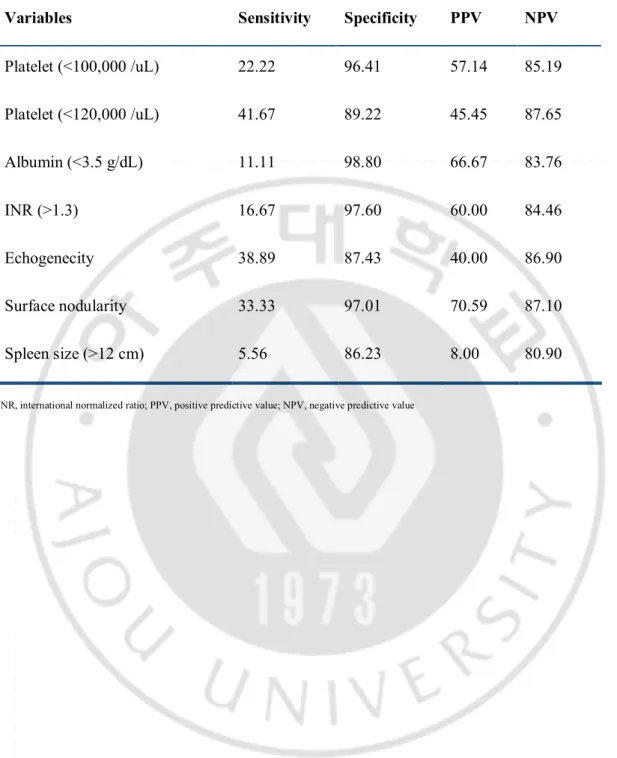
To determine the diagnostic usefulness of each variable, cross-analysis was performed (Table 4). The sensitivity of each variable had a low range (5.56%-41.67%), but the specificity was > 85%. A platelet count < 100,000 / uL, an albumin level < 3.5 g / dL, a prothrombin time (INR) > 1.3, and surface nodularity had a specificity >95%. With a platelet count cut-off value <120,000 / uL, sensitivity and specificity for the diagnosis of cirrhosis were 41% and 89%, respectively. Specificity increased to 96% and sensitivity decreased to 22% with a platelet count cut-off < 100,000 / uL.
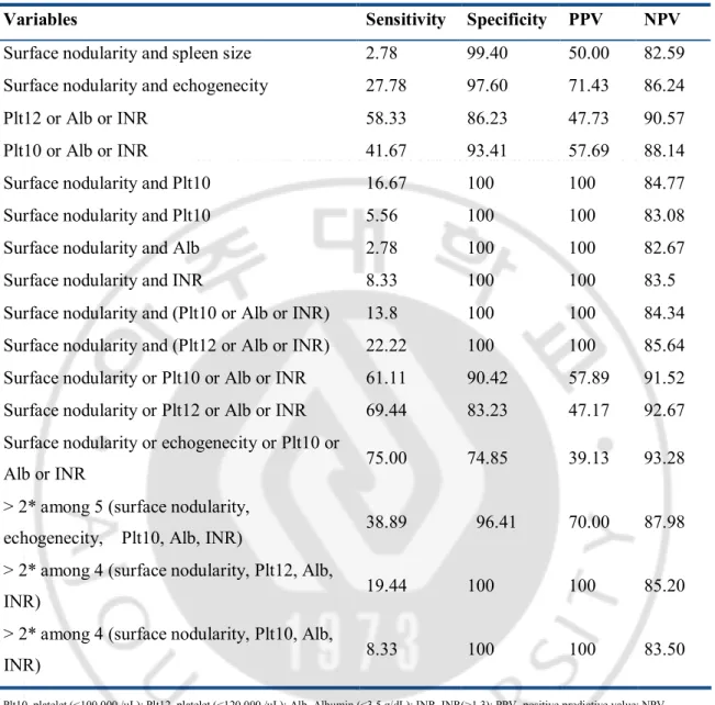
Cross-analysis was performed in combination with high specificity variables on the basis of the above results (Table 5). The combination of surface nodularity and parenchyma echogenecity satisfying two ultrasonographic variables showed a specificity of 97.6 % and a sensitivity of 27.7%. Any 1 of 3 blood variables (platelet count < 100,000 / uL, albumin level < 3.5 g / dL, and INR > 1.3) provided a sensitivity of 41% and a specificity of 93% for the detection of compensated cirrhosis. The combination of surface nodularity and 1 of 3 blood variables provided 100% of specificity, but low sensitivity (range, 2%-22%).
Based on these findings of at least one of 4 variables (surface nodularity, platelet counts < 100,000 / uL, albumin levels < 3.5 g / dL, INR > 1.3), cirrhosis was accurately identified with 90% specificity and 61% sensitivity. With a platelet cut off value of 120,000 / uL, the sensitivity increased to 69% but specificity decreased to 83%. And the combinations satisfying two among many variables showed 40% or less of sensitivity.
- 12 -
Table 4. Diagnostic accuracy, sensitivity, and specificity of compensated liver cirrhosis by different variables
Variables Sensitivity Specificity PPV NPV Platelet (<100,000 /uL) 22.22 96.41 57.14 85.19 Platelet (<120,000 /uL) 41.67 89.22 45.45 87.65 Albumin (<3.5 g/dL) 11.11 98.80 66.67 83.76 INR (>1.3) 16.67 97.60 60.00 84.46 Echogenecity 38.89 87.43 40.00 86.90 Surface nodularity 33.33 97.01 70.59 87.10 Spleen size (>12 cm) 5.56 86.23 8.00 80.90
INR, international normalized ratio; PPV, positive predictive value; NPV, negative predictive value
Table 5. Diagnostic accuracy, sensitivity, and specificity of compensated liver cirrhosis by combination variables
Variables Sensitivity Specificity PPV NPV Surface nodularity and spleen size 2.78 99.40 50.00 82.59 Surface nodularity and echogenecity 27.78 97.60 71.43 86.24
Plt12 or Alb or INR 58.33 86.23 47.73 90.57
Plt10 or Alb or INR 41.67 93.41 57.69 88.14
Surface nodularity and Plt10 16.67 100 100 84.77
Surface nodularity and Plt10 5.56 100 100 83.08
Surface nodularity and Alb 2.78 100 100 82.67
Surface nodularity and INR 8.33 100 100 83.5
Surface nodularity and (Plt10 or Alb or INR) 13.8 100 100 84.34 Surface nodularity and (Plt12 or Alb or INR) 22.22 100 100 85.64 Surface nodularity or Plt10 or Alb or INR 61.11 90.42 57.89 91.52 Surface nodularity or Plt12 or Alb or INR 69.44 83.23 47.17 92.67 Surface nodularity or echogenecity or Plt10 or
Alb or INR 75.00 74.85 39.13 93.28
> 2* among 5 (surface nodularity,
echogenecity, Plt10, Alb, INR) 38.89 96.41 70.00 87.98 > 2* among 4 (surface nodularity, Plt12, Alb,
INR) 19.44 100 100 85.20
> 2* among 4 (surface nodularity, Plt10, Alb,
INR) 8.33 100 100 83.50
Plt10, platelet (<100,000 /uL); Plt12, platelet (<120,000 /uL); Alb, Albumin (<3.5 g/dL); INR, INR(>1.3); PPV, positive predictive value; NPV, negative predictive value; 2*, 2 variables
- 14 -
IV. DISCUSSION
Early diagnosis of liver cirrhosis in chronic hepatitis patients is critical because it can predict and reduce complications of cirrhosis and the occurrence of hepatocellular carcinoma. Guidelines for the diagnosis of compensated liver cirrhosis are rarely reported worldwide(Fontaine et al., 2007). In the absence of specific non-invasive diagnostic guidelines, clinicians use a variety of criteria. It is considered that these phenomena are caused by the limitations of non-invasive methods in the diagnosis of liver cirrhosis. Therefore, non-invasive diagnostic instructions of liver cirrhosis are very important to treat the patients and perform the research.
For a non-invasive diagnosis of cirrhosis, ideal conditions require that the results are reliable, diagnostic tools are easy to perform, are inexpensive and commonly used, and deemed worthy by a number of checks (Sebastiani, 2009). Methods satisfying these conditions include ultrasonographic imaging and blood tests. In the diagnosis of compensated liver cirrhosis, liver biopsy is the most accurate diagnostic method. However, a liver biopsy is performed in few patients in clinical practice. In most patients, the blood tests and abdominal ultrasonography are performed to diagnose compensated liver cirrhosis. The accuracy of these non-invasive diagnostic tests can vary depending on the physician.
In addition to ultrasonographic imaging, computed tomography and nuclear magnetic resonance imaging have been used to diagnose cirrhosis in some studies, but there was no distinct advantage over ultrasonography, and computed tomography and nuclear magnetic resonance imaging was not generally available (Kudo et al., 2008).
Because ultrasonography has been widely used for the observation of chronic hepatitis and hepatocellular carcinoma screening, the role of abdominal ultrasonography has been reported in the diagnosis of compensated cirrhosis. Gaiani et al. investigated the accuracy of an ultrasonographic score derived from liver, spleen, and portal vein features in predicting the final diagnosis in 212 patients with compensated chronic liver disease undergoing percutaneous liver biopsy. They identified liver surface nodularity and portal flow velocity as factors independently associated with cirrhosis (p <0.005), and a score based on these two variables correctly identified cirrhosis in 82.2% of cases (82.2% sensitivity and 79.9% specificity)(Gaiani et al., 1997). Liver surface nodularity was associated with the diagnosis of cirrhosis in other studies. Enlargement of the caudate lobe had a high specificity, but low sensitivity (26%-41%). Spleen size and echogenicity pattern also had a high specificity, but low sensitivity for diagnosing cirrhosis(Di Lelio et al., 1989; Colli et al., 2003; Shen et al., 2006).
In the current study, the ultrasonographic findings, such as liver surface nodularity, internal echogenicity pattern, and spleen size, were compared with pathologically-confirmed liver cirrhosis. In agreement with previous studies, liver surface nodularity had very high specificity (97%), but low sensitivity (33%). The sensitivity and specificity of the other two variables were lower than those of liver surface nodularity. The role of ultrasonographic examination in the diagnosis of early cirrhosis is limited due to low specificity, although sonographic variables could be combined. Therefore, ultrasonographic findings alone are insufficient as a screening test for liver cirrhosis(Ong et al., 2003).
In addition to ultrasonography, there are many non-invasive ways using blood tests to diagnose liver cirrhosis. The serum fibrosis markers can be divided into direct and indirect markers. Direct markers are involved in the formation of extracellular matrix during fibrosis,
- 16 -
and indirect markers reflect hepatic dysfunction. Several studies on the diagnosis of liver cirrhosis using direct fibrosis markers, such as collagen and hyaluronic acid, had a low sensitivity and specificity (approximately 80%), and fibrosis markers are expensive and not available in clinical practice(Guechot et al., 1995; Mehta et al., 2008; Murawaki et al., 2001). Indirect fibrosis markers include an indirect AST-to-ALT ratio (AAR) and an AST-to-platelet ratio index (APRI), Forn's index, and FIB-4. However, indirect fibrosis markers were not sufficient as a single indicator in diagnosing liver cirrhosis in many studies11, 26-28. (Forns, Ampurdanes, 2002; Lackner et al., 2005; Wai et al., 2003).
Sebastian et al. published results showing that a fibroscan and blood tests can reduce the liver biopsy, but a fibroscan is an expensive piece of equipment. A fibroscan is not available for obese patients, and is not available for clinical practice(Sebastiani, 2009).
We evaluated the diagnostic accuracy of ultrasonography and routine blood tests in the diagnosis of compensated cirrhosis and established the diagnostic criteria. We investigated the diagnostic accuracy of the model consisting of four variables (surface nodularity, platelet count, albumin level, and INR). Based on the findings of at least 1 of 4 variables, we identified cirrhosis accurately (90% specificity and 61% sensitivity). Variables used in this study included serum albumin and prothrombin time (INR) indicating the protein synthetic activity of liver and platelet count, reflecting portal hypertension. As these variables, except the platelet count, are used in the Child-Pugh classification, the specificity of the items appears to be high.
The albumin level and INR had > 95% specificity in identifying cirrhosis, but the sensitivity was very low. The platelet count had better sensitivity while maintaining high specificity compared to the albumin level and INR. A platelet count < 100,000 / uL has been widely
accepted as the cut-off level of portal hypertension 29. In this study, a platelet count < 100,000 / uL had 22% sensitivity and 96% specificity in identifying compensated cirrhosis. When we defined a platelet count cut-off of < 1200,000 / uL, the sensitivity increased to 41% with a minor reduction in specificity (89%).
This study has several strengths. First, liver cirrhosis can be predicted without special equipment in any hospital. Second, three experienced physicians reviewed ultrasonographic findings of 30 patients to objectify the subjective sonographic findings. Third, we suggested the standard criteria which are not unified in diagnosing liver cirrhosis on the basis of Korean data. This study had several limitations. First, a retrospective study may have a bias in selecting patients and the number of patients was relatively small. Second, the study design was cross-sectional. The results of blood tests can vary depending on the patient condition. Thus, sequential measurements or mean values would be better as a representative value.
- 18 -
V. CONCLUSION
In conclusion, the model consisting of surface nodularity, platelet count < 100,000 / uL, albumin level < 3.5 g / dL, and prothrombin time (INR) > 1.3 may be useful for the identification of compensated cirrhosis with a high degree of accuracy in daily practice.
REFERENCES
1. Anthony PP, Ishak KG, Nayak NC, Poulsen HE, Scheuer PJ, Sobin LH: The morphology of cirrhosis: definition, nomenclature, and classification. Bull World Health Organ 55:521-540,1977
2. Maynard JE: Hepatitis B: global importance and need for control. Vaccine 8 Suppl:S18-20; discussion S21-13,1990
3. Alter MJ: Epidemiology of hepatitis C. Hepatology 26:62S-65S,1997
4. Sorrell MF, Belongia EA, Costa J, Gareen IF, Grem JL, Inadomi JM, Kern ER, McHugh JA, Petersen GM, Rein MF, Strader DB, Trotter HT: National Institutes of Health consensus development conference statement: management of hepatitis B. Hepatology 49:S4-S12,2009
5. Paik SW: [Goals of treatment, indication, and treatment for chronic hepatitis C]. Korean J
Gastroenterol 51:368-371,2008
6. Bravo AA, Sheth SG, Chopra S: Liver biopsy. N Engl J Med 344:495-500,2001
7. Piccinino F, Sagnelli E, Pasquale G, Giusti G: Complications following percutaneous liver biopsy. A multicentre retrospective study on 68,276 biopsies. J Hepatol 2:165-173,1986
8. Abdi W, Millan JC, Mezey E: Sampling variability on percutaneous liver biopsy. Arch
- 20 -
9. Bedossa P, Dargere D, Paradis V: Sampling variability of liver fibrosis in chronic hepatitis C. Hepatology 38:1449-1457,2003
10. Laharie D, Zerbib F, Adhoute X, Boue-Lahorgue X, Foucher J, Castera L, Rullier A, Bertet J, Couzigou P, Amouretti M, de Ledinghen V: Diagnosis of liver fibrosis by transient elastography (FibroScan) and non-invasive methods in Crohn's disease patients treated with methotrexate. Aliment Pharmacol Ther 23:1621-1628,2006
11. Forns X, Ampurdanes S, Llovet JM, Aponte J, Quinto L, Martinez-Bauer E, Bruguera M, Sanchez-Tapias JM, Rodes J: Identification of chronic hepatitis C patients without hepatic fibrosis by a simple predictive model. Hepatology 36:986-992,2002
12. Chen YP, Dai L, Wang JL, Zhu YF, Feng XR, Hou JL: Model consisting of ultrasonographic and simple blood indexes accurately identify compensated hepatitis B cirrhosis. J Gastroenterol Hepatol 23:1228-1234,2008
13. Park YN, Kim Hg, Chon CY, Park JB, Sohn JH, Yang SH, Yu ES, Lee MS, Jang JJ, Chang HK, Jeong JJ, Kang DY, Kim YI, Park CI: Histological Grading and Staging of Chronic Hepatitis Standardized Guideline Proposed by the Korean Study Group for the Pathology of Digestive Diseases. Korean J Pathol 33:337-346,1999
14. Poynard T, Bedossa P, Opolon P: Natural history of liver fibrosis progression in patients with chronic hepatitis C. The OBSVIRC, METAVIR, CLINIVIR, and DOSVIRC groups.
15. Fontaine H, Petitprez K, Roudot-Thoraval F, Trinchet JC: Guidelines for the diagnosis of uncomplicated cirrhosis. Gastroenterol Clin Biol 31:504-509,2007
16. Sebastiani G: Non-invasive assessment of liver fibrosis in chronic liver diseases: implementation in clinical practice and decisional algorithms. World J Gastroenterol 15:2190-2203,2009
17. Kudo M, Zheng RQ, Kim SR, Okabe Y, Osaki Y, Iijima H, Itani T, Kasugai H, Kanematsu M, Ito K, Usuki N, Shimamatsu K, Kage M, Kojiro M: Diagnostic accuracy of imaging for liver cirrhosis compared to histologically proven liver cirrhosis. A multicenter collaborative study. Intervirology 51 Suppl 1:17-26,2008
18. Gaiani S, Gramantieri L, Venturoli N, Piscaglia F, Siringo S, D'Errico A, Zironi G, Grigioni W, Bolondi L: What is the criterion for differentiating chronic hepatitis from compensated cirrhosis? A prospective study comparing ultrasonography and percutaneous liver biopsy. J Hepatol 27:979-985,1997
19. Di Lelio A, Cestari C, Lomazzi A, Beretta L: Cirrhosis: diagnosis with sonographic study of the liver surface. Radiology 172:389-392,1989
20. Colli A, Fraquelli M, Andreoletti M, Marino B, Zuccoli E, Conte D: Severe liver fibrosis or cirrhosis: accuracy of US for detection--analysis of 300 cases. Radiology 227:89-94,2003
- 22 -
21. Shen L, Li JQ, Zeng MD, Lu LG, Fan ST, Bao H: Correlation between ultrasonographic and pathologic diagnosis of liver fibrosis due to chronic virus hepatitis. World J
Gastroenterol 12:1292-1295,2006
22. Ong TZ, Tan HJ: Ultrasonography is not reliable in diagnosing liver cirrhosis in clinical practice. Singapore Med J 44:293-295,2003
23. Guechot J, Poupon RE, Poupon R: Serum hyaluronan as a marker of liver fibrosis. J
Hepatol 22:103-106,1995
24. Mehta P, Ploutz-Snyder R, Nandi J, Rawlins SR, Sanderson SO, Levine RA: Diagnostic accuracy of serum hyaluronic acid, FIBROSpect II, and YKL-40 for discriminating fibrosis stages in chronic hepatitis C. Am J Gastroenterol 103:928-936,2008
25. Murawaki Y, Ikuta Y, Okamoto K, Koda M, Kawasaki H: Diagnostic value of serum markers of connective tissue turnover for predicting histological staging and grading in patients with chronic hepatitis C. J Gastroenterol 36:399-406,2001
26. Lackner C, Struber G, Liegl B, Leibl S, Ofner P, Bankuti C, Bauer B, Stauber RE: Comparison and validation of simple noninvasive tests for prediction of fibrosis in chronic hepatitis C. Hepatology 41:1376-1382,2005
27. Wai CT, Greenson JK, Fontana RJ, Kalbfleisch JD, Marrero JA, Conjeevaram HS, Lok AS: A simple noninvasive index can predict both significant fibrosis and cirrhosis in patients with chronic hepatitis C. Hepatology 38:518-526,2003
국문 요약
-만성
바이러스성 간염 환자에서 초음파 및 일반 혈액 검사를
이용한 대상 간경변증 예측
아주대학교 대학원의학과 이홍섭 (지도교수: 조 성 원) 배경: 현재 간경변증을 진단하기 위해서 간 조직 검사가 표준 방법으로 되어있으나 간 조직 검사는 침습적인 방법으로 일부의 환자에서만 시행되고 있다. 보통 일반 혈액 검사와 초음파 검사를 시행하여 간경변증을 진단하고 있다. 이에 이번 연구는 만성 B 형 및 C 형 바이러스 간염 환자를 대상으로 초음파 검사와 일반혈액검사를 통한 대상성 간경변증의 진단 정확도를 알아보고자 하였다. 대상 및 방법: 아주대병원에 내원하여 복부 초음파 검사 와 혈액 검사 및 조직 검사를 시행한 만성 B 형 및 C 형 바이러스 간염 환자 203 명을 대상으로, 초음파 검사 와 일반혈액검사를 통한 대상성 간경변증의 진단 정확도를 알아보기 위해서 간 조직 검사 결과와 다변량 및 교차 분석 실시하였다. 결과: 로지스틱 회귀 분석 결과 간표면 결절성이 간경변증의 진단에 의미 있게 영향을 미치며 교차비가 12.645 로 높았고 혈소판수가 100,000/uL 이하로 잡았을 때는 유의한 값을 얻지 못했으나 120,000/uL 이하인 것과 알부민 수치가 3.5 g/dL 이하로 했을 때는 유의하게 영향을 미치고 각각의 교차비는 4.575 와 12.409 였다. 교차분석 결과 혈소판 100,000/uL 이하, 알부민 3.5g/dL 이하, 프로트롬빈시간(INR)- 24 - 1.3 이상, 간 표면 결절화가 있는 경우는 각각 특이도가 95% 이상으로 매우 높았다. 교차 분석에서 특이도가 높았던 초음파상 간 표면 결절성이 있고, 100,000 /uL 이하의 혈소판, 3.5 g/dL 이하의 알부민, 1.3 이상의 프로트롬빈시간(INR) 중에 하나만 만족하는 교차분석 에서 90.42%의 높은 특이도를 보이면서도 61.11%의 비교적 높은 민감도를 보였다. 결론: 만성 간질환 환자에서 초음파로 간 표면 결절성 소견이 보이거나 혈소판이 100,000/uL 이하이거나 알부민 3.5g/dL 이하, 또는 프로트롬빈시간(INR)이 1.3 이상인 경우는 비교적 높은 특이도로 대상성 간경변증을 진단할 수 있었다. 핵심어 : 간경변증, 복부초음파, 진단, 혈액 검사, 간조직 생검