Introduction
Diabetes is becoming increasingly prevalent worldwide due to ag-ing, physical inactivity, westernized eating habits, population growth, and obesity; consequently, the incidences of diabetic feet are increasing. Diabetic patients are predicted to have a 25% prob-ability of having at least one diabetic foot ulcer during their life-time. Furthermore, a lower limb amputation due to a diabetic foot
Background: A diabetic foot is the most common cause of non-traumatic lower extremity ampu-tations (LEA). The study seeks to assess the risk factors of amputation in patients with diabetic foot ulcers (DFU).
Methods: The study was conducted on 351 patients with DFUs from January 2010 to December 2018. Their demographic characteristics, disease history, laboratory data, ankle-brachial index, Wagner classification, osteomyelitis, sarcopenia index, and ulcer sizes were considered as vari-ables to predict outcome. A chi-square test and multivariate logistic regression analysis were performed to test the relationship of the data gathered. Additionally, the subjects were divided into two groups based on their amputation surgery.
Results: Out of the 351 subjects, 170 required LEA. The mean age of the subjects was 61 years and the mean duration of diabetes was 15 years; there was no significant difference between the two groups in terms of these averages. Osteomyelitis (hazard ratio [HR], 6.164; 95% confidence interval [CI], 3.561−10.671), lesion on percutaneous transluminal angioplasty (HR, 2.494; 95% CI, 1.087−5.721), estimated glomerular filtration rate (eGFR; HR, 0.99; 95% CI, 0.981−0.999), ulcer size (HR, 1.247; 95% CI, 1.107−1.405), and forefoot ulcer location (HR, 2.475; 95% CI, 0.224−0.73) were associated with risk of amputation.
Conclusion: Osteomyelitis, peripheral artery disease, chronic kidney disease, ulcer size, and fore-foot ulcer location were risk factors for amputation in diabetic fore-foot patients. Further investiga-tion would contribute to the establishment of a diabetic foot risk stratificainvestiga-tion system for Kore-ans, allowing for optimal individualized treatment.
Keywords: Amputation; Diabetic foot ulcer; Diabetes mellitus; Risk factors
Risk factors affecting amputation in diabetic foot
Jun Ho Lee
1, Ji Sung Yoon
2, Hyoung Woo Lee
2, Kyu Chang Won
2, Jun Sung Moon
2, Seung Min Chung
3,
Yin Young Lee
31Republic of Korea Army, Hwacheon, Korea
2Division of Endocrinology and Metabolism, Department of Internal Medicine, Yeungnam University College of Medicine, Daegu, Korea 3Division of Endocrinology and Metabolism, Department of Internal Medicine, Yeungnam University Hospital, Daegu, Korea
Yeungnam Univ J Med 2020;37(4):314-320 https://doi.org/10.12701/yujm.2020.00129 Received: March 4, 2020 Revised: April 9, 2020 Accepted: April 14, 2020 Corresponding author: Ji Sung Yoon
Division of Endocrinology and Metabolism, Department of Internal Medicine, Yeungnam University College of Medicine, 170 Hyunchung-ro, Nam-gu, Daegu 42415, Korea
Tel: +82-53-620-4049 Fax: +82-53-623-8006 E-mail: jsyoon9@ynu.ac.kr
is carried out every 30 seconds worldwide, with rates being 30 to 40 times higher for diabetic patients than it is for individuals with-out the disease [1-3].
The cost of diabetic foot treatment accounts for approximately 25% of a diabetic patient’s total hospital costs [4]. To reduce this burden, clinicians should focus on prevention as well as treatment of diabetic foot disease. Needless to say, amputation in patients with diabetic foot disease debilitates their ability to perform
ev-Copyright © 2020 Yeungnam University College of Medicine
This is an Open Access article distributed under the terms of the Creative Commons Attribution Non-Commercial License (http://creativecommons.org/licenses/by-nc/4.0/) which permits unrestricted non-commercial use, distribution, and reproduction in any medium, provided the original work is properly cited.
eryday tasks, which negatively affects their quality of life ; there-fore, knowledge of the risk factors of diabetic foot amputation is an important issue [5]. Minute observation and prophylactic ac-tion for patients at high risk of having a diabetic foot and early de-tection of foot complications could reduce the occurrence of ul-cerations and amputations [6].
The development of a diabetic foot ulcer has multifactorial causes, and its principal factors include: diabetic peripheral neu-ropathy, infection, peripheral arterial disease, and socioeconomic status [7]. Moreover, various features such as age, smoking, foot deformities, poor glycemic control, ulcer size, hypertension, white blood cell count, and lipid abnormalities have also been reported as risk factors for diabetic foot amputation [8-11]. However, pre-vious studies on the risk factors of diabetic feet indicated inconsis-tent results. Therefore, the aim of this study was to determine the risk factors of amputation in Korean diabetic foot patients who re-ceived standard treatment from one institution.
Materials and methods
1. Subjects and data collectionThis study was approved by the Institutional Review Board (IRB) of the Yeungnam University Hospital (IRB No: 2019-03-040). It followed the Declaration of Helsinki on medical protocol and eth-ics. The patients’ personal information was withheld from the re-searchers.
This case control study involved 425 subjects who were admit-ted to the Yeungnam University Hospital due to a diabetic foot from January 1, 2010 to December 31, 2018. Due to lack of data, 74 of the 425 subjects were excluded, leaving 351 valid subjects. They were divided into two groups based on their amputation surgery.
2. Clinical information of patients
Medical records, including admission notes, were examined to obtain information on the patients and laboratory results collect-ed during the first day of admission. The independent variables were selected based on previous studies to determine the risk fac-tors of diabetic feet and amputation. Amputation was defined as surgery, which goes beyond the toe level. Minor debridement of soft tissue was not considered as amputation surgery.
Hypertension was defined as the use of anti-hypertensive medi-cation or previously documented diagnoses. Ankle-brachial index (ABI) was calculated by dividing the systolic blood pressure of the ankle divided by the systolic blood pressure of the upper arm of the affected side. The mean value of the ABI of the two groups were compared and subjects with ABI of less than 0.9 were found
to be abnormal.
Diabetic peripheral polyneuropathy was identified through the consultation records of the neurology department, electrophysiol-ogy studies, the Semmes Weinstein monofilament test, clinical scores (i.e., the Michigan Neuropathy Screening Instrument ques-tionnaire), and medical record reviews. Diabetic retinopathy was identified through ophthalmologic records, including any history of photocoagulation and vitrectomy. Coronary artery disease was defined as any history of myocardial infarction, unstable angina, percutaneous transluminal coronary angioplasty, or coronary ar-tery bypass surgery. Stroke was defined as any history of cerebral infarction or transient cerebral ischemia. Chronic kidney disease was defined as the estimated glomerular filtration rate < 60 mL/ min/1.73 m2
, which was calculated using the Modification of Diet in Renal Disease formula: 186×(creatinine)−1.154
× (age)−0.203
× (0.742, if female). The sarcopenia index (SI) was used to estimate skeletal muscle mass and was derived from the formula serum creatinine value/cystatin C value [12].
Information of previous amputation history was based on or-thopedic surgery and plastic surgery department records. The ul-cer size was defined as the longest diameter, in centimeters, from one end of an ulcer margin to the other; this was assessed by an endocrinologist at admission. The Wagner classification catego-rized the diabetic foot ulcers based on the depth and the presence of osteomyelitis or gangrene. A grade ranging from 0 to 5 was as-signed to pre-ulcerative lesions; partial superficial ulcers; exten-sions into tendons, ligaments, fascia, or joint capsules without os-teomyelitis; deep ulcers with osos-teomyelitis; partial forefoot necro-sis; and extensive foot gangrene, respectively [13]. The location of the foot ulcers was classified as forefoot, midfoot, and hindfoot. Percutaneous transluminal angioplasty (PTA) was assessed through records of the vascular department while osteomyelitis was assessed through scans such as an magnetic resonance imag-ing or 3-phase bone scan.
3. Statistical analysis
IBM SPSS version 20.0 (IBM Corp, Armonk, NY, USA) was used for statistical analysis and a Student t-test was conducted to
com-pare the quantitative variables of the two groups. A chi-square test was used to analyze the categorial variables, and the risk factors for amputation were determined through stepwise multiple logistic regression analysis. All p-values less than 0.05 were considered
statistically significant.
Results
Table 1. The mean age of the subjects and the mean duration of diabetes was 61 and 15 years, respectively; there was no significant difference between the two groups. Among the amputation group, 119 subjects underwent surgery below the ankle, while 51 subjects had above-the-knee or below-the-knee amputations.
The amputation group generally had significantly lower high density lipoprotein cholesterol (HDL-C) and eGFR, but a larger ulcer size. The chi-square test indicated that the amputation group had a higher incidence of previous amputation history (Table 2). The use of statins, antiplatelet drugs, anti-hypertensive drugs, and insulin did not show any significant difference between the two groups.
Among the 351 subjects, 193 showed positive wound culture results. The most common pathogens were Staphylococcus aureus,
followed by Pseudomonas aeruginosa, and Escherichia coli. The two
groups did not exhibit any differences in terms of pathogens. According to the Wagner classification, the chi-square test indi-cated a significant difference between the two groups (p=0.0001),
whereas the multivariate regression analysis did not (Table 2). The non-amputation group consisted of 110, 61, and 10 sub-jects with forefoot, midfoot, and hindfoot ulcers, respectively. On the other hand, the amputation group had 129, 37, and four sub-jects with forefoot, midfoot, and hindfoot ulcers, respectively.
The ABI of 126 subjects were each obtained from the non-am-putation and the amnon-am-putation group and 26 and 46 of them had decreased ABI value, respectively. The chi-square test (p=0.005)
showed a significance between the two groups and the decreased ABI indicates an increased risk of amputation. Due to insufficient data, the ABI was excluded from the regression analysis.
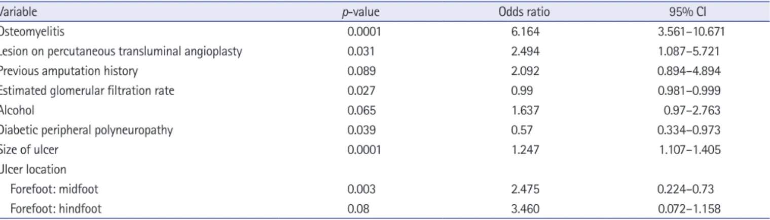
By means of multivariate regression analysis, the risk factors of amputation were identified as the presence of osteomyelitis of OR 6.164, a lesion on PTA of OR 2.494, a forefoot location of OR 2.475, an ulcer size of OR 1.247, and a kidney function of OR 0.99 (Table 3).
Table 1. Univariate analysis of subjects with or without amputation surgery
Variable Non-amputation group (n= 181) Amputation group (n= 170) p-valuea)
Age (yr) 61.8± 12.6 62.4± 10.9 0.642
Body mass index (kg/m2
) 23.2± 4.3 23.0± 3.6 0.682
SBP (mmHg) 130.7± 17.5 130.8± 20.5 0.959
DBP (mmHg) 78.9± 10.7 77.1± 12.8 0.152
Length of hospitalization (day) 51.1± 54.9 53.7± 58.4 0.660
Duration of diabetes (yr) 15.4± 9.5 15.1± 10.0 0.821
Ankle-brachial index 1.1± 0.2 1.0± 0.3 0.027 Size of ulcer (cm) 2.6± 2.2 3.6± 2.6 0.0001 Sarcopenia index 101.7± 101.5 99.9± 95.5 0.861 WBC (x103/μL) 10.0± 5.3 11.2± 6.4 0.540 Hemoglobin (g/dL) 11.3± 2.0 11.1± 2.1 0.526 ESR (mm/hr) 60.2± 38.5 66.8± 36.3 0.100 hsCRP (mg/L) 6.7± 9.5 7.2± 9.2 0.603 Glycated hemoglobin (%) 8.9± 2.2 9.0± 2.3 0.565 Albumin (g/dL) 3.5± 0.8 3.4± 0.8 0.293 Total cholesterol (mg/dL) 158.0± 50.7 155.8± 61.6 0.730 Triglyceride (mg/dL) 139.9± 100.5 161.6± 172.0 0.173 HDL-C (mg/dL) 38.2± 15.4 33.8± 16.1 0.015 LDL-C (mg/dL) 93.1± 36.3 90.5± 41.1 0.533
Blood urea nitrogen (mg/dL) 23.2± 14.7 22.9± 16.7 0.843
Creatinine (mg/dL) 2.0± 4.3 1.8± 2.1 0.569
eGFR (mL/min/1.73 m2) 68.7± 29.0 62.2± 28.6 0.035
ACR (mg/mmol) 761.5± 1,690.5 978.6± 2,394.2 0.531
Cystatin C (mg/dL) 1.8± 1.2 2.0± 1.49 0.212
Values are presented as mean±standard deviation.
SBP, systolic blood pressure; DBP, diastolic blood pressure; WBC, white blood cell; ESR, erythrocyte sedimentation rate; hsCRP, high-sensitivity C-reactive protein; HDL-C, high density lipoprotein cholesterol; LDL-C, low density lipoprotein cholesterol; eGFR, estimated glomerular filtration rate; ACR, albumin creatinine ratio.
a)
Discussion
While various risk factors of diabetic foot amputation may have been identified in previous research, this study found that osteo-myelitis, ulcer size, chronic kidney disease, forefoot location, and peripheral arterial disease were associated with diabetic foot am-putation. This diversity could be brought about by differences in the genetic profiles, treatment protocols, study designs, and cul-tural features of the study subjects.
Osteomyelitis is one of the most critical factors in diabetic foot treatment process. The removal of infected bones is crucial, as
pri-or studies have suggested superipri-or results from surgical therapy compared with medical therapy [14,15]. Moreover, surgical de-bridement was discovered to be necessary to control chronic bone infection because antimicrobial therapy alone showed low success rates in the event of osteomyelitis [16]. The presence of osteomy-elitis was the most significant risk factor (OR, 6.164) for diabetic foot amputation in this study.
Arterial insufficiency causes an impairment of bloodstream (i.e., a shortage of nutrition, antibodies, and white blood cells), which leads to poor wound outcome. Claudication, diminished or absent lower extremity pulses, lower ankle blood pressure, ABI, and
trans-Table 2. Major risk factors of amputation
Variable Non-amputation (n= 181) Amputation (n= 170) p-valuea)
Osteomyelitis 77 (42.5) 142 (83.5) 0.0001
Lesion on percutaneous transluminal angioplasty 10 (5.5) 30 (17.6) 0.0001
Diabetic peripheral polyneuropathy 120 (66.2) 97 (57.0) 0.095
Chronic kidney disease (eGFR< 60 mL/min/1.73 m2) 49 (27.0) 66 (36.4) 0.019
ABI decreased (ABI< 0.9) 26 (n= 126, 20.6) 46 (n= 126, 36.5) 0.005
Previous amputation history 12 (6.6) 22 (12.9) 0.046
Hypertension 113 (62.4) 103 (60.6) 0.723 Smoking 79 (43.6) 82 (48.2) 0.358 Alcohol 60 (33.1) 70 (41.2) 0.108 Wagner classification 0.0001 Grade 0−1 72 (39.7) 30 (17.6) Garde 2 47 (26.0) 31 (18.2) Grade 3 36 (19.9) 52 (30.6) Grade 4−5 26 (14.4) 57 (33.5) Ulcer location 0.002 Forefoot 110 (60.8) 129 (75.9) Midfoot 61 (33.7) 37 (21.8) Hindfoot 10 (5.5) 4 (2.3)
Values are presented as number (%).
eGFR, estimated glomerular filtration rate; ABI, ankle-brachial index.
a)p-value based on chi-square test and Fisher exact test.
Table 3. Multivariate logistic regression analysis of diabetic foot amputation
Variable p-value Odds ratio 95% CI
Osteomyelitis 0.0001 6.164 3.561–10.671
Lesion on percutaneous transluminal angioplasty 0.031 2.494 1.087–5.721
Previous amputation history 0.089 2.092 0.894–4.894
Estimated glomerular filtration rate 0.027 0.99 0.981–0.999
Alcohol 0.065 1.637 0.97–2.763
Diabetic peripheral polyneuropathy 0.039 0.57 0.334–0.973
Size of ulcer 0.0001 1.247 1.107–1.405
Ulcer location
Forefoot: midfoot 0.003 2.475 0.224–0.73
Forefoot: hindfoot 0.08 3.460 0.072–1.158
cutaneous oxygen pressure of the foot (foot TcPo2) were important risk factors of developing foot ulcer and amputation surgery, indi-cating peripheral artery disease in diabetic patients [17,18]. In this study, the presence of PTA lesions and lower ABIs were associated with an increased risk of amputation surgery. However, Sun et al. [19] demonstrated that a lower ABI was closely associated with risk of amputation only in Wagner grade 3 wounds, not in grades 2 or 4. They assumed that a Wagner grade 4 grade wound would have cat-astrophic necrosis, which limits the influence of circulation; and vice versa for grade 2 wounds. This is why further comprehensive studies (i.e., with a subdivision of subjects with a classification of di-abetic foot severity) focusing on the relationship between peripher-al artery disease and prognosis are needed.
Ulcers located on the forefoot area were found to be a greater risk factor for diabetic foot amputation compared with midfoot or hindfoot located ulcers. Due to its distal position, it would be rea-sonable to assume that the forefoot area has the least sufficient blood supply in the foot region, resulting in a shortage of oxygen, white blood cell, and nutrition.
Several studies have shown that a large foot ulcer size and a high Wagner classification grade considerably increased the risk of am-putation [9,20]. This study found that a higher Wagner classifica-tion grade and ulcer size were significantly associated with the risk of amputation through a chi-square test and regression analysis, respectively. This was in accordance with the findings of previous observational studies that more extensive wounds require more extensive surgical procedures such as amputation [19,21].
Complications including diabetic microangiopathy often arise as a patient’s diabetes progresses. Diabetic kidney disease is anoth-er complication, and it is known as a useful markanoth-er for the genanoth-er- gener-alized vascular status of patients with diabetes. Additionally, pa-tients with nephropathy are also prone to developing peripheral vascular disease [22]. Several studies have shown that the inci-dence of diabetic foot disease is more frequent among patients with albuminuria [23-25]. However, given the insufficient data on albuminuria, this study was unable to properly examine the rela-tionship between albuminuria and amputation surgery.
Current dialysis for end-stage renal disease and chronic kidney disease were also identified as risk factors for diabetic feet and ma-jor amputation [21,26]. These results confirm that a lower eGFR and the presence of chronic kidney disease are associated with a higher risk of diabetic foot amputation.
Recently, sarcopenia has been demonstrated as an important risk factor of diabetic foot disease, which also influenced progno-sis; however, the measurement of skeletal muscles entails expen-sive and complex imaging techniques [27]. Thus far, the gold standard of measuring skeletal muscle mass has been either body
composition analysis or computed tomography. The SI is a novel biomarker for estimating muscle mass, which uses two molecules: cystatin C, which originates from all nucleated cells and creatinine from skeletal muscle cells. The SI is significantly correlated with abdominal computed tomography and showed superior out-comes compared with serum creatinine on its own [12,28,29]. The study sought to assess the value of the SI as a marker to pre-dict the prognosis of diabetic feet and compare it with other al-ready established prognostic factors. In this study, the SI did not show statistically significant results. Since several studies have demonstrated the relationship between the SI and the prognosis of diabetic foot ulcers, this could be regarded as a limitation of the calculation formula.
The strength of this study is in its ethnic-specific design, where-in the chosen subjects share a common cultural, dietary lifestyle. Moreover, all of the subjects also underwent standard treatment protocols due to the single center-based recruitment process. These features greatly reduced biases present in previous studies that were brought about by the subjects’ heterogeneous character-istics as well as the diverse treatment protocols among multi-centers.
Conducting a larger scale study similar to this could lead to the establishment of a diabetic foot risk stratification system for Kore-ans.
The study has several limitations. Because this was a hospital-ization-based single center design, a selection bias could not be excluded and the subjects may not have reflected the loco-region-al population. So further multicenter studies are needed. This study was retrospective design, therefore independent variables were could not be fully assessed, and causality between each fac-tors and outcome cannot be definitely established.
Osteomyelitis, a large ulcer size, nephropathy, forefoot location, and peripheral artery disease were identified as risk factors for am-putation in hospitalized diabetic foot ulcer patients. Understand-ing their influence on amputation outcomes is necessary to devel-op risk stratification system, management, and treatment proto-cols for patients with diabetic feet. Through risk categorization, a multidisciplinary team for diabetic feet could receive timely assis-tance on decision-making (e.g., admission timing and invasive procedures), providing the best possible treatment for individual-ized patients.
Acknowledgments
Conflicts of interestNo potential conflict of interest relevant to this article was report-ed.
Author contributions
Conceptualization: JHL, JSY, HWL, KCW, JSM, YYL; Data cura-tion: JHL, SMC; Formal analysis: JSY, KCW, JSM, YYL; Meth-odology: JHL, JSY, JSM, YYL; Investigation: JHL, JSY, HWL, KCW, YYL; Project administration: JHL; Supervision: JSY, KCW, JSM; Resources: JSY; Visualization: JHL, SMC; Writ-ing-original draft: JHL; Writing-review & editing: JHL, JSM.
ORCID
Jun Ho Lee, https://orcid.org/0000-0002-5551-862X Ji Sung Yoon, https://orcid.org/0000-0003-3091-3700 Hyoung Woo Lee, https://orcid.org/0000-0002-0773-1581 Kyu Chang Won, https://orcid.org/0000-0001-5945-3395 Jun Sung Moon, https://orcid.org/0000-0003-1569-3068 Seung Min Chung, https://orcid.org/0000-0003-3336-7557 Yin Young Lee, https://orcid.org/0000-0003-3684-8735
References
1. Boulton AJ, Vileikyte L, Ragnarson-Tennvall G, Apelqvist J. The global burden of diabetic foot disease. Lancet 2005;366:1719– 24.
2. Singh N, Armstrong DG, Lipsky BA. Preventing foot ulcers in patients with diabetes. JAMA 2005;293:217–28.
3. Bakker K, Foster AV, van Houtum WH, Riley P; International Working Group on the Diabetic Foot. Diabetes and foot care: time to act. Brussels (BE): International Diabetes Federation and the International Working Group on the Diabetic Foot; 2005.
4. Songer TJ. The role of cost-effectiveness analysis and health in-surance in diabetes care. Diabetes Res Clin Pract 2001;54(Sup-pl 1):S7–11.
5. Monteiro-Soares M, Boyko EJ, Ribeiro J, Ribeiro I, Dinis-Ri-beiro M. Risk stratification systems for diabetic foot ulcers: a systematic review. Diabetologia 2011;54:1190–9.
6. Boulton AJ, Kirsner RS, Vileikyte L. Clinical practice: neuro-pathic diabetic foot ulcers. N Engl J Med 2004;351:48–55.
7. Markakis K, Bowling FL, Boulton AJ. The diabetic foot in 2015: an overview. Diabetes Metab Res Rev 2016;32(Suppl 1):169–78.
8. Fleischer AE, Wrobel JS, Leonards A, Berg S, Evans DP, Baron RL, et al. Post-treatment leukocytosis predicts an unfavorable clinical response in patients with moderate to severe diabetic foot infections. J Foot Ankle Surg 2011;50:541–6.
9. Oyibo SO, Jude EB, Tarawneh I, Nguyen HC, Armstrong DG, Harkless LB, et al. The effects of ulcer size and site, patient's age, sex and type and duration of diabetes on the outcome of
diabet-ic foot ulcers. Diabet Med 2001;18:133–8.
10. Chaturvedi N, Stevens LK, Fuller JH, Lee ET, Lu M. Risk fac-tors, ethnic differences and mortality associated with lower- ex-tremity gangrene and amputation in diabetes. The WHO Mul-tinational Study of Vascular Disease in Diabetes. Diabetologia 2001;44(Suppl 2):S65–71.
11. Resnick HE, Carter EA, Sosenko JM, Henly SJ, Fabsitz RR, Ness FK, et al. Incidence of lower-extremity amputation in American Indians: the Strong Heart Study. Diabetes Care 2004; 27:1885–91.
12. Kashani KB, Frazee EN, Kukralova L, Sarvottam K, Herasevich V, Young PM, et al. Evaluating muscle mass by using markers of kidney function: development of the sarcopenia index. Crit Care Med 2017;45:e23–9.
13. Monteiro-Soares M, Martins-Mendes D, Vaz-Carneiro A, Sam-paio S, Dinis-Ribeiro M. Classification systems for lower ex-tremity amputation prediction in subjects with active diabetic foot ulcer: a systematic review and meta-analysis. Diabetes Me-tab Res Rev 2014;30:610–22.
14. Aragon-Sanchez FJ, Cabrera-Galvan JJ, Quintana-Marrero Y, Hernandez-Herrero MJ, Lazaro-Martinez JL, Garcia-Morales E, et al. Outcomes of surgical treatment of diabetic foot osteomy-elitis: a series of 185 patients with histopathological confirma-tion of bone involvement. Diabetologia 2008;51:1962–70.
15. Ha Van G, Siney H, Danan JP, Sachon C, Grimaldi A. Treatment of osteomyelitis in the diabetic foot. Contribution of conserva-tive surgery. Diabetes Care 1996;19:1257–60.
16. Jeffcoate WJ, Lipsky BA. Controversies in diagnosing and man-aging osteomyelitis of the foot in diabetes. Clin Infect Dis 2004; 39(Suppl 2):S115–22.
17. Boyko EJ, Ahroni JH, Stensel V, Forsberg RC, Davignon DR, Smith DG. A prospective study of risk factors for diabetic foot ulcer. The Seattle Diabetic Foot Study. Diabetes Care 1999;22: 1036–42.
18. Adler AI, Boyko EJ, Ahroni JH, Smith DG. Lower-extremity amputation in diabetes: the independent effects of peripheral vascular disease, sensory neuropathy, and foot ulcers. Diabetes Care 1999;22:1029–35.
19. Sun JH, Tsai JS, Huang CH, Lin CH, Yang HM, Chan YS, et al. Risk factors for lower extremity amputation in diabetic foot dis-ease categorized by Wagner classification. Diabetes Res Clin Pract 2012;95:358–63.
20. Chuan F, Tang K, Jiang P, Zhou B, He X. Reliability and validity of the perfusion, extent, depth, infection and sensation (PEDIS) classification system and score in patients with diabetic foot ul-cer. PLoS One 2015;10:e0124739.
Risk factors associated with amputation-free survival in patient with diabetic foot ulcers. Yonsei Med J 2014;55:1373–8.
22. Chuengsamarn S, Rattanamongkolgul S, Jirawatnotai S. Associ-ation between serum uric acid level and microalbuminuria to chronic vascular complications in Thai patients with type 2 dia-betes. J Diabetes Complications 2014;28:124–9.
23. Al-Maskari F, El-Sadig M. Prevalence of risk factors for diabetic foot complications. BMC Fam Pract 2007;8:59.
24. Pradeepa R, Anjana RM, Unnikrishnan R, Ganesan A, Mohan V, Rema M. Risk factors for microvascular complications of dia-betes among South Indian subjects with type 2 diadia-betes: the Chennai Urban Rural Epidemiology Study (CURES) Eye Study-5. Diabetes Technol Ther 2010;12:755–61.
25. Aragon-Sanchez J, Lazaro-Martinez JL, Garcia-Alvarez Y, Mo-rales EG, Hernandez-Herrero MJ. Albuminuria is a predictive factor of in-hospital mortality in patients with diabetes admitted
for foot disease. Diabetes Res Clin Pract 2014;104:e23–5.
26. Winkley K, Stahl D, Chalder T, Edmonds ME, Ismail K. Risk factors associated with adverse outcomes in a population-based prospective cohort study of people with their first diabetic foot ulcer. J Diabetes Complications 2007;21:341–9.
27. Cheng Q, Hu J, Yang P, Cao X, Deng X, Yang Q, et al. Sarcopenia is independently associated with diabetic foot disease. Sci Rep 2017;7:8372.
28. Shlipak MG, Mattes MD, Peralta CA. Update on cystatin C: in-corporation into clinical practice. Am J Kidney Dis 2013;62: 595–603.
29. Kyhse-Andersen J, Schmidt C, Nordin G, Andersson B, Nils-son-Ehle P, Lindstrom V, et al. Serum cystatin C, determined by a rapid, automated particle-enhanced turbidimetric method, is a better marker than serum creatinine for glomerular filtration rate. Clin Chem 1994;40:1921–6.