https://doi.org/10.5624/isd.20200146
Introduction
The availability of sufficient bone volume is an absolute requirement for placing dental implants. In maxillary post e r ior regions, where bone atrophy and pneumatization of maxillary sinuses are common sequelae of tooth loss, the placement of implants following a standard surgical proto col may be contraindicated. Implant site development by sinus floor elevation has proven to be a highly predictable surgical modality for increasing bone availability in the vertical dimension for dental implant placement.1 However, clinicians must be aware of the intraoperative and post operative complications that may arise when performing sinus floor elevation procedures. One such complication is
trauma to a blood vessel, which may lead to severe hemor rhage.2
The vascular supply of the maxillary sinuses comes from the posterior superior alveolar(PSA) artery, the infraorbital artery, the greater palatine artery, and the sphenopalatine artery. The PSA and the infraorbital arteries are the branches of the maxillary artery that supply the lateral sinus wall and Schneiderian membrane. Both arteries have extraosseous and intraosseous branches, which may anastomose. Out side of the maxillary bone, the PSA artery runs laterally to the convexity of the maxillary tuberosity, and lies in close proximity to the bone and the periosteum.3,4
Conebeam computed tomography(CBCT) is a valuable diagnostic tool commonly used to plan advanced surgical procedures, such as implant placement and site development in posterior maxillary segments. CBCT images provide highly useful information, including bone morphology in 3 spatial dimensions, the presence of concomitant pathoses,
Evaluation of the posterior superior alveolar artery canal by cone-beam computed
tomography in a sample of the Egyptian population
Marco Malak Fayek
1,*, Maha Eshak Amer
1, Ahmed Mohamed Bakry
11Department of Oral and Maxillofacial Radiology, Faculty of Dentistry, Minia University, Minia, Egypt
ABSTRACT
Purpose: This study was conducted to evaluate the accuracy of conebeam computed tomography(CBCT) in detecting the posterior superior alveolar(PSA) artery canal in a sample of the Egyptian population.
Materials and Methods: CBCT images of 600 maxillary sinuses of patients were examined for the presence or
absence of the PSA artery along the lateral wall of the maxillary sinus, and for the diameter and type of the canal in relation to age and sex. The distances from the canal to the alveolar crest and sinus floor were also measured. Each canal was assessed to determine whether it was bifid.
Results: The PSA artery canal could be detected in 92.0% of the sinuses. The mean distance from the inferior
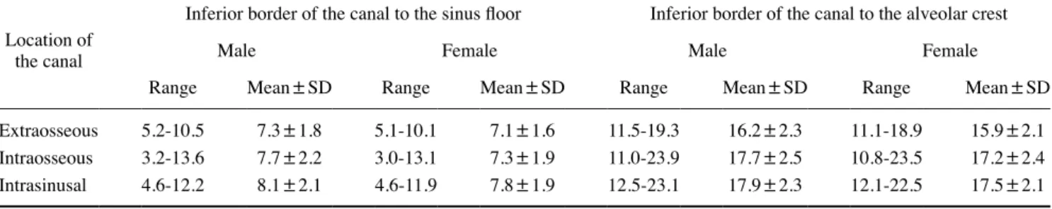
border of the PSA artery canal to the sinus floor was 8.2±2.2mm(range, 3.213.6mm) in males and 7.3±2.1mm (range, 3.013.1mm) in females. The mean distance from the inferior border of the PSA artery canal to the alveolar crest was 18.2±2.7mm(range, 11.023.9mm) in males and 17.4±2.3mm(range, 10.8-23.5mm) in females. The mean diameter of the PSA artery canal was larger in male subjects. The PSA artery canal was bifid in 8.7% of cases. The most frequently observed location of the PSA artery canal was intraosseous(82.2%).
Conclusion: CBCT was confirmed to be a valuable tool for evaluation and localization of the PSA artery before
maxillary sinus lift surgery to avoid intraoperative bleeding.(Imaging Sci Dent 2021; 51: 35-40)
KEY WORDS: ConeBeam Computed Tomography; Maxillary Sinus; Sinus Floor Augmentation; Dental Implants
Copyright ⓒ 2021 by Korean Academy of Oral and Maxillofacial Radiology
This is an Open Access article distributed under the terms of the Creative Commons Attribution NonCommercial License(http://creativecommons.org/licenses/bync/3.0) which permits unrestricted noncommercial use, distribution, and reproduction in any medium, provided the original work is properly cited.
Imaging Science in Dentistry·pISSN 2233-7822 eISSN 2233-7830
Received June 11, 2020; Revised August 17, 2020; Accepted September 9, 2020 *Correspondence to : Dr. Marco Malak Fayek
Department of Oral and Maxillofacial Radiology, Faculty of Dentistry, Minia University, Minia 61111, Egypt
and the location of important anatomical landmarks, inclu ding the PSA artery canal.1 This study was performed to evaluate the presence of the PSA artery canal on CBCT scans in a sample of the Egyptian population.
Materials and Methods
This was a retrospective study using archived CBCT radio graphs of 600 maxillary sinuses from 300 patients(right and left sides), including 152 males and 148 females, who ranged in age from 8 to 77 years. These patients under went CBCT imaging for various purposes, such as dental implant surgery or orthodontic treatment, and the images were obtained from the database of the outpatient clinic of Department of Oral and Maxillofacial Radiology, Faculty of Dentistry, Minia University. The proposal for this study was approved by Research Ethics Committee of the Faculty of Dentistry, Minia University(No. 54) on April 30, 2018.
All scans were obtained using a Scanora® 3Dx CBCT dental unit(Soredex, Tuusula, Finland). The CBCT images were obtained with a slice thickness of 0.5mm. The mea surements were made to the nearest 0.01mm using the caliper tool in OnDemand software(Cybermed Inc., Seoul, Korea). The presence or absence of the PSA artery canal was evaluated using coronalsection scans.
This study only included highquality images showing the complete maxillary sinus area(one or both) in Egyptian patients. Cases with sinus pathology such as a cyst, tumor, or developmental anomaly that caused an alteration in the morphology of the sinus and images from patients who had previously undergone maxillary sinus surgery or experi enced trauma to the walls of the sinuses were excluded. Lowquality images and those that did not show the com plete sinus were also excluded.
The images were all evaluated and assessed for presence or absence of the PSA artery canal along the lateral wall of the maxillary sinus. The location of the PSA artery canal (intraosseous, intrasinusal, or extraosseous) was noted,5 and measurements were made of the diameter of the PSA artery canal(the greatest diameter of the PSA artery was mea sured),6 the distance from the inferior border of the PSA artery canal to the alveolar crest(the closest distance to the ridge was measured),7 the distance from the inferior border of the PSA artery canal to the floor of the maxillary sinus, and the number of missing maxillary posterior teeth. The relationship of the PSA artery canal with age and sex was analyzed, it was noted whether each canal was bifid or not.
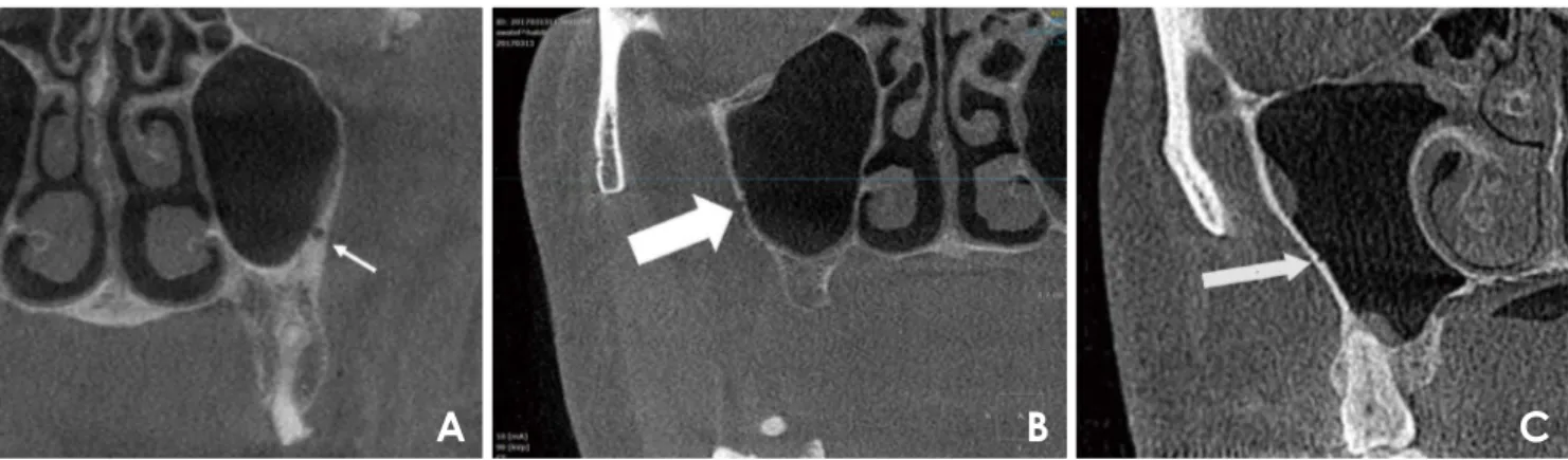
The PSA artery canal locations were classified as follows: 1) intraosseous: inside the lateral wall; 2) extraosseous or superficial: on the outer cortex of the lateral sinus wall; and 3) intrasinusal: below the membrane(Fig. 1). The dia meter of the PSA artery canal was measured using the cali per tool in OnDemand software. The diameter of the canal was recorded as the greatest distance between the inner sides of the cortical borders. Since the artery was visible in more than 1 area, the greatest value between the most posterior and the most anterior position of the canal was recorded as the diameter of the canal(Fig. 2). The diameter was divided into three categories: 1) diameter<1mm, 2) diameter 12mm, and 3) diameter>2mm. The perpen dicular distance from the inferior border of the PSA artery canal to the alveolar crest(the closest distance to the ridge) was measured(Fig. 3). The perpendicular distance from the inferior border of the PSA artery canal to the sinus floor was also measured(Fig. 3).
The patients’ CBCT scans were saved in the Digital Ima ging and Communications in Medicine format. Axial, sagit tal, and coronal images were reformatted using OnDemand
Fig. 1. Conebeam computed tomographic coronal sections show the location of the posterior superior alveolar artery canal. A. Intraosse
ous. B. Extraosseous. C. Intrasinusal.
software. Appropriate background lighting and a color LCD computer screen were used to process the scans. All data about age, sex, number of missing teeth, and the examined side were collected to detect any possible relationships bet ween them and the PSA artery canal.
The data were analyzed using SPSS version 24(IBM Corp., Armonk, NY, USA). Numerical data were expressed as mean and standard deviation. Qualitative data were exp ressed as frequency and percentage. The chisquare test was used to examine relationships between qualitative variables. The KruskalWallis test was used to test the significance of differences in quantitative data. P values<0.05 were con sidered to indicate statistical significance.
Results
The maxillary sinuses were evaluated in a sample of pati ents with a mean age of 34.5±5.2 years(age range, 8-77 years; mean age, 39.2±9.5 years for males and 42±8.7 years for females). The PSA artery canal was observed in 92.0% of the sinuses, with 82.2% of the arteries being intraosseous, 13.8% intrasinusal, and 4.0% extraosseous. The canal diameters were less than 1 mm in 55.8% of the cases, 12mm in 40.2%, and more than 2mm in 4.0%. The diameters ranged from 0.3 to 2.6mm, and the mean dia meter was 1.0±0.5mm(Table 1). The mean distance from the infer ior border of the PSA artery canal to the sinus floor was 8.2±2.2mm(range, 3.213.6mm) in males and 7.3±2.1 mm(range, 3.013.1mm) in females. The mean distance from the inferior border of the PSA artery canal to the alveolar crest was 18.2±2.7mm(range, 11.023.9mm) in males and 17.4±2.3mm(range, 10.8-23.5mm) in fe males(Table 2). The most frequently observed location of PSA artery canal was intraosseous(82.2%). Extraosseous canals showed the largest diameter(1.3mm). The mean di ameter of the PSA artery canal was larger in male subjects (1.0mm) than in female subjects, but the difference was not statistically significant. A slight inverse relationship was found between age and the distance from the inferior border
Table 1. Diameter of the posterior superior alveolar artery canal(unit: mm)
Location of the canal
Male Female Total
Range Mean±SD Range Mean±SD Range Mean±SD
Extraosseous 0.8-2.6 1.4±0.5 0.72.3 1.2±0.5 0.72.6 1.3±0.6
Intraosseous 0.4-2.5 1.1±0.4 0.32.2 0.9±0.3 0.32.4 0.9±0.4
Intrasinusal 0.5-2.2 0.9±0.4 0.42.0 0.8±0.3 0.42.1 0.9±0.4
SD: standard deviation
Fig. 2. Measurement of the diameter of the posterior superior alve
olar artery.
Fig. 3. The perpendicular distance from the inferior border of the
posterior superior alveolar artery canal to the alveolar crest(A) and from the inferior border of the posterior superior alveolar artery canal to the sinus floor(B).
of the PSA artery canal to the ridge. A moderate positive re lationship was found between the distance from the inferior border of PSA artery canal to the sinus floor and the num ber of missing maxillary posterior teeth, which ranged from 1 to 5 missing teeth. The canal was bifid in only 8.7% of cases(28 in male subjects and 24 in female subjects), and bifid canals were only found in the intraosseous type.
Discussion
In this study, the presence of the PSA artery canal was evaluated using CBCT, and it was detected in 92.0% of the 600 sinuses examined. This finding is slightly higher than the prevalence reported by Ilgüy et al.(89.3%),5 Tehranchi et al.(87%),8 and Apostolakis and Bissoon(82%).9 However, our result is slightly lower than that reported by Anamali et al.,1 who found that the prevalence of the PSA artery canal was 94%; these differences might be due to differences in the populations and methodologies among studies, as well as the large sample size that was examined in this study. However, Solar et al.3 and Rosano et al.10 demonstrated that an endosseous anastomosis of the PSA artery and infraor bital artery was present in 100% of cadaveric specimens. This finding indicates that not detecting the PSA artery on CBCT scans does not necessarily mean that it is absent; instead, it might not be visible due to its small diameter or low image resolution.
Moreover, many studies reported lower prevalence rates; for instance, in studies using computed tomography(CT), Elian et al.7 reported a prevalence of 52.9%, Mardinger et al.6 reported a prevalence of 55%, Kim et al.11 reported a prevalence of 52%, Güncü et al.12 reported a prevalence of 64%, and Rosano et al.10 reported a prevalence of 47%. These discrepancies can be attributed to the higher spatial resolution of CBCT than traditional CT imaging, which helps to detect small canals. Furthermore, in a number of previous studies, the identification of the canal was done on images printed on film, such as in the study by Mardinger
et al.,6 which might further limit the ability of the observer to identify the canal.
The most frequent position of the artery in relation to the lateral sinus wall was intraosseous(82.2%), followed by intra sinusal(13.8%) and then extraosseous(4.0%). This finding regarding the position of the PSA artery agrees with those obtained by previous studies.5,12-14
In this study, the diameter of the PSA artery canal was less than 1mm in 55.8% of cases, between 1-2mm in 40.2% of cases, and more than 2mm in 4.0% of cases. These findings align with those of Güncü et al.12, Mardinger et al.6, Rosano et al.,10 and Ilgüy et al.5 However, these findings disagree with those reported by Mardinger et al.6 and Kim et al.,11 who found that only 13.9% of vessels had diameters<1mm using CT scans; this discrepancy could be due to measurement differences between CT and CBCT and the ability of CBCT to detect narrow arteries.
Although the mean diameter of the PSA artery was larger in males than in females, the difference was not statistically significant. These findings agree with those reported by Ilgüy et al.,5 Kim et al.11, and Güncü et al.12
A moderate inverse correlation was found between the patient’s age and the distance from the inferior border of the PSA artery canal to the alveolar crest, which may reflect decreasing bone quantity and quality with age, as older subjects exhibited less volume, regardless of sex and eden tulism. Furthermore, in postmenopausal women, deficiency of estrogen hormone accelerates skeletal bone loss and might result in rapid alveolar bone resorption.15,16
The moderate positive relationship between the distance from the inferior border of the PSA artery canal to the sinus floor with the number of missing posterior teeth might be explained by the possibility that after tooth extraction pneu matization, the sinus size increased at the expense of the alveolar crest.17 Supporting this possibility, Sharan and Madjar18 confirmed the inferior expansion of sinus volume after second molar extraction and after the extraction of 2 or more adjacent posterior teeth.18
Table 2. Distance from the inferior border of posterior superior alveolar artery canal to the sinus floor and to the alveolar crest(unit: mm)
Location of the canal
Inferior border of the canal to the sinus floor Inferior border of the canal to the alveolar crest
Male Female Male Female
Range Mean±SD Range Mean±SD Range Mean±SD Range Mean±SD
Extraosseous 5.2-10.5 7.3±1.8 5.1-10.1 7.1±1.6 11.5-19.3 16.2±2.3 11.1-18.9 15.9±2.1
Intraosseous 3.213.6 7.7±2.2 3.013.1 7.3±1.9 11.023.9 17.7±2.5 10.8-23.5 17.2±2.4
Intrasinusal 4.612.2 8.1±2.1 4.611.9 7.8±1.9 12.5-23.1 17.9±2.3 12.1-22.5 17.5±2.1
In this study, the mean distance from the inferior border of the PSA artery canal to the sinus floor was 7.7±2.2mm, with a higher distance in males than females. These findings agree with those of Güncü et al.12 and Mardinger et al,6 but not with those reported by Ilgüy et al.5 This difference might be explained by anatomic variations in the positions of arteries; furthermore, it has been reported that the maxillary sinus volume was greater in males than in females.19,20
In the present study, the PSA artery canal was bifid in 8.7% of cases, which has not been reported in other studies. Some possible explanations are that 1) the bifid canals were all detected using a small field of view with better resolu tion; 2) other studies6,7,1012,21 used CT, which has lower spatial resolution than that of CBCT used in this study; 3) the sample size of this study was larger than those of pre vious studies; and 4) our cases were examined by dental and maxillofacial radiologists using a computer monitor and the multiplanar capabilities provided by the device’s software, and the radiologists were allowed to identify the canal while rotating the volume using all the capabilities of the software, including changing the window settings, zooming, and filtering to control the sharpness of images. In contrast, in a number of previous studies, canals were identified on images printed on film, which might further limit the observer’s ability to identify the canal, as in the studies conducted by Mardinger et al.6 and Ella et al.21
The narrowest diameters of the canals were found in patients younger than 20 years, and the widest in cases betw een 2040 years; then, the diameters decreased with age, and showed statistically significant differences. An explanation for this may be that the volume of the sinus in creases from birth to the age of 20, and then pneumatization stops and the sinus reaches its final position; subsequently, bone remodeling and narrowing of the canal with age cause the canal diameter to decrease.19,20,22
The intraosseous type of the PSA artery canal was most frequently observed, followed by the intrasinusal type and then the extraosseous type. The extraosseous type had the largest diameter, followed by the intraosseous type and then the intrasinusal type. In patients missing a high number of maxillary posterior teeth, the distance from the inferior border of PSA artery canal to the ridge was shorter. The PSA artery canal was bifid in some cases.
In conclusion, preoperative imaging with CBCT seemed to be helpful for assessing the location and diameter of the PSA artery in order to minimize the risk of membrane per foration and bleeding complications.
Conflicts of Interest: None
References
1. Anamali S, Avila-Ortiz G, Elangovan S, Qian F, Ruprecht A, Finkelstein M, et al. Prevalence of the posterior superior alveolar canal in cone beam computed tomography scans. Clin Oral Imp lants Res 2015; 26: e8-12.
2. Testori T, Rosano G, Taschieri S, Del Fabbro M. Ligation of an unusually large vessel during maxillary sinus floor augmenta tion. A case report. Eur J Oral Implantol 2010; 3: 255-8. 3. Solar P, Geyerhofer U, Traxler H, Windisch A, Ulm C, Watzek
G. Blood supply to the maxillary sinus relevant to sinus floor elevation procedures. Clin Oral Implants Res 1999; 10: 3444. 4. Traxler H, Windisch A, Geyerhofer U, Surd R, Solar P, Firbas
W. Arterial blood supply of the maxillary sinus. Clin Anat 1999; 12: 41721.
5. Ilgüy D, Ilgüy M, Dolekoglu S, Fisekcioglu E. Evaluation of the posterior superior alveolar artery and the maxillary sinus with CBCT. Braz Oral Res 2013; 27: 4317.
6. Mardinger O, Abba M, Hirshberg A, SchwartzArad D. Preval ence, diameter and course of the maxillary intraosseous vas cular canal with relation to sinus augmentation procedure: a radiographic study. Int J Oral Maxillofac Surg 2007; 36: 735-8. 7. Elian N, Wallace S, Cho SC, Jalbout ZN, Froum S. Distribution
of the maxillary artery as it relates to sinus floor augmentation. Int J Oral Maxillofac Implants 2005; 20: 784-7.
8. Tehranchi M, Taleghani F, Shahab S, Nouri A. Prevalence and location of the posterior superior alveolar artery using cone beam computed tomography. Imaging Sci Dent 2017; 47: 39 44.
9. Apostolakis D, Bissoon AK. Radiographic evaluation of the superior alveolar canal: measurements of its diameter and of its position in relation to the maxillary sinus floor: a cone beam computerized tomography study. Clin Oral Implants Res 2014; 25: 553-9.
10. Rosano G, Taschieri S, Gaudy JF, Weinstein T, Del Fabbro M. Maxillary sinus vascular anatomy and its relation to sinus lift surgery. Clin Oral Implants Res 2011; 22: 711-5.
11. Kim JH, Ryu JS, Kim KD, Hwang SH, Moon HS. A radio graphic study of the posterior superior alveolar artery. Implant Dent 2011; 20: 30610.
12. Güncü GN, Yildirim YD, Wang HL, Tözüm TF. Location of posterior superior alveolar artery and evaluation of maxillary sinus anatomy with computerized tomography: a clinical study. Clin Oral Implants Res 2011; 22: 11647.
13. Kang SJ, Shin SI, Herr Y, Kwon YH, Kim GT, Chung JH. Ana tomical structures in the maxillary sinus related to lateral sinus elevation: a cone beam computed tomographic analysis. Clin Oral Implants Res 2013; 24 Suppl A100: 75-81.
14. DaneshSani SA, Movahed A, ElChaar ES, Chong Chan K, Amintavakoli N. Radiographic evaluation of maxillary sinus lateral wall and posterior superior alveolar artery anatomy: a conebeam computed tomographic study. Clin Implant Dent Relat Res 2017; 19: 151-60.
15. Velasco-Torres M, Padial-Molina M, Avila-Ortiz G, García-Del gado R, O̓Valle F, Catena A, et al. Maxillary sinus dimensions decrease as age and tooth loss increase. Implant Dent 2017; 26: 288-95.
16. Ural Ç Bereket C, Şener I, Aktan AM, Akpinar YZ. Bone height measurement of maxillary and mandibular bones in panoramic radiographs of edentulous patients. J Clin Exp Dent 2011; 3: e5-9.
17. Trimarchi M, Lombardi D, Tomenzoli D, Farina D, Nicolai P. Pneumosinus dilatans of the maxillary sinus: a case report and review of the literature. Eur Arch Otorhinolaryngol 2003; 260: 386-9.
18. Sharan A, Madjar D. Maxillary sinus pneumatization following extractions: a radiographic study. Int J Oral Maxillofac Implants 2008; 23: 48-56.
19. Rani SU, Rao GV, Kumar DR, Sravya T, Sivaranjani Y, Kumar MP. Age and gender assessment through threedimensional
morphometric analysis of maxillary sinus using magnetic reso nance imaging. J Forensic Dent Sci 2017; 9: 46.
20. Takahashi Y, Watanabe T, Iimura A, Takahashi O. A study of the maxillary sinus volume in elderly persons using Japanese cadavers. Okajimas Folia Anat Jpn 2016; 93: 217.
21. Ella B, Sédarat C, Noble Rda C, Normand E, Lauverjat Y, Si berchicot F, et al. Vascular connections of the lateral wall of the sinus: surgical effect in sinus augmentation. Int J Oral Maxillofac Implants 2008; 23: 1047-52.
22. Compston J. Agerelated changes in bone remodelling and structure in men: histomorphometric studies. J Osteoporos 2011; 2011: 108324.