0270-7306/07/$08.00⫹0 doi:10.1128/MCB.01919-06
Copyright © 2007, American Society for Microbiology. All Rights Reserved.
Luteinizing Hormone-Dependent Activation of the Epidermal Growth
Factor Network Is Essential for Ovulation
䌤
Minnie Hsieh,
1Daekee Lee,
2‡ Sara Panigone,
1† Kathleen Horner,
1Ruby Chen,
1Alekos Theologis,
1David C. Lee,
3§ David W. Threadgill,
2and Marco Conti
1*
Division of Reproductive Biology, Department of Obstetrics and Gynecology, Stanford University School of Medicine, Stanford, California 943051; Department of Genetics, University of North Carolina, Chapel Hill, North Carolina 275992; and
Department of Biochemistry and Biophysics, University of North Carolina, Chapel Hill, North Carolina 275993
Received 10 October 2006/Returned for modification 1 November 2006/Accepted 14 December 2006
In the preovulatory ovarian follicle, mammalian oocytes are maintained in prophase meiotic arrest until the luteinizing hormone (LH) surge induces reentry into the first meiotic division. Dramatic changes in the somatic cells surrounding the oocytes and in the follicular wall are also induced by LH and are necessary for ovulation. Here, we provide genetic evidence that LH-dependent transactivation of the epidermal growth factor receptor (EGFR) is indispensable for oocyte reentry into the meiotic cell cycle, for the synthesis of the extracellular matrix surrounding the oocyte that causes cumulus expansion, and for follicle rupture in vivo. Mice deficient in either amphiregulin or epiregulin, two EGFR ligands, display delayed or reduced oocyte maturation and cumulus expansion. In compound-mutant mice in which loss of one EGFR ligand is associated with decreased signaling from a hypomorphic allele of the EGFR, LH no longer signals oocyte meiotic resumption. Moreover, induction of genes involved in cumulus expansion and follicle rupture is compromised in these mice, resulting in impaired ovulation. Thus, these studies demonstrate that LH induction of epidermal growth factor-like growth factors and EGFR transactivation are essential for the regulation of a critical physiological process such as ovulation and provide new strategies for manipulation of fertility.
The luteinizing hormone (LH) surge plays a central role in promoting a cascade of events in ovarian preovulatory follicles that are essential for the ovulation of a fertilizable oocyte. Acting through LH-chorionic gonadotropin (LH-CG) recep-tors (LHRs) (LHR is a member of the G protein-coupled receptor superfamily encoded by Lhcgr), LH induces repro-gramming of the gene expression profiles of follicular somatic cells (theca and granulosa cells), changes in the secretory prop-erties of the cumulus cells surrounding the oocyte and cumulus expansion, oocyte reentry into the meiotic cell cycle, and fol-licle rupture (7, 41). LHRs are highly expressed on the gran-ulosa cells lining the antral cavity of preovulatory follicles (mu-ral granulosa cells) and on the external theca cells that are in continuity with the surrounding stroma. However, within pre-ovulatory follicles, oocytes and cumulus cells that are pro-foundly affected by the LH surge express few or no LHRs and fail to respond when directly exposed to LH in vitro (37).
To explain how LH signals are propagated from the periph-ery toward the cumulus oocyte complex (COC), a model has been proposed whereby factors released by mural granulosa
cells function in an autocrine and paracrine manner to trans-duce the LH effects within the follicle (34). Secretion of bio-active growth factors from the oocyte to affect somatic cells is well established (27, 30); conversely, the paracrine signals orig-inating from the somatic cells and affecting oocytes have long been sought but are largely unknown. Recently, we have pro-posed that intrafollicular release of members of the epidermal growth factor (EGF)-like family (34) may fulfill this role. LH or human CG (hCG) induces the rapid and transient expres-sion of Areg, Ereg, and Btc (encoding amphiregulin [AREG], epiregulin [EREG], and betacellulin [BTC], respectively) in mural granulosa cells of mouse (34) and rat preovulatory fol-licles (1, 48) and in human granulosa cell cultures (9). AREG, EREG, and BTC belong to the family of EGF-like growth factors synthesized as integral membrane precursors. The li-gands are shed from the cell surface by proteolytic cleavage of the ectodomain and bind to homo- or heterodimers of the EGF receptor (EGFR) family of receptor tyrosine kinases (18, 58). AREG binds specifically to EGFR (also called ERBB1), while EREG and BTC can bind both EGFR and ERBB4 (19, 42, 43). Importantly, data from culture systems have docu-mented that occupancy of G protein-coupled receptors (GPCRs) often leads to shedding of EGF-like growth factors and transactivation of EGFRs (13, 38, 46, 51).
Most of the current data on EGF-like growth factor actions in the ovary are derived from in vitro studies. In cultured preovulatory follicles, exogenous AREG or EREG induces oocyte maturation as efficiently as LH, whereas BTC is only partially effective (34). In vitro, AREG, EREG, and BTC each induce the expression of Ptgs2 (prostaglandin synthase-2 or cyclooxygenase-2 [COX2]), Tnfaip6 (tumor necrosis factor al-pha-induced protein [Tnfaip6]), and Has2 (hyaluronan
syn-* Corresponding author. Mailing address: Division of Reproductive Biology, Department of Obstetrics and Gynecology, Stanford Univer-sity School of Medicine, 300 Pasteur Drive, Stanford, CA 94305-5317. Phone: (650) 725-2452. Fax: (650) 725-7102. E-mail: marco.conti @stanford.edu.
† Present address: Laboratory of Endocrinological Research, Isti-tuto Auxologico Italiano (IRCCS), Via Zucchi 18, Cusano Milanino (MI) 20095, Italy.
‡ Present address: Division of Molecular Life Sciences, Ewha Womans University, Seoul 120-720, South Korea.
§ Present address: The Office of the Vice President for Research, University of Georgia, Athens, GA 30602.
䌤Published ahead of print on 28 December 2006.
1914
on October 21, 2016 by Ewha Womans Univ
http://mcb.asm.org/
thase 2 [HAS2]), genes that are necessary for synthesis and stabilization of the extracellular matrix by cumulus cells, and are potent stimulators of cumulus expansion. Cultured COCs expand in response to direct stimulation by the EGF-like fac-tors but not by LH. Inhibition of EGFR tyrosine kinase activity by AG1478 blocks LH-induced oocyte maturation and cumulus expansion in cultured follicles. Together, these studies strongly suggest that these EGF-like growth factors are sufficient to promote many of the LH effects in vitro.
Nevertheless, a physiological role for these growth factors in vivo has not been established. Mice null for either Areg or Ereg have been previously reported to be fertile (21, 26). Mice null for Egfr die at peri-implantation, at mid-gestation, or soon after birth, depending on the strain, thus precluding analysis of fertility (50, 52). Mice that express a hypomorphic allele of
Egfrwa2(waved-2) are viable, but an effect on fertility is
con-troversial (8, 25). Egfrwa2 homozygous mice that express a
receptor with reduced tyrosine kinase activity due to a single amino acid substitution in the kinase domain (V743G) have been considered fertile. In other reports, although reduced litter sizes have been described for the Egfrwa2homozygous
mice, this was thought to be due to impaired glial cell control of LH-releasing hormone secretion (39). Therefore, evidence for a physiological role of this signaling network in LH-induced ovulation in vivo is lacking.
To determine the involvement of the EGF network in LH-induced ovulation, we have investigated the effect of single and multiple disruptions of the EGF network on events preceding and following the time of ovulation in mice in a controlled model of hormone-induced follicle maturation. Because dou-ble or triple knockouts for Areg, Ereg, and Btc would be difficult to produce due to the tight linkage of the three genes on the same chromosome (36), alternative approaches were sought to further disrupt the EGF network in mice. To this end, double-mutant mice null for Areg and homozygous for the Egfrwa2
allele were generated (Areg⫺/⫺Egfrwa2/wa2). Here, we provide
genetic evidence that LH-induced oocyte reentry into the mei-otic cell cycle, expression of specific genes during somatic cell differentiation, and ovulation are all dependent on efficient activation of signaling through the EGFR.
MATERIALS AND METHODS
Animals and hormone treatments.Amphiregulin-null (Areg⫺/⫺) mice (C57BL/6J⫻ 129Sv) were generated in the laboratory of David C. Lee (21, 26). Epiregulin-null
mice (Eregtm1Dwt, herein referred to as Ereg⫺/⫺) (21, 26) and Egfrwa2 mice
(C57BL/6J) were developed in the laboratory of David W. Threadgill. Areg⫺/⫺
mice were mated with wa2 heterozygous mice to generate double heterozygous
mice (Areg⫹/⫺Egfrwa2/⫹). Double heterozygous mice were then mated to
pro-duce offspring of the desired genotypes (Areg⫹/⫹Egfr⫹/⫹, Areg⫹/⫹Egfrwa2/wa2,
Areg⫺/⫺Egfrwa2/wa2
).
Areg⫺/⫺mice were genotyped by PCR analysis using specific primers: AR-F1
(5⬘-CTTTCCAGCTTTCTCCACCTCAAG-3⬘), AR-R1 (5⬘-ACAGTAACCTCT
GTTGCATGCCAC-3⬘), and PGK2 (5⬘-CTGCACGAGACTAGTGAGACGTG
C-3⬘). The sizes for the Areg wild-type (WT) (AR-F1/AR-R1) and null (AR-F1/ PGK2) PCR-amplified products are 330 bp and 600 bp, respectively. Reverse transcription-PCR (RT-PCR) analysis using Areg-specific primers (34) confirmed
the absence of Areg mRNA in Areg⫺/⫺ovaries. Egfrwa2mice were genotyped by
PCR amplification of the region containing the point mutation that results in a valine to glycine substitution at residue 743 by use of the specific primer pair Wa2-F (5⬘-TACCCAGAAAGGGATATGCG-3⬘) and Wa2-R (5⬘-GGAGCCA
ATGTTGTCCTTGT-3⬘). The PCR product was digested with the FokI
restric-tion enzyme to detect the mutarestric-tion (Egfr⫹/⫹, 230 bp; Egfrwa2/wa2
, 100 bp and 130 bp).
Immature female mice (22 to 24 days old) were injected intraperitoneally with 5 IU of pregnant mare serum gonadotropin (PMSG; Calbiochem, La Jolla, CA) to stimulate ovarian follicular growth to the preovulatory stage. After 44 h, mice were injected intraperitoneally with an ovulatory dose of 5 IU of hCG (Sigma-Aldrich, St. Louis, MO) (17). Ovaries were isolated at selected times after hormone priming and processed for histological analyses or were used as de-scribed below and in the figure legends. For histological analyses, ovaries were fixed in Bouin’s fixative (Polysciences, Inc., Warrington, PA) overnight at 4°C,
dehydrated and embedded in paraplast, sectioned serially at 6m onto
Super-frost Plus slides (Fisher Scientific, Pittsburgh, PA), stained with hematoxylin and eosin, and analyzed by light microscopy. For superovulation experiments, oocytes were recovered from the oviducts 13 to 14 h or 22 to 24 h after hCG and counted (17). Superovulated ovaries were also fixed as described above for histological analyses. Because the numbers of ovulated oocytes for a given genotype did not differ at either time point, the data were pooled. All animals were treated in accordance with the guidelines of the Stanford University Administrative Panel on Laboratory Animal Care.
Evaluation of oocyte meiotic resumption.Ovaries were isolated from imma-ture female mice 0, 1, 2, 3, 4, and 5 h after hCG stimulation in vivo and placed in Liebovitz’s L-15 medium (Invitrogen, Grand Island, NY) containing 5% fetal
bovine serum (Invitrogen) and 100 U penicillin and 100g streptomycin
(Sigma-Aldrich) (L-15 complete medium). Preovulatory follicles were punctured using 26-1/2-gauge needles (Becton Dickinson & Co., Franklin Lakes, NJ) to release COCs. COCs were collected using a Drummond sequencing pipette, and oocytes were denuded of cumulus cells by gentle repeat pipetting in a microcentrifuge tube. COCs collected at 4 h or later were briefly cultured in L-15 complete
medium containing 1g/ml hyaluronidase to facilitate denuding of oocytes.
Denuded oocytes were examined under a stereomicroscope for evidence of dissolution of the oocyte nuclear membrane (a process called germinal vesicle breakdown [GVBD]), a hallmark of oocyte meiotic resumption. Oocytes within large antral follicles were also examined through serial sections of hematoxylin-and eosin-stained ovaries fixed 9 h after hCG (see below) for GVBD. The percentage of GVBD oocytes was recorded and averaged at each time point.
To determine the time course of spontaneous oocyte maturation in vitro,
COCs were released from Areg⫹/⫹Egfr⫹/⫹ and Areg⫺/⫺ Egfrwa2/wa2
PMSG-primed mouse ovaries by needle puncture and oocytes were denuded as
de-scribed above. Oocytes were cultured in 30-l droplets of L-15 complete medium
under mineral oil at 37°C in air and were examined at regular time intervals for evidence of complete GVBD. The total number of oocytes that completed GVBD was recorded at each time point and is reported as percent GVBD. The time that oocytes were first released from preovulatory follicles by needle punc-ture was designated time 0.
Evaluation of cumulus expansion.Mouse ovaries were isolated 9 h after hCG stimulation, fixed in Bouin’s fixative (Polysciences Inc.), and processed as de-scribed above. COCs within large antral follicles were examined through serial
sections of hematoxylin- and eosin-stained ovaries. A subjective 0 to⫹4 scoring
system was used to evaluate the degree of cumulus expansion for each complex
(55). Unexpanded complexes received a score of 0 to⫹1. Complexes in which
the outer layers of cumulus cells had begun to expand received a score of⫹2. A
score of⫹3 was indicative of complexes in which all layers except the corona
radiata (the layer immediately adjacent to the oocyte) had expanded, and
max-imally expanded complexes were scored⫹4. Ovaries from at least four mice were
analyzed for each genotype.
Semiquantitative RT-PCR.RNA was extracted from whole mouse ovaries by use of TRIzol reagent (Invitrogen) and from COCs by use of an RNeasy Micro kit (QIAGEN, Valencia, CA) as directed by the respective manufacturers. For analysis of Ptgs2, Tnfaip6, and Has2 expression, the 3 h time point was selected because increased expression of these genes was previously observed in cultured ovarian follicles (33) and in cumulus cells (unpublished observations). Total RNA (300 ng from whole ovaries and 100 ng from COCs) was reverse
tran-scribed using 1⫻ thermocycle buffer, 3.75 mM MgCl2, 1 mM deoxynucleoside
triphosphates (Promega Corp., Madison, WI), 500 ng poly(dT) (Amersham Pharmacia, Newark, NJ), and 2.5 U avian myeloblastosis virus–reverse transcrip-tase (Promega Corp.) at 42°C for 75 min and 95°C for 5 min. After completion of the RT reactions, 1/4 of each reaction was used for PCR amplification of the
genes of interest. The following were added: 0.1M of each primer, 0.25 Ci
[32P]deoxy-CTP (Perkin Elmer, Boston, MA), and 0.625 U Taq polymerase in
1⫻ thermocycle buffer–2 mM MgCl2(Promega Corp.). PCR conditions were
94°C for 2 min followed by cycles of 94°C for 30 s, 60°C for 45 s, and 72°C for 1 min and a final extension at 72°C for 7 min. PCR amplifications using specific primers were performed for the indicated number of cycles: Ptgs2 (COX2), 25 cycles (32); Tnfaip6 (Tnfaip6), 24 cycles (32); Has2 (HAS2), 28 cycles (6); Lhcgr (LHR), sense, 25 cycles (6); and Rpl19 (L19), 20 cycles (35, 45). The amplified
on October 21, 2016 by Ewha Womans Univ
http://mcb.asm.org/
cDNA products were resolved on a 5% polyacrylamide gel and quantified using a Storm 840 PhosphorImager and ImageQuant software (Molecular Dynamics, Inc., Sunnyvale, CA).
Preovulatory follicle culture and cAMP radioimmunoassay.PMSG-primed mouse ovaries were placed in L-15 complete medium, and preovulatory follicles were microdissected under a stereomicroscope using fine forceps. The isolated follicles were moved through fresh L-15 complete medium and three changes of minimal essential medium (MEM) supplemented with 10% fetal bovine serum
(Invitrogen) and 100 U penicillin and 100g streptomycin (Sigma-Aldrich).
Follicles were then cultured in 1 ml complete MEM sealed under conditions of
95% O2–5% CO2in glass scintillation vials at 37°C. After equilibration, follicles
were cultured for 30 min in the absence or presence of 5 IU recombinant LH (rLH) (Luveris; Serono, Rockland, MA). At the end of the incubation, follicles were first transferred into 2 ml Dulbecco’s phosphate-buffered saline (DPBS)
with Ca2⫹and Mg2⫹containing 0.5 mM 3-isobutyl-1-methylxanthine
(Sigma-Aldrich) and then moved into a small volume of DPBS–0.5 mM 3-isobutyl-1-methylxanthine for needle puncture. Samples were collected in 1.5 ml Eppendorf tubes and incubated for 5 min at 95°C, and then 1 ml 95% ethanol–0.1% trichloroacetic acid was added. After 10 min centrifugation, supernatants were transferred to new tubes, dried in a speed vacuum, and resuspended in 1 ml DPBS. An aliquot of each sample was diluted in DPBS and acetylated, and cyclic AMP (cAMP) content was measured by radioimmunoassay (14).
Immunoprecipitation and Western blot analyses.Preovulatory follicles from PMSG-primed mouse ovaries were isolated in L-15 complete medium and cul-tured in 1 ml complete MEM as described above. Thirty-five follicles per treat-ment group were cultured in the absence or presence of rLH for 2 h. At the end
of the incubation, follicles were transferred into DPBS without Ca2⫹and Mg2⫹
to remove residual medium. The follicles were then homogenized in 300l of
ice-cold radioimmunoprecipitation assay buffer (50 mM Tris [pH 7.5], 150 mM NaCl, 1% NP-40, 0.1% sodium dodecyl sulfate [SDS], 0.25% deoxycholic acid) containing an EDTA-free protease inhibitor cocktail (Roche, Indianapolis, IN), 1 mM EDTA, 1 mM NaF, 1 mM sodium pyrophosphate, and 1 mM sodium orthovanadate to extract proteins. After centrifugation for 5 min at maximum
speed at 4°C, the supernatants were collected and stored at⫺80°C.
Protein concentrations were quantified using a bicinchoninic acid protein assay kit (Pierce, Rockford, IL). A constant amount of proteins was
immunoprecipi-tated with 4g sheep polyclonal anti-EGFR antibody (Upstate, Lake Placid,
NY) overnight at 4°C. The next day, samples were incubated with protein G-Sepharose for 3 h at 4°C and then washed twice in phosphate-buffered saline containing protease inhibitors as described above. Proteins were eluted from the immunoprecipitated pellet by resuspending in 3⫻ SDS buffer and boiling for 5 min.
Immunoprecipitated protein samples were run on an SDS-polyacrylamide electrophoresis gel and transferred onto Immobilon-P membranes (Millipore Corp., Bedford, MA) by electrophoresis. Membranes were blocked in 5% nonfat dry milk (Bio-Rad, Hercules, CA) for 1 h at room temperature, washed in Tris-buffered saline containing 0.1% Tween 20 (TBST), and incubated overnight at 4°C with rabbit polyclonal anti-phospho-EGFR antibody (pY1068; Cell Sig-naling Technology, Beverly, MA) diluted 1:1,000 in TBST containing 5% bovine serum albumin. Next, the membranes were washed in TBST and then incubated for 1 h at room temperature with anti-rabbit immunoglobulin peroxidase-linked antibody (Amersham) diluted 1:40,000 in TBST containing 0.5% nonfat dry milk (Bio-Rad). After membranes were washed in TBST, ECL Plus reagent (Amer-sham) was used to detect the immunoreactive phospho-EGFR and the signal was visualized by autoradiography.
The same membranes were incubated for 30 min at 37°C with agitation in a
solution containing 62.5 mM Tris (pH 6.8), 2% SDS, and 100 mM
-mercapto-ethanol to remove bound antibodies. After being washed in TBST and blocked in 1% nonfat dry milk, membranes were incubated for 2 h at room temperature with rabbit polyclonal anti-EGFR antibody (Santa Cruz Biotechnology, Santa Cruz, CA) diluted 1:200 in TBST containing 0.2% nonfat dry milk (Bio-Rad). Next, membranes were washed in TBST, incubated for 1 h at room temperature with anti-rabbit immunoglobulin G peroxidase-linked antibody (Amersham) di-luted 1:5,000 in TBST containing 0.2% nonfat dry milk (Bio-Rad), and detected as above.
In situ hybridization.A riboprobe in vitro transcription system kit (Promega)
was used to synthesize [35
S]UTP-labeled antisense and sense RNA probes from
Ptgs2 and Cyp11a1 cDNAs that were subcloned in the pCR2-TOPO and
pBlue-script vectors, respectively. In situ hybridization was performed as previously described (34).
Statistical analyses.All data are shown as the means⫾ standard errors of the means (SEM). Data were analyzed by one-way analysis of variance and
Bonfer-roni’s multiple comparison test or by Student’s t test where appropriate. P⬍ 0.05
was considered statistically significant.
RESULTS
Delayed onset of oocyte meiotic maturation and reduced cumulus expansion in amphiregulin- and epiregulin-null fe-male mice.To address the role of the EGF network in vivo, we investigated how meiotic resumption and differentiation of so-matic cells surrounding the oocyte proceed in genetic models where the EGF network is progressively disrupted. To this aim, immature female mice homozygously null for the Areg allele (Areg⫺/⫺) or the Ereg allele (Ereg⫺/⫺) were primed with PMSG to promote follicle development to the preovulatory stage. After 44 h, mice were stimulated with hCG to induce terminal differentiation and ovulation, and the responses that ensued were investigated. In this model, normally oocyte meiotic resump-tion occurs by 4 h, cumulus expansion by 9 h, and ovularesump-tion approximately 12 h after hCG stimulation. This well-established and extensively used model of exogenous administration of go-nadotropins was chosen to avoid interference from any possible extragonadal effects of the EGF network.
In wild-type and heterozygous (Areg⫹/⫺) littermates of
Areg⫺/⫺mice, oocytes reenter the meiotic cell cycle between 2 and 4 h after hCG (Fig. 1A). In Areg⫺/⫺mice, meiotic reentry still occurs but with a delayed time course (Fig. 1A), suggesting that the transfer of signals from the somatic to the germ cell compartment occurs with decreased efficiency. By 9 h, most of the oocytes in the Areg⫺/⫺ antral follicles had undergone GVBD (Table 1). When oocytes in antral follicles of epiregu-lin-null (Ereg⫺/⫺) mouse ovaries were monitored for meiotic resumption 8 h after hCG, only⬃60% of those examined had resumed meiosis (Table 1).
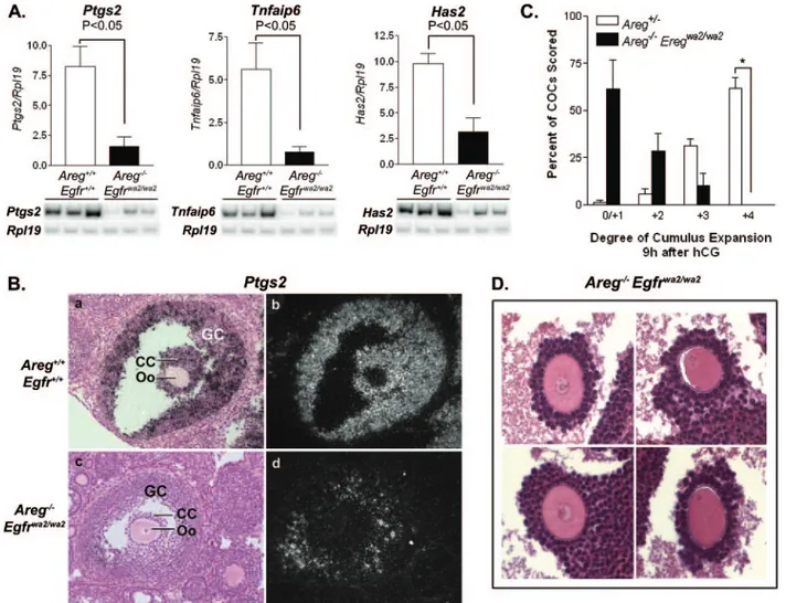
The expression of genes important for cumulus matrix for-mation and stability was examined in COCs from Areg⫺/⫺ ovaries collected 3 h after hCG and compared to that in COCs from Areg⫹/⫺mice. By semiquantitative RT-PCR, reduced ex-pression of Ptgs2, Tnfaip6, and Has2 was detected in Areg⫺/⫺ COCs (Fig. 1B). This inefficient activation of genes involved in matrix production was confirmed by visual scoring of the state of expansion of the cumulus in vivo. COCs in preovulatory follicles were examined through serial sections of hematoxylin-and eosin-stained Areg⫹/⫺and Areg⫺/⫺mouse ovaries isolated 9 h after hCG. In Areg⫹/⫺ ovaries, the majority of COCs examined appeared well expanded (Fig. 1C and D). However, the degree of expansion for COCs from Areg⫺/⫺ovaries was reduced (Fig. 1C and D). A similar or more pronounced effect was observed with the Ereg⫺/⫺ mice (Fig. 1D and data not shown).
The results presented above indicate that removal of a single EGF-like growth factor has an impact on events critical during the periovulatory period. However, the effects are small and disruption of meiotic resumption and cumulus expansion is incomplete likely because of the redundancy in the EGF net-work.
Development of ovarian follicles in the preovulatory stage in Aregⴚ/ⴚEgfrwa2/wa2
mice.To explore the possibility of a redun-dant function, we investigated the effect of multiple defects in the EGF network on events during ovulation. To further dis-rupt the EGF network, Areg⫺/⫺mice were bred to mice
on October 21, 2016 by Ewha Womans Univ
http://mcb.asm.org/
rying the hypomorphic Egfrwa2allele to generate
double-mu-tant mice (Areg⫺/⫺Egfrwa2/wa2). These mice were compared to
mice that were wild type with respect to the two alleles. In view of the reports of extragonadal effects of the EGFR (23, 39) and the possible impact of EGF on follicle develop-ment (12, 29, 47), several paradigms were used to determine whether early follicular development was affected in the
Areg⫺/⫺ Egfrwa2/wa2mice. Double-mutant mice had no overt
defect in growth and viability, ovaries after PMSG priming were of the normal size, and uteri appeared swollen, indicating a normal steroidogenic output from the gonad (data not shown). Ovaries from PMSG-primed Areg⫺/⫺Egfrwa2/wa2mice
appeared morphologically identical to those of wild-type con-trols and contained follicles at different stages of development, including many at the preovulatory stage (Fig. 2A). In these antral follicles, the granulosa cell layer had thickness compa-rable to wild-type controls. Moreover, oocytes isolated from
FIG. 1. Delayed onset of oocyte meiotic maturation and reduced cumulus expansion in Areg⫺/⫺mice. (A) Time course of breakdown of the oocyte nuclear membrane (GVBD) in wild-type and Areg⫺/⫺mice. In vivo-stimulated ovaries were collected at the indicated times after hCG, and large antral follicles were punctured to release the oocytes. Data are shown as means⫾ SEM, and three to seven females were used at each time point. The percentage of oocytes that resumed meiosis was significantly lower in Areg⫺/⫺mice at 3 h and 4 h after hCG than in WT/Areg⫹/⫺females. (B) Pattern of gene expression in Areg⫺/⫺COCs. Semiquantitative RT-PCR analysis was performed to evaluate Ptgs2, Tnfaip6, and Has2 mRNA expression levels in COCs isolated from Areg⫹/⫺and Areg⫺/⫺mice stimulated with hCG for 3 h. Data are shown as the means⫾ SEM of the relative expression levels for Ptgs2, Tnfaip6, or Has2 normalized to Rpl19 (Areg⫹/⫺, n⫽ 7; Areg⫺/⫺, n ⫽ 10). (C) Morphological analysis of cumulus expansion in Areg⫺/⫺mice. The degree of cumulus expansion was evaluated for COCs in preovulatory follicles of ovaries stimulated with hCG for 9 h by use of a subjective 0 to⫹4 scoring system (55). Ovaries from four different females were analyzed for each genotype. The data are shown as means⫾ SEM.*, P⬍ 0.001. (D) Histology of COCs in Areg⫹/⫺and Areg⫺/⫺preovulatory follicles 9 h after hCG and Ereg⫺/⫺follicles 8 h after hCG. In these selected COCs, the metaphase spindle is visible in the oocytes. Areg⫹/⫺COCs are well expanded, whereas COCs from Areg⫺/⫺and Ereg⫺/⫺mice appear moderately or poorly expanded.
TABLE 1. Oocyte meiotic resumption in mouse ovaries upon hCG stimulationa Genotype No. of h after hCG Total no. of oocytes in preovulatory follicles examined % GVBD Areg⫹/⫺ 9 85 98.8⫾ 1.4 Areg⫺/⫺ 9 74 83.2⫾ 9.1 Ereg⫺/⫺ 8 45 62.2b
Areg⫹/⫹Egfrwa2/wa2 9 79 29.3⫾ 14.1 Areg⫺/⫺Egfrwa2/wa2 9 115 24.0⫾ 13.5
aFor all mice except the Ereg⫺/⫺mice, the percentage of oocytes that had
undergone GVBD was calculated for each mouse (n⫽ 4 except Areg⫹/⫹Egfrwa2/wa2
mice, for which n⫽ 6) and averaged. Data are shown as means ⫾ SEM.
bThe percentage of GVBD oocytes in Ereg⫺/⫺ovaries was calculated by
dividing the number of oocytes in GVBD by the total number of oocytes exam-ined in sections of ovaries from 10 mice. Because of the method of collection of the specimens, percent GVBD for individual mice could not be assessed.
on October 21, 2016 by Ewha Womans Univ
http://mcb.asm.org/
preovulatory follicles of PMSG-primed Areg⫺/⫺ Egfrwa2/wa2
mouse ovaries underwent in vitro spontaneous maturation at a rate similar to that seen with oocytes from wild-type mice (Fig. 2B), demonstrating that oocyte meiotic competence is normal in these mice. By semiquantitative RT-PCR analysis, the re-sults of induction of Lhcgr expression, a hallmark of granulosa cell differentiation towards a preovulatory phenotype, were comparable between PMSG-primed Areg⫺/⫺ Egfrwa2/wa2 and
wild-type ovaries (Fig. 2C). When preovulatory follicles were cultured in the presence of rLH for 30 min, the increase in cAMP induced in Areg⫺/⫺Egfrwa2/wa2follicles was comparable
to that in wild-type follicles (Fig. 2D). Taken together, these findings demonstrate that disruption of the EGF network does not produce detectable changes in the growth and initial
dif-ferentiation of somatic and germ cells of the follicle and that the ability to respond to LH with an increase in cAMP is retained.
Impaired EGFR transactivation by LH in double-mutant mice.To examine the effect of LH on activation of the EGFR, preovulatory follicles from wild-type (Areg⫹/⫹ Egfr⫹/⫹),
Areg⫹/⫹Egfrwa2/wa2, or Areg⫺/⫺Egfrwa2/wa2mice were cultured
in the absence or presence of rLH for 2 h. Immunoprecipita-tion and Western blot analyses were performed to detect EGFR phosphorylation. LH induced a progressive increase in EGFR phosphorylation in wild-type follicles, with a maximum reached after 2 h of stimulation (Fig. 3A and data not shown). However, EGFR phosphorylation levels were reduced by more than 50% in Areg⫹/⫹Egfrwa2/wa2follicles by comparison and FIG. 2. Development of ovarian follicles up to the preovulatory stage in Areg⫺/⫺Egfrwa2/wa2mice. (A) Histological analyses of PMSG-primed Areg⫺/⫺ Egfrwa2/wa2mouse ovaries showed that many large antral follicles develop in response to the hormone stimulation. (B) Spontaneous maturation of oocytes (scored as percent GVBD) isolated from preovulatory follicles of PMSG-primed Areg⫺/⫺Egfrwa2/wa2mice occurs in culture in vitro at a rate similar to that seen with wild-type oocytes (n⫽ 4 for each genotype). (C) Similar levels of Lhcgr mRNA are expressed in Areg⫺/⫺ Egfrwa2/wa2and wild-type ovaries as determined by semiquantitative RT-PCR. Data are means⫾ SEM of the relative expression levels for Lhcgr normalized to Rpl19 (n⫽ 3 for each genotype). (D) Preovulatory follicles from Areg⫺/⫺Egfrwa2/wa2mice cultured in the presence of rLH for 30 min respond with a normal increase in cAMP. Data represent the means⫾ SEM of the results of three separate experiments.
on October 21, 2016 by Ewha Womans Univ
http://mcb.asm.org/
were further decreased in Areg⫺/⫺ Egfrwa2/wa2 follicles, with
only⬃20% of that detected in the wild-type follicles (Fig. 3A). Thus, a major disruption in LH-induced transactivation of the EGFR was indeed present in the mutant mice. When double-mutant follicles were incubated with high doses of EREG and AREG, tyrosine phosphorylation of EGFR was restored to wild-type levels (data not shown), suggesting that a
supraphysi-ological signal is able to partially restore the response of the mutated EGFR.
Impaired oocyte meiotic resumption in mice with disrupted EGFR signaling.Double-mutant immature mice were primed with PMSG, and the effect of hCG on oocyte meiosis was monitored at 4 h after injection, a time when significant effects were detected in the Areg⫺/⫺ mice. In the double mutants, most of the oocytes (90%) had not resumed meiosis (Fig. 3B). Furthermore, this loss of function was proportional to the disruption of the EGF network, as Areg⫺/⫺ oocytes were the least affected, Areg⫹/⫹ Egfrwa2/wa2 oocytes showed a greater
than 50% decrease in GVBD, and combination of the two lesions produced the maximal effect (Fig. 3B). By 9 h post-hCG, only⬃25% of the double-mutant oocytes examined had resumed meiosis (Table 1), demonstrating that major disrup-tion of EGFR signaling prevents signaling for oocyte matura-tion. This in vivo finding was confirmed by culture of preovu-latory follicles from the Areg⫺/⫺Egfrwa2/wa2mice. Stimulation
with rLH failed to induce GVBD in oocytes from these follicles (0 oocytes out of 21 monitored). Conversely, exposure of these follicles to high concentrations of AREG and EREG induced maturation of approximately 50% of the oocytes (data not shown).
Decreased gene expression and expansion of cumulus oocyte complexes in Aregⴚ/ⴚ Egfrwa2/wa2
mouse ovaries. When the expression of Ptgs2, Tnfaip6, and Has2 was examined in COCs isolated 3 h after hCG from wild-type and Areg⫺/⫺Egfrwa2/wa2
mice by semiquantitative RT-PCR analyses, the mRNA levels for these genes were found to be greatly decreased in the double mutants (Fig. 4A). Ptgs2 mRNA localization was exam-ined further by in situ hybridization in mouse ovaries stimu-lated with hCG for 3 h. Decreased signal was observed in the mural granulosa cells and was below the threshold of detection in cumulus cells of double-mutant preovulatory follicles com-pared to the high signal detected in both cellular compart-ments in wild-type preovulatory follicles (Fig. 4B). The failure to express Ptgs2, Tnfaip6, and Has2 in COCs is consistent with the morphological analysis of the degree of cumulus expansion at 9 h after in vivo hCG injection. In contrast to the well-expanded COCs observed in wild-type mouse ovaries (not shown) or Areg⫹/⫺mouse ovaries (Fig. 1C and 1D; included in Fig. 4C for comparison), COCs in Areg⫺/⫺Egfrwa2/wa2
preovu-latory follicles were mostly unexpanded or poorly expanded (Fig. 4C and 4D).
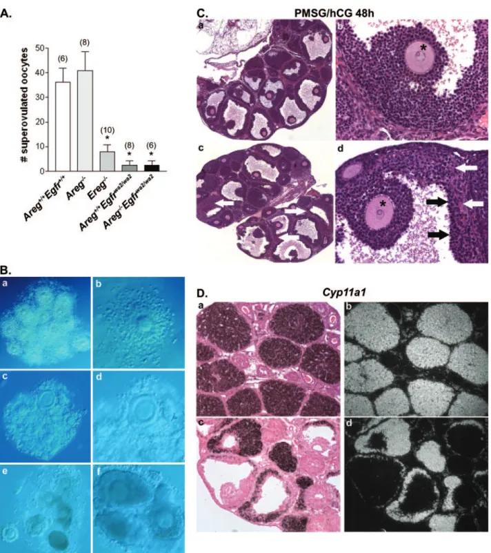
Impaired ovulation and luteinization in Aregⴚ/ⴚEgfrwa2/wa2 females.To determine the in vivo effect of disrupted signaling through the EGFR on ovulation, PMSG-primed mice were stimulated with hCG to induce ovulation of oocytes. After 13 to 24 h, the oviducts were carefully opened to release the ovulated cluster of COCs. Areg⫺/⫺females released numbers of oocytes (Fig. 5A) into the oviducts similar to those seen with wild-type and Areg⫹/⫺ littermates (WT/Areg⫹/⫺, 40⫾ 10.56,
n ⫽ 5). Interestingly, immature Ereg⫺/⫺ mice ovulated few oocytes (Fig. 5A) in comparison to wild-type and Ereg⫹/⫺ lit-termates (WT/Ereg⫹/⫺, 46.15⫾ 3.93, n ⫽ 20) when superovu-lated. Conversely, there was no statistically significant dif-ference between adult Ereg⫺/⫺ and wild-type females in ovulation rates after natural matings (data not shown). Very few or no oocytes could be recovered from the oviducts of
Areg⫹/⫹Egfrwa2/wa2and Areg⫺/⫺Egfrwa2/wa2mice (Fig. 5A). FIG. 3. Impaired EGFR activation and oocyte meiotic resumption
in Areg⫺/⫺Egfrwa2/wa2mice. (A) LH-induced EGFR phosphorylation in Areg⫹/⫹Egfr⫹/⫹, Areg⫹/⫹Egfrwa2/wa2, and Areg⫺/⫺ Egfrwa2/wa2 pre-ovulatory follicles. Proteins extracted from follicles cultured in the absence or presence of 5 IU rLH for 2 h were immunoprecipitated using an anti-EGFR antibody, and Western blotting was performed using an EGFR-specific phosphoantibody. To control for protein load-ing, the same membrane was reprobed using an anti-EGFR antibody. Data represent the means⫾ SEM of the results of three separate exper-iments. P⬍ 0.05 compared to wild-type (a) and Areg⫹/⫹Egfrwa2/wa2(b) mouse results. A representative blot is shown. (B) Resumption of oocyte maturation (scored as GVBD) after 4 h hCG stimulation in vivo was evaluated for immature mice of the indicated genotypes. Results represent the means⫾ SEM of the results for each group. The number of mice per group is indicated above each bar. The letters a and b correspond to P⬍ 0.05 and P ⬍ 0.001, respectively, compared to wild-type results, and c indicates P⬍ 0.05 compared to Areg⫺/⫺results.
on October 21, 2016 by Ewha Womans Univ
http://mcb.asm.org/
On the rare occasion in which ovulated oocytes were de-tected in the mutant mice, most complexes appeared poorly expanded or compact in comparison to the well-expanded complexes from wild-type mice (Fig. 5B).
Histological analyses of the ovaries from the superovulated double-mutant females revealed the presence of large antral follicles, many containing entrapped, compact COCs with oocytes still in the GV stage (data not shown). In the few follicles that had ovulated, terminal differentiation (luteiniza-tion) of somatic cells appeared normal, with an increase in cytoplasmic volume associated with increased steroidogenic capacity of luteinized cells and vascularization of the newly formed corpus luteum (not shown). Moreover, at 48 h after hCG, a time when corpora lutea are typically fully formed in wild-type ovaries, few fully luteinized structures were observed in the Areg⫺/⫺Egfrwa2/wa2ovaries. Double-mutant ovaries
con-tained many antral follicles that morphologically did not ap-pear luteinized or that apap-peared partially luteinized with en-trapped oocytes surrounded by compact cumuli (Fig. 5C). In situ hybridization analysis of Cyp11a1 expression and localiza-tion was performed to confirm the differentialocaliza-tion of somatic cells into luteal cells. Cyp11a1 encodes the P450 cholesterol side-chain cleavage enzyme that is highly expressed by luteal cells for the synthesis of progesterone. Consistent with the above-described histology, Cyp11a1 expression was detected in partially luteinized and luteinized structures but not in granu-losa cells of antral follicles that had not undergone terminal differentiation (Fig. 5D). Cyp11a1 expression was detected in the theca cell layers surrounding nonluteinized antral follicles. Detailed analyses of fecundity and fertility were not per-formed for the Areg⫺/⫺Egfrwa2/wa2mice, since interpretation
of the results might be complicated by potential extragonadal
FIG. 4. Defective cumulus expansion in Areg⫺/⫺Egfrwa2/wa2mice. (A) Semiquantitative RT-PCR results show significantly decreased expression levels for Ptgs2, Tnfaip6, and Has2 in COCs from Areg⫺/⫺Egfrwa2/wa2mouse ovaries 3 h after hCG compared to wild-type control results. Data represent means⫾ SEM of the expression levels for Ptgs2, Tnfaip6, or Has2 normalized to Rpl19 (n ⫽ 3 for each genotype). (B) In situ hybridization analysis shows high expression and localization of Ptgs2 mRNA in mural granulosa cells (GC) and cumulus cells (CC) of preovulatory follicles from wild-type ovaries 3 h after hCG (a and b). The signal is decreased in GCs and is below the threshold of detection in CCs of Areg⫺/⫺ Egfrwa2/wa2follicles (c and d) (Oo, oocyte). (C) The degree of cumulus expansion for COCs in Areg⫺/⫺Egfrwa2/wa2mouse ovaries 9 h after hCG was evaluated as described in Materials and Methods. A large proportion of COCs were poorly expanded or unexpanded. Data represent the mean percentage of COCs scored⫾ SEM (n ⫽ 4 females;*, P⬍ 0.001). The degree of cumulus expansion in Areg⫹/⫺mice (see Fig. 1C) is included for comparison. (D) Histology of COCs with GV and GVBD oocytes (left and right panels, respectively) from Areg⫺/⫺Egfrwa2/wa2mouse ovaries 9 h after hCG.
on October 21, 2016 by Ewha Womans Univ
http://mcb.asm.org/
FIG. 5. Impaired ovulation and luteinization in Areg⫺/⫺Egfrwa2/wa2mice. (A) Superovulation of wild-type and mutant mice. Ereg⫺/⫺, Areg⫹/⫹
Egfrwa2/wa2, and Areg⫺/⫺Egfrwa2/wa2mice ovulated significantly fewer oocytes than wild-type and Areg⫺/⫺females. The number of females used per
group is indicated in parentheses above the bars. Data represent means⫾ SEM;*indicates P⬍ 0.001 compared to either wild-type (Areg⫹/⫹ Egfr⫹/⫹) or Areg⫺/⫺ values. (B) Images of ovulated COCs produced using Hoffman interference microscopy. Panels a and b show ovulated, well-expanded complexes from a representative wild-type mouse. In contrast, both expanded and poorly expanded complexes were observed in the few COCs that were ovulated by Areg⫹/⫹Egfrwa2/wa2mice (c; at higher magnification in panel d). Structures resembling ovulated COCs from Areg⫺/⫺Egfrwa2/wa2mice (e and f) appeared poorly expanded. (C) Histology of Areg⫺/⫺Egfrwa2/wa2ovaries 48 h after hCG stimulation. Many antral follicles contain entrapped, unexpanded COCs (panels a and c) with oocytes still in the GV stage (shown at higher magnification in panels b and d). Somatic cells from antral follicles appeared either nonluteinized (a and b) or partially luteinized (c and d), with morphologically nonluteinized granulosa cells (black arrows) lining the antrum and luteal cells (white arrows) in the outer cell layers. The asterisks mark entrapped GV oocytes. (D) In situ hybridization analysis localizes Cyp11a1 mRNA in the corpora lutea of a wild-type ovary (a and b) and in luteinized cells of antral follicles from an Areg⫺/⫺Egfrwa2/wa2ovary (c and d) 48 h after hCG stimulation. Signal for Cyp11a1 is absent from non-terminally differentiated somatic cells.
on October 21, 2016 by Ewha Womans Univ
http://mcb.asm.org/
defects in adult mice, i.e., the regulation of LH-releasing hor-mone release from the hypothalamic-pituitary axis (23, 39). Nevertheless, it should be noted that one adult double-mutant female gave birth to two small litters in the first 2 months of mating (four and three pups, respectively) but produced no additional litters over the following 6 months. A second adult female produced two small litters (three pups and one pup, respectively), while a third female had no litters, after 4.5 and 6 months of mating, respectively. Thus, a major disruption in fecundity and perhaps fertility was likely present in these mice. When wild-type and Areg⫺/⫺ female mice were mated with wild-type males over a 6-month period, a small but significant decrease in the average number of pups born per litter was observed for the Areg⫺/⫺ females compared to wild-type fe-males (5.97⫾ 0.36 pups/litter, n ⫽ 5, and 7.83 ⫾ 0.29 pups/ litter, n⫽ 6, respectively; P ⬍ 0.001). The average numbers of litters born per month were not significantly different between
Areg⫺/⫺(1.03⫾ 0.07, n ⫽ 5) and wild-type (1.05 ⫾ 0.08, n ⫽ 6) mice.
DISCUSSION
With this report, we provide conclusive evidence that LH-induced activation of EGFR signaling is essential for regulation of oocyte maturation and cumulus cell functions. Progressive ge-netic disruption of the EGF network in the periovulatory fol-licle prevents LH induction of oocyte meiotic reentry and tran-scription of genes involved in cumulus expansion. More importantly, ovulation is compromised when growth factor production and EGFR signaling is disrupted. Finally, the ge-netic model that we have established indicates that activation of the EGF network affects mural granulosa cells as well as the transfer of the signal to cumulus cells, demonstrating both autocrine and paracrine roles for these growth factors in the ovulatory follicle.
In immature mice deficient in the AREG ligand, oocyte meiotic resumption and cumulus expansion are delayed or reduced. Although immature Areg⫺/⫺ females released num-bers of oocytes into the oviducts similar to those seen with wild-type females when superovulated, adult Areg⫺/⫺ female mice produced fewer pups per litter than wild-type females, demonstrating that removal of a single ligand is sufficient to impact fertility. Because the disruption of fertility in Areg⫺/⫺ mice is modest, it is likely that redundancy of other EGF-like growth factors compensates for the loss of AREG production. Interestingly, Ereg ablation appears to have a larger impact on oocyte maturation and ovulation in hormone-primed imma-ture mice than Areg ablation. This may be due in part to differences in the genetic backgrounds of the mice, Ereg⫺/⫺ mice being on a C57BL/6J background while Areg⫺/⫺ and
Areg⫺/⫺ Egfrwa2/wa2 mice are on a mixed background
(C57BL/6J ⫻ 129Sv). It is also possible that the divergent effects are due to the activities of the two ligands or to the fact that EREG interacts with both EGFR and ERBB4 whereas AREG interacts only with EGFR (19, 43). Although a role for ERBB4 cannot be ruled out, the significantly reduced ovula-tion rates in Areg⫹/⫹Egfrwa2/wa2and Areg⫺/⫺Egfrwa2/wa2mice
strongly suggests that the EGFR is the predominant receptor involved.
In immature Areg⫺/⫺ Egfrwa2/wa2 mice, more pronounced
disruption of ovarian function is observed. Although antral follicles appear to develop normally, respond to LH with a robust increase in cAMP accumulation, and contain oocytes that are competent to undergo spontaneous maturation, trans-fer of the LH signal to the oocyte for induction of oocyte maturation is prevented in these mice. In the double mutants, LH-induced EGFR phosphorylation is significantly decreased compared to wild-type results, suggesting that this phosphory-lation is critical for transphosphory-lation of the LH signal into oocyte meiotic resumption. Whereas the presence of EGFR in oo-cytes is debated, several experiments suggest that transactiva-tion of EGFR occurs in the cumulus cells (11, 40). Indeed, EGF-like growth factors induce oocyte maturation in COCs but have no effect on denuded oocytes (5, 34). Thus, impaired EGFR signaling likely precludes transfer of a signal from cu-mulus cells to the oocyte, although direct signaling cannot be excluded at this stage. The nature of this signal is at present unknown. Recent findings have implicated a class of GPCRs belonging to the lipid receptor subfamily with constitutive ac-tivity in the maintenance of meiotic arrest in oocytes (15, 20a, 28). Several studies indicate that EGF has profound effects on lipid metabolism and signaling (2, 22). Thus, it is possible that the signal emanating from the EGFR involves modulation of the activity of these GPCRs.
Cumulus expansion is greatly impaired in the double-mutant mice. Significantly reduced expression of Ptgs2, Tnfaip6, and
Has2, genes important for formation and stabilization of the
cumulus matrix, in COCs of Areg⫺/⫺Egfrwa2/wa2mice precedes
and is consistent with the observed defects in cumulus expan-sion. These findings document that cumulus cell activation does not occur in the absence of a functional EGF network. By in situ hybridization, signal for Ptgs2 mRNA is notably de-creased in both mural granulosa and cumulus cells of preovu-latory follicles in Areg⫺/⫺Egfrwa2/wa2mouse ovaries 3 h after
hCG. This observation indicates that LH induction of Ptgs2 expression is dependent on activation of EGFR signaling not only in cumulus cells but also in mural granulosa cells. Thus, EGF-like growth factor signaling through the EGFR appears to have both autocrine and paracrine roles in transducing LH effects in the preovulatory follicle for ovulation. Recently, an increase in Areg and Ereg mRNAs has been detected 4 to 8 h after hCG stimulation in the cumulus (49). This induction is reduced in Ptgs2-null mice and is in part induced by AREG itself. Therefore, it is likely that the autocrine loop activating
Ptgs2 expression in mural granulosa cells, which is required for
prostaglandin production, contributes to the propagation of the stimulus to cumulus cells. Thus, a positive feedback further amplifies the effect of these growth factors, extending the du-ration of activation in the cumulus.
Ovulation is compromised in the Areg⫹/⫹ Egfrwa2/wa2 and Areg⫺/⫺ Egfrwa2/wa2 mice. This is documented by the results
showing that few or no oocytes were recovered from the ovi-ducts and by the concomitant observation of entrapped oocytes in follicles 24 to 48 h after hCG. The rare follicles that were able to ovulate appeared to be fully luteinized, suggesting that a subset of follicles is able to escape the EGF network disruption, a finding consistent with the residual fertility and fecundity of the double-mutant mice. This may be due to the incomplete inactivation of Egfrwa2or utilization of
compen-satory pathways in a subset of follicles similar to what was
on October 21, 2016 by Ewha Womans Univ
http://mcb.asm.org/
observed in a subset of intestinal tumors arising in an
Egfrwa2 homozygous background (44). Additional caution
should be used in interpreting the data, because disruption in the hypothalamic-pituitary axis has been described for
Egfrwa2and astroglial cell-specific EGFR mutant mice (23,
39). Therefore, decreased fertility in adult females may be due to ovarian and/or extraovarian defects.
In superovulated Areg⫺/⫺ Egfrwa2/wa2mice, the majority of
antral follicles were unruptured, with entrapped GV oocytes surrounded by compact cumulus cells and partial or no lutein-ization of somatic cells. The few COCs that were ovulated displayed defective cumulus expansion. Follicle rupture may be the most sensitive event, because it requires many cooperating steps to occur. Remodeling of the extracellular matrices, the cell layers in the follicle wall, and the ovarian surface epithe-lium at the apex of the follicle is required for follicle rupture (31, 41, 53). Constriction of a layer of smooth-muscle cells in the follicular wall at the time of rupture is also likely important (20). In addition, in mouse models with impaired cumulus expansion, follicle rupture is impaired though not always com-pletely abolished depending on the model (3, 10, 16, 33, 56, 57) and as observed here. Previous attempts to define the effect of the EGF network in vivo by intrabursal injection of an EGFR kinase inhibitor provided an initial clue to the role of this network in follicle rupture (1). However, at high doses of inhibitor, no significant effect on ovulation was observed. Here, our studies demonstrate the critical role of the EGF network in ovulation using a genetic approach. We propose that defective follicle rupture may be due in part to the greatly reduced expression of Ptgs2, and likely other genes, in the follicle. Indeed, the phenotype of the Areg⫺/⫺Egfrwa2/wa2mice
resem-bles that of Ptgs2-null mice (4, 24) that are anovulatory and that have few or no corpora lutea. Nrip1 (previously known as RIP140)-null mice that display decreased expression of the EGF-like growth factors Ptgs2, Tnfaip6, and Has2 also have impaired cumulus expansion and follicle rupture (54). Simi-larly, a defect in gene expression and follicle rupture has been previously reported in the Pde4d-null mice (33) together with a decrease in EGF-like growth factor induction (34). However, both genetic mouse models exhibit normal oocyte maturation and luteinization, in contrast to the Areg⫺/⫺Egfrwa2/wa2mouse
results. Why oocyte maturation and luteinization are not dis-rupted in Nrip1- and Pde4d-null mice is unclear. It is possible that the residual activity of a wild-type EGFR in follicular cells is sufficient to support these processes.
Taken together, the findings presented above demonstrate that LH induction of EGF-like growth factors and activation of EGFR signaling is essential for ovulation of mature oocytes and somatic cell terminal differentiation. To our knowledge, this is the first in vivo demonstration that a critical physiolog-ical process such as ovulation is dependent on GPCR transac-tivation of EGFR signaling. It also establishes a paradigm whereby LH actions are propagated by activation of the EGF network, which in turn acts in an autocrine and paracrine fashion within this functional unit of the ovary. These findings have important implications for the development of new strat-egies for the control of fertility and for the improvement of in vitro culture conditions for the maturation of oocytes and follicles.
ACKNOWLEDGMENTS
We thank Anita Payne, Greg Barsh, and You-Qiang Su for helpful comments on the manuscript.
S.P.’s contribution was performed in partial fulfillment of her grad-uate work as a visiting student from the University of Milan, Milan, Italy.
This work was supported by National Research Service Award F32 HD049966 (M.H.) from the National Institute of Child Health and Human Development of the National Institutes of Health through a cooperative agreement (U54-HD31389 to M.C.) as part of the Spe-cialized Cooperative Centers Program in Reproduction Research and by a grant from Organon (M.C.). D.C.L. is supported by grants CA43793 and CA61896 and D.W.T. by grants HD039896 and CA092479.
REFERENCES
1. Ashkenazi, H., X. Cao, S. Motola, M. Popliker, M. Conti, and A. Tsafriri. 2005. Epidermal growth factor family members: endogenous mediators of the ovulatory response. Endocrinology 146:77–84.
2. Daub, H., F. U. Weiss, C. Wallasch, and A. Ullrich. 1996. Role of transac-tivation of the EGF receptor in signalling by G-protein-coupled receptors. Nature 379:557–560.
3. Davis, B. J., D. E. Lennard, C. A. Lee, H. F. Tiano, S. G. Morham, W. C.
Wetsel, and R. Langenbach.1999. Anovulation in cyclooxygenase-2-deficient
mice is restored by prostaglandin E2 and interleukin-1. Endocrinology
140:2685–2695.
4. Dinchuk, J. E., B. D. Car, R. J. Focht, J. J. Johnston, B. D. Jaffee, M. B.
Covington, N. R. Contel, V. M. Eng, R. J. Collins, P. M. Czerniak, et al.1995. Renal abnormalities and an altered inflammatory response in mice lacking cyclooxygenase II. Nature 378:406–409.
5. Downs, S. M., S. A. Daniel, and J. J. Eppig. 1988. Induction of maturation in cumulus cell-enclosed mouse oocytes by follicle-stimulating hormone and epidermal growth factor: evidence for a positive stimulus of somatic cell origin. J. Exp. Zool. 245:86–96.
6. Elvin, J. A., A. T. Clark, P. Wang, N. M. Wolfman, and M. M. Matzuk. 1999. Paracrine actions of growth differentiation factor-9 in the mammalian ovary. Mol. Endocrinol. 13:1035–1048.
7. Eppig, J. J. 1991. Intercommunication between mammalian oocytes and companion somatic cells. Bioessays 13:569–574.
8. Fowler, K., F. Walker, W. Alexander, M. Hibbs, E. Nice, R. Bohmer, G.
Mann, C. Thumwood, R. Maglitto, J. Danks, R. Chetty, A. Burgess, and A. Dunn.1995. A mutation in the epidermal growth factor receptor in waved-2 mice has a profound effect on receptor biochemistry that results in impaired lactation. Proc. Natl. Acad. Sci. USA 92:1465–1469.
9. Freimann, S., I. Ben-Ami, A. Dantes, R. Ron-El, and A. Amsterdam. 2004. EGF-like factor epiregulin and amphiregulin expression is regulated by gonadotropins/cAMP in human ovarian follicular cells. Biochem. Biophys. Res. Commun. 324:829–834.
10. Fu¨lo¨p, C., S. Szanto, D. Mukhopadhyay, T. Bardos, R. V. Kamath, M. S.
Rugg, A. J. Day, A. Salustri, V. C. Hascall, T. T. Glant, and K. Mikecz.2003. Impaired cumulus mucification and female sterility in tumor necrosis factor-induced protein-6 deficient mice. Development 130:2253–2261.
11. Gall, L., N. Chene, M. Dahirel, S. Ruffini, and C. Boulesteix. 2004. Expres-sion of epidermal growth factor receptor in the goat cumulus-oocyte com-plex. Mol. Reprod. Dev. 67:439–445.
12. Gospodarowicz, D., and H. Bialecki. 1979. Fibroblast and epidermal growth factors are mitogenic agents for cultured granulosa cells of rodent, porcine, and human origin. Endocrinology 104:757–764.
13. Gschwind, A., S. Hart, O. M. Fischer, and A. Ullrich. 2003. TACE cleavage of proamphiregulin regulates GPCR-induced proliferation and motility of cancer cells. EMBO J. 22:2411–2421.
14. Harper, J. F., and G. Brooker. 1975. Femtomole sensitive radioimmunoassay
for cyclic AMP and cyclic GMP after 2⬘0 acetylation by acetic anhydride in
aqueous solution. J. Cyclic Nucleotide Res. 1:207–218.
15. Hinckley, M., S. Vaccari, K. Horner, R. Chen, and M. Conti. 2005. The G-protein-coupled receptors GPR3 and GPR12 are involved in cAMP sig-naling and maintenance of meiotic arrest in rodent oocytes. Dev. Biol.
287:249–261.
16. Hizaki, H., E. Segi, Y. Sugimoto, M. Hirose, T. Saji, F. Ushikubi, T.
Matsuoka, Y. Noda, T. Tanaka, N. Yoshida, S. Narumiya, and A. Ichikawa.
1999. Abortive expansion of the cumulus and impaired fertility in mice lacking the prostaglandin E receptor subtype EP(2). Proc. Natl. Acad. Sci. USA 96:10501–10506.
17. Hogan, B., R. Beddington, F. Costantini, and E. Lacy. 1994. Manipulating the mouse embryo: a laboratory manual, 2nd ed. Cold Spring Harbor Lab-oratory Press, Cold Spring Harbor, NY.
18. Holbro, T., and N. E. Hynes. 2004. ErbB receptors: directing key signaling networks throughout life. Annu. Rev. Pharmacol. Toxicol. 44:195–217. 19. Johnson, G. R., B. Kannan, M. Shoyab, and K. Stromberg. 1993.
Amphi-regulin induces tyrosine phosphorylation of the epidermal growth factor receptor and p185erbB2. Evidence that amphiregulin acts exclusively
on October 21, 2016 by Ewha Womans Univ
http://mcb.asm.org/
through the epidermal growth factor receptor at the surface of human epithelial cells. J. Biol. Chem. 268:2924–2931.
20. Ko, C., M. C. Gieske, L. Al-Alem, Y. Hahn, W. Su, M. C. Gong, M. Iglarz,
and Y. Koo.2006. Endothelin-2 in ovarian follicle rupture. Endocrinology
147:1770–1779.
20a.Ledent, C., I. Demeestere, D. Blum, J. Petermans, T. Hamalainen, G. Smits,
and G. Vassart.2005. Premature ovarian aging in mice deficient for Gpr3. Proc. Natl. Acad. Sci. USA 102:8922–8926.
21. Lee, D., R. S. Pearsall, S. Das, S. K. Dey, V. L. Godfrey, and D. W. Threadgill. 2004. Epiregulin is not essential for development of intestinal tumors but is required for protection from intestinal damage. Mol. Cell. Biol. 24:8907– 8916.
22. Le Stunff, H., A. Mikami, P. Giussani, J. P. Hobson, P. S. Jolly, S. Milstien,
and S. Spiegel.2004. Role of sphingosine-1-phosphate phosphatase 1 in epidermal growth factor-induced chemotaxis. J. Biol. Chem. 279:34290– 34297.
23. Li, B., Z. Yang, J. Hou, A. McCracken, M. A. Jennings, and M. Y. Ma. 2003. Compromised reproductive function in adult female mice selectively ex-pressing mutant ErbB-1 tyrosine kinase receptors in astroglia. Mol. Endo-crinol. 17:2365–2376.
24. Lim, H., B. C. Paria, S. K. Das, J. E. Dinchuk, R. Langenbach, J. M.
Trzaskos, and S. K. Dey.1997. Multiple female reproductive failures in cyclooxygenase 2-deficient mice. Cell 91:197–208.
25. Luetteke, N. C., H. K. Phillips, T. H. Qiu, N. G. Copeland, H. S. Earp, N. A.
Jenkins, and D. C. Lee.1994. The mouse waved-2 phenotype results from a point mutation in the EGF receptor tyrosine kinase. Genes Dev. 8:399–413. 26. Luetteke, N. C., T. H. Qiu, S. E. Fenton, K. L. Troyer, R. F. Riedel, A. Chang,
and D. C. Lee.1999. Targeted inactivation of the EGF and amphiregulin genes reveals distinct roles for EGF receptor ligands in mouse mammary gland development. Development 126:2739–2750.
27. Matzuk, M. M., K. H. Burns, M. M. Viveiros, and J. J. Eppig. 2002. Inter-cellular communication in the mammalian ovary: oocytes carry the conver-sation. Science 296:2178–2180.
28. Mehlmann, L. M., Y. Saeki, S. Tanaka, T. J. Brennan, A. V. Evsikov, F. L.
Pendola, B. B. Knowles, J. J. Eppig, and L. A. Jaffe.2004. The Gs-linked receptor GPR3 maintains meiotic arrest in mammalian oocytes. Science
306:1947–1950.
29. Mondschein, J. S., and D. W. Schomberg. 1981. Growth factors modulate gonadotropin receptor induction in granulosa cell cultures. Science 211: 1179–1180.
30. Moore, R. K., and S. Shimasaki. 2005. Molecular biology and physiological role of the oocyte factor, BMP-15. Mol. Cell. Endocrinol. 234:67–73. 31. Murdoch, W. J., and M. L. Gottsch. 2003. Proteolytic mechanisms in the
ovulatory folliculo-luteal transformation. Connect. Tissue Res. 44:50–57. 32. Ochsner, S. A., A. J. Day, M. S. Rugg, R. M. Breyer, R. H. Gomer, and J. S.
Richards.2003. Disrupted function of tumor necrosis factor-alpha-stimu-lated gene 6 blocks cumulus cell-oocyte complex expansion. Endocrinology
144:4376–4384.
33. Park, J. Y., F. Richard, S. Y. Chun, J. H. Park, E. Law, K. Horner, S. L. Jin,
and M. Conti.2003. Phosphodiesterase regulation is critical for the differ-entiation and pattern of gene expression in granulosa cells of the ovarian follicle. Mol. Endocrinol. 17:1117–1130.
34. Park, J. Y., Y. Q. Su, M. Ariga, E. Law, S. L. Jin, and M. Conti. 2004. EGF-like growth factors as mediators of LH action in the ovulatory follicle. Science 303:682–684.
35. Park, O. K., and K. E. Mayo. 1991. Transient expression of progesterone receptor messenger RNA in ovarian granulosa cells after the preovulatory luteinizing hormone surge. Mol. Endocrinol. 5:967–978.
36. Pathak, B. G., D. J. Gilbert, C. A. Harrison, N. C. Luetteke, X. Chen, M.
Klagsbrun, G. D. Plowman, N. G. Copeland, N. A. Jenkins, and D. C. Lee.
1995. Mouse chromosomal location of three EGF receptor ligands: amphi-regulin (Areg), betacellulin (Btc), and heparin-binding EGF (Hegfl). Genomics
28:116–118.
37. Peng, X. R., A. J. Hsueh, P. S. LaPolt, L. Bjersing, and T. Ny. 1991. Local-ization of luteinizing hormone receptor messenger ribonucleic acid expres-sion in ovarian cell types during follicle development and ovulation. Endo-crinology 129:3200–3207.
38. Prenzel, N., E. Zwick, H. Daub, M. Leserer, R. Abraham, C. Wallasch, and
A. Ullrich.1999. EGF receptor transactivation by G-protein-coupled recep-tors requires metalloproteinase cleavage of proHB-EGF. Nature 402:884– 888.
39. Prevot, V., A. Lomniczi, G. Corfas, and S. R. Ojeda. 2005. erbB-1 and erbB-4 receptors act in concert to facilitate female sexual development and mature reproductive function. Endocrinology 146:1465–1472.
40. Prochazka, R., P. Kalab, and E. Nagyova. 2003. Epidermal growth factor-receptor tyrosine kinase activity regulates expansion of porcine oocyte-cumulus cell complexes in vitro. Biol. Reprod. 68:797–803.
41. Richards, J. S., D. L. Russell, S. Ochsner, and L. L. Espey. 2002. Ovulation: new dimensions and new regulators of the inflammatory-like response. Annu. Rev. Physiol. 64:69–92.
42. Riese, D. J., Jr., Y. Bermingham, T. M. van Raaij, S. Buckley, G. D. Plowman,
and D. F. Stern.1996. Betacellulin activates the epidermal growth factor receptor and erbB-4, and induces cellular response patterns distinct from those stimulated by epidermal growth factor or neuregulin-beta. Oncogene
12:345–353.
43. Riese, D. J., Jr., T. Komurasaki, G. D. Plowman, and D. F. Stern. 1998. Activation of ErbB4 by the bifunctional epidermal growth factor family hormone epiregulin is regulated by ErbB2. J. Biol. Chem. 273:11288–11294. 44. Roberts, R. B., L. Min, M. K. Washington, S. J. Olsen, S. H. Settle, R. J.
Coffey, and D. W. Threadgill.2002. Importance of epidermal growth factor receptor signaling in establishment of adenomas and maintenance of carci-nomas during intestinal tumorigenesis. Proc. Natl. Acad. Sci. USA 99:1521– 1526.
45. Robker, R. L., D. L. Russell, L. L. Espey, J. P. Lydon, B. W. O’Malley, and
J. S. Richards.2000. Progesterone-regulated genes in the ovulation process: ADAMTS-1 and cathepsin L proteases. Proc. Natl. Acad. Sci. USA 97:4689– 4694.
46. Scha¨fer, B., A. Gschwind, and A. Ullrich.2004. Multiple G-protein-coupled
receptor signals converge on the epidermal growth factor receptor to pro-mote migration and invasion. Oncogene 23:991–999.
47. Schomberg, D. W., J. V. May, and J. S. Mondschein. 1983. Interactions between hormones and growth factors in the regulation of granulosa cell differentiation in vitro. J. Steroid Biochem. 19:291–295.
48. Sekiguchi, T., T. Mizutani, K. Yamada, T. Kajitani, T. Yazawa, M. Yoshino,
and K. Miyamoto.2004. Expression of epiregulin and amphiregulin in the rat ovary. J. Mol. Endocrinol. 33:281–291.
49. Shimada, M., I. Hernandez-Gonzalez, I. Gonzalez-Robayna, and J. S.
Richards. 2006. Paracrine and autocrine regulation of epidermal growth factor-like factors in cumulus oocyte complexes and granulosa cells: key roles for prostaglandin synthase 2 and progesterone receptor. Mol. Endocrinol.
20:1352–1365.
50. Sibilia, M., and E. F. Wagner. 1995. Strain-dependent epithelial defects in mice lacking the EGF receptor. Science 269:234–238.
51. Sukocheva, O., C. Wadham, A. Holmes, N. Albanese, E. Verrier, F. Feng, A.
Bernal, C. K. Derian, A. Ullrich, M. A. Vadas, and P. Xia.2006. Estrogen transactivates EGFR via the sphingosine 1-phosphate receptor Edg-3: the role of sphingosine kinase-1. J. Cell Biol. 173:301–310.
52. Threadgill, D. W., A. A. Dlugosz, L. A. Hansen, T. Tennenbaum, U. Lichti,
D. Yee, C. LaMantia, T. Mourton, K. Herrup, R. C. Harris, et al.1995. Targeted disruption of mouse EGF receptor: effect of genetic background on mutant phenotype. Science 269:230–234.
53. Tsafriri, A., and R. Reich. 1999. Molecular aspects of ammalian ovulation. Exp. Clin. Endocrinol. Diabetes 107:1–11.
54. Tullet, J. M. A., V. Pocock, J. H. Steel, R. White, S. Milligan, and M. G.
Parker.2005. Multiple signalling defects in the absence of RIP140 impair both cumulus expansion and follicle rupture. Endocrinology 146:4127–4137. 55. Vanderhyden, B. C., P. J. Caron, R. Buccione, and J. J. Eppig. 1990. Devel-opmental pattern of the secretion of cumulus expansion-enabling factor by mouse oocytes and the role of oocytes in promoting granulosa cell differen-tiation. Dev. Biol. 140:307–317.
56. Varani, S., J. A. Elvin, C. Yan, J. DeMayo, F. J. DeMayo, H. F. Horton, M. C.
Byrne, and M. M. Matzuk.2002. Knockout of pentraxin 3, a downstream target of growth differentiation factor-9, causes female subfertility. Mol. Endocrinol. 16:1154–1167.
57. Yan, C., P. Wang, J. DeMayo, F. J. DeMayo, J. A. Elvin, C. Carino, S. V.
Prasad, S. S. Skinner, B. S. Dunbar, J. L. Dube, A. J. Celeste, and M. M. Matzuk.2001. Synergistic roles of bone morphogenetic protein 15 and growth differentiation factor 9 in ovarian function. Mol. Endocrinol. 15:854– 866.
58. Yarden, Y., and M. X. Sliwkowski. 2001. Untangling the ErbB signalling network. Nat. Rev. Mol. Cell Biol. 2:127–137.