ISSN 2234-3806 • eISSN 2234-3814
https://doi.org/10.3343/alm.2018.38.2.110
Utility of Conventional Culture and MALDI-TOF MS for
Identification of Microbial Communities in
Bronchoalveolar Lavage Fluid in Comparison with the
GS Junior Next Generation Sequencing System
Ji Yeon Sung, M.D.1, Younjee Hwang, B.S.2, Mi Hwa Shin, B.S.3, Moo Suk Park, M.D.3, Sang Hoon Lee, M.D.4,
Dongeun Yong, M.D.1, and Kyungwon Lee, M.D.1
Research Institute of Bacterial Resistance and Department of Laboratory Medicine1, Yonsei University College of Medicine, Seoul; Brain Korea 21 PLUS
Project for Medical Science2, Yonsei University, Seoul; Division of Pulmonary and Critical Care Medicine3, Department of Internal Medicine, Severance
Hospital, Yonsei University College of Medicine, Seoul; Department of Internal Medicine4, Seoul National University College of Medicine, Division of
Pulmonary and Critical Care Medicine, Seoul National University Bundang Hospital, Seongnam, Korea Background: Diverse microbiota exist in the lower respiratory tract. Although next
genera-tion sequencing (NGS) is the most widely used microbiome analysis technique, it is diffi-cult to implement NGS in clinical microbiology laboratories. Therefore, we evaluated the performance of conventional culture methods together with matrix-assisted laser desorp-tion/ionization time-of-flight mass spectrometry (MALDI-TOF MS) in identifying microbiota in bronchoalveolar lavage (BAL) fluid.
Methods: BAL fluid samples (n=27) were obtained from patients undergoing diagnostic
bronchoscopy for lung mass evaluation. Bacterial and fungal culture was performed with conventional media used in clinical microbiology laboratories. On an average, 20 isolated colonies were picked from each agar plate and identified by MALDI-TOF MS. Microbiome analysis using 16S rRNA NGS was conducted for comparison.
Results: Streptococcus spp. and Neisseria spp. were most frequently cultured from the
BAL fluid samples. In two samples, Enterobacteriaceae grew predominantly on MacCon-key agar. Actinomyces and Veillonella spp. were commonly identified anaerobes; gut bac-teria, such as Lactobacillus, Bifidobacterium, and Clostridium, and fungi were also iso-lated. NGS revealed more diverse bacterial communities than culture, and Prevotella spp. were mainly identified solely by NGS. Some bacteria, such as Staphylococcus spp., Clos-tridium spp., and Bifidobacterium spp., were identified solely by culture, indicating that culture may be more sensitive for detecting certain bacteria.
Conclusions: Culture and NGS of BAL fluid samples revealed common bacteria with some
different microbial communities. Despite some limitations, culture combined with MALDI-TOF MS might play a complementary role in microbiome analysis using 16S rRNA NGS.
Key Words: Bronchoalveolar lavage, Microbiota, Matrix-assisted laser desorption-ionization
mass spectrometry, Culture, Next generation sequencing
Received: February 14, 2017 Revision received: April 14, 2017 Accepted: October 10, 2017 Corresponding author: Dongeun Yong
Department of Laboratory Medicine and Research Institute of Bacterial Resistance, Yonsei University College of Medicine 50-1 Yonsei-ro, Seodaemun-gu, Seoul 03722, Korea
Tel: +82-2-2228-2442 Fax: +82-2-364-1583 E-mail: DEYONG@yuhs.ac
© Korean Society for Laboratory Medicine This is an Open Access article distributed under the terms of the Creative Commons Attribution Non-Commercial License (http://creativecom-mons.org/licenses/by-nc/4.0) which permits unrestricted non-commercial use, distribution, and reproduction in any medium, provided the original work is properly cited.
2017-03-16 https://crossmark-cdn.crossref.org/widget/v2.0/logos/CROSSMARK_Color_square.svg
INTRODUCTION
Increasing data have demonstrated that colonizing microorgan-isms might dictate human health and disease [1]. Although the 2012 Human Microbiome Project did not include the lower re-spiratory tract in its surveys, advances in sequencing technology have revealed a diverse population of non-routinely cultured bacteria in the lung, which has traditionally been thought to be sterile [2-5]. Most of these studies are based on 16S rRNA next generation sequencing (NGS), a highly sensitive method for an-alyzing the microbiome [6]. Because the human microbiota consists of many ‘unculturable’ bacteria, culture-independent metagenomic approaches have driven significant advances in microbiome research. However, the bacterial species informa-tion obtainable from these metagenomic approaches might be limited, and there are several steps, including nucleic acid ex-traction, PCR amplification, sequencing (method and depth), and bioinformatic analysis, that may produce variations in the results. Read length is also one of the most important factors when considering clinical application. Hence, the cultivation of microbes provides more reliable information regarding the phe-notype and ecological role, allowing a greater understanding of microbe-host interactions [7].
Recently, a number of new approaches for gut microbiota cul-tivation have emerged with potential for use in microbiome re-search [8]; these have mainly been applied to investigate the gut microbiome [9-11]. Although these novel approaches have shown that culture can capture a significant subset of the spe-cies identified using culture-independent methods [8], it is not easy to apply these techniques in clinical laboratories. In addi-tion, limited information is available regarding culture-based mi-crobiome analysis of the respiratory tract compared with that of the gut. We used culture conditions commonly adopted in con-ventional clinical microbiology laboratories to explore the micro-biota in bronchoalveolar lavage (BAL) fluid compared with the microbiome results obtained via 16S rRNA NGS.
METHODS
1. Sample collection
BAL fluid samples were collected from patients undergoing bronchoscopy procedures for lung mass evaluation at Sever-ance Hospital, Seoul, Korea from May to November 2015. The bronchoscope was inserted through the mouth, and BAL was collected according to a standardized protocol at the opposite side of the lung mass; 10 mL BAL fluid was acquired from the
patients with approximately 30 mL sterile 0.9% saline. Sample was collected in accordance with the Institutional Review Board of Severance Hospital (IRB No. 4-2014-1014); informed con-sent was obtained from the patients.
2. Culture conditions
BAL fluid samples were cultured on the day within 6 hours of collection. Undiluted BAL fluid (100 μL) was inoculated onto an agar plate using a spreader. Different agar plates with aerobic and anaerobic conditions were used: blood agar (aerobic, 35°C, 5% CO2), chocolate agar (aerobic, 35°C, 5% CO2), MacConkey agar (aerobic, 35°C, 5% CO2), Brucella agar (anaerobic, 35°C), phenylethyl alcohol agar (anaerobic, 35°C), and Sabouraud dex-trose agar (aerobic, 30°C, ambient air). Bacterial colonies were isolated after 48 hours of culture. Sabouraud dextrose agar (SDA) plates used for fungal culture were observed for up to four weeks of incubation.
3. Bacterial identification
An average of 20 colonies with different morphologies and sizes were selected from each agar plate and applied to a Microflex LT matrix-assisted laser desorption/ionization time-of-flight mass spectrometry (MALDI-TOF MS) system with Biotyper software 3.1 (Bruker Daltonics, Bremen, Germany) for simultaneous identification and subculture. When an accurate identification was not available (score <1.7) using only α-cyano-4-hydroxycin-namic acid, formic acid was added. Strains that could not be reliably identified by MALDI-TOF MS were subjected to further analysis; the subcultured colonies were analyzed using 16S rRNA sequencing for bacteria and ITS sequencing for fungi per-formed with primers 27F-1492R and ITS4, respectively (Table 1). To detect Staphylococcus spp., Klebsiella spp., and Entero-bacter spp. in samples from which Entero-bacterial species were iso-lated solely on culture plates, we performed PCR amplification using organism-specific primers as previously described [12-14] (Table 1).
4. High-throughput sequencing
DNA was extracted from BAL fluid within 6 hours of sample col-lection by using the DNeasy Blood & Tissue kit (Qiagen, Venlo, Limburg, Netherlands), according to the manufacturer’s proto-col. DNA extraction was performed simultaneously with bacte-rial culture. The DNA samples were stored at –70°C until further analysis. The 16S rRNA gene from each DNA sample was am-plified using barcoded fusion primers targeting the V1 to V3 re-gions (27F-518R), because this region has been reported to be
reliable for accurate taxonomic classification of bacterial sequen-ces [15] (Table 1). Amplification was carried out under the lowing conditions: initial denaturation at 95°C for 5 minutes; fol-lowed by 30 cycles of denaturation at 95°C for 30 seconds, primer annealing at 55°C for 30 seconds, and extension at 72°C for 30 seconds; and a final elongation step at 72°C for 7 minutes. PCR products were confirmed using 2% agarose gel electropho-resis and visualized with a Gel Doc system (BioRad, Hercules, CA, USA). The amplified products were purified using the QIA-quick PCR purification kit (Qiagen). Equal concentrations of pu-rified products were pooled together, and short fragments (non-target products) were remov ed using the AMPure beads kit (Agen-court Bioscience, Beverly, MA, USA). Quality and product size were assessed on a Bioanalyzer 2100 (Agilent, Palo Alto, CA, USA) using a DNA 7500 chip. Mixed amplicons were obtained by emul-sion PCR and then deposited on picotiter plates. Sequencing was carried out at Chunlab, Inc. (Seoul, Korea) using the GS Junior Sequencing system (Roche, Branford, CT, USA) accord-ing to the manufacturer’s instructions. Reads obtained from dif-ferent samples were sorted using the unique barcodes on each PCR product. The barcode, linker, and primer sequences were removed from the original sequencing reads. Any reads con-taining two or more ambiguous nucleotides, low-quality score (average score <25), or reads shorter than 300 bp were dis-carded. Potential chimera sequences were detected using the UCHIME algorithm [16]. Following removal of the chimera se-quences, the taxonomic classification of each read was assigned against the EzTaxon-e database (http://eztaxon-e.ezbiocloud. net) [17].
The operational taxonomic units (OTUs) identified at the
ge-nus level were listed with the number of reads, reflecting the relative frequency of detection. Next, the number of cultured colonies was compared with the number of OTUs determined by NGS in the same sample to analyze the difference in identifi-cation by NGS and culture.
RESULTS
1. Culture and MALDI-TOF MS identification
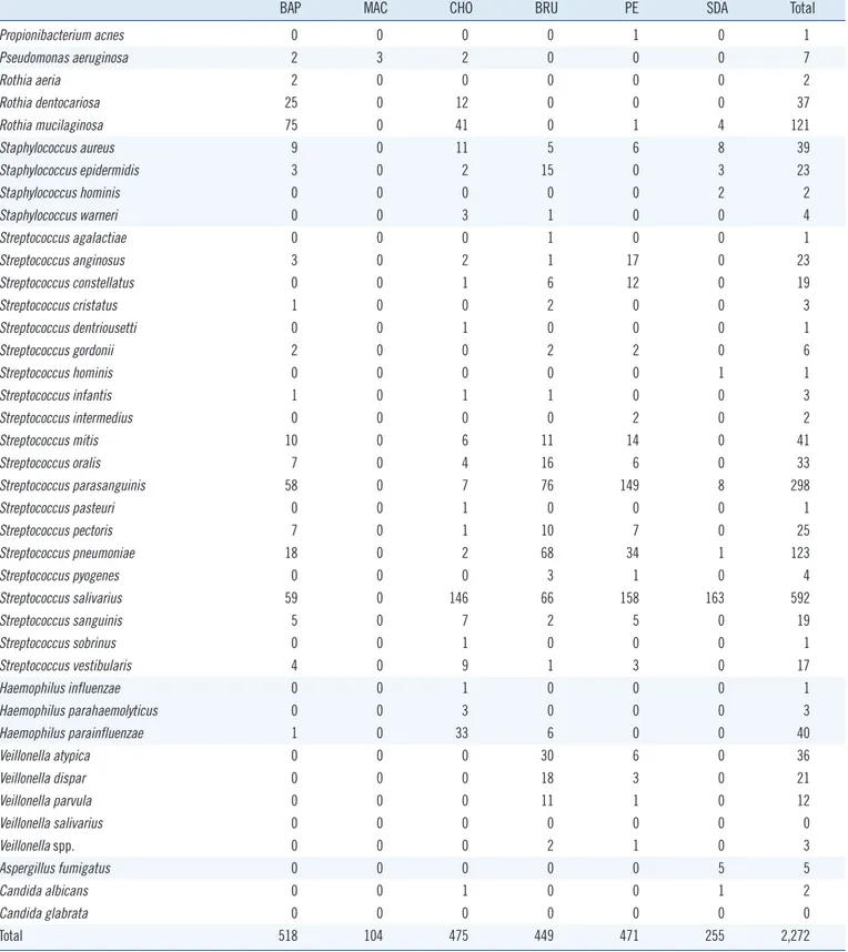
A total of 2,272 microbial colonies were isolated from 27 BAL fluid samples and identified using culture and MALDI-TOF MS. The number of genera identified per sample ranged from 4 to 14, with an average of 8. The total number of genera identified was 29; these included 85 species. Streptococcus spp. were the predominant isolates with a total number of 1,213 colonies (Table 2). Culture on blood and MacConkey agar resulted in the highest and the lowest number of microorganisms, respectively. Forty (1.76%, 40/2,272) colonies were not identified by MALDI-TOF MS. These colonies were subcultured and re-identified us-ing MALDI-TOF MS with the aid of formic acid, yieldus-ing mainly Streptococcus spp. (47.5%, 19/40). Four colonies (0.18%, 4/2,272) could not be identified with repeated MALDI-TOF MS; these were identified as Actinomyces odontolyticus, Actinomyces gravenit-zii, Fusobacterium periodonticum, and Aspergillus fumigatus by sequencing using 16S rRNA and ITS gene amplification.
2. 16S rRNA NGS identification
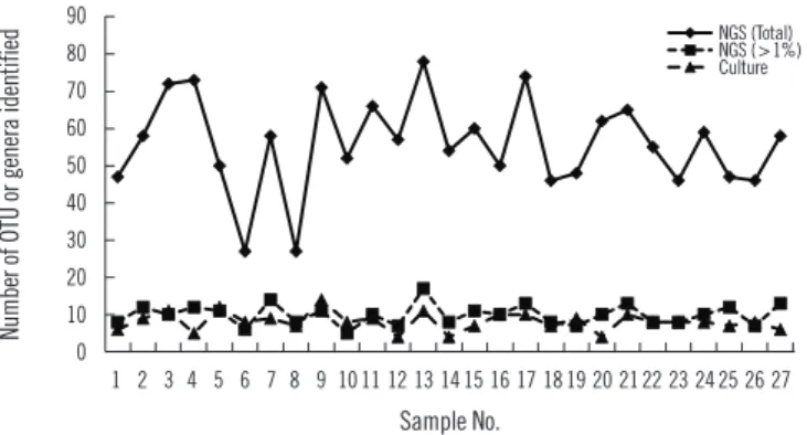
Sequencing using the GS Junior Sequencing system generated 10,332 valid reads per sample on average (range 6,761–19,764). At the genus level, a total of 288 OTUs were identified with a mean of 55.8 (range 27–78) per sample. Considering those with a relative abundance greater than 1%, the mean number of OTUs identified at the genus level was 10.0 (range 6–17) per sample (Fig. 1), which is comparable to the number of bacterial genera identified using conventional culture and MALDI-TOF MS. Pre-votella spp. was the most frequent and abundant OTU identified at the genus level; it was identified in all 27 samples (relative abundance 6.5–54.5% with a mean of 29.2%). The next most prevalent OTUs (mean relative abundance) were Streptococcus (11.5%), Neisseria (10.5%), Haemophilus (10.1%), and Veil-lonella (9.1%).
3. Comparison of microbial identification using culture and
MALDI-TOF MS vs NGS
NGS revealed more OTUs compared with the number of genera identified by culture (Fig. 1). The relative abundance of bacte-Table 1. Primers used for 16s rRNA and ITS PCR and sequencing
Primers Sequences (5´-3´) Target
27F GAGTTTGATCMTGGCTCAG Universal bacteria [32]
518R WTTACCGCGGCTGCTGG
27F AGAGTTTGATCMTGGCTCAG Universal bacteria [32]
1492R TACGGYTACCTTGTTACGACTT
ITS4_F TCCGTAGGTGAACCTGCGG Universal fungus [33]
ITS4_R TCCTCCGCTTATTGATATGC
tuf_F GCCAGTTGAGGACGTATTCT Staphylococcus [13]
tuf_R CCATTTCAGTACCTTCTGGTAA
Hemolysin_F CGACCTGATTGCATTCGCCAC Klebsiella modified from [14]
Hemolysin_R TGGTCAACCCAACGATCCTG
rpoB_F CAGGTCGTCACGGTAACAAG Enterobacter [12]
Table 2. Number of bacterial and fungal colonies on the culture media, identified by MALDI-TOF MS
BAP MAC CHO BRU PE SDA Total
Actinomyces gravenitzii 0 0 4 7 0 0 11 Actinomyces odontolyticus 1 0 14 20 17 0 52 Actinomyces oris 6 0 2 0 1 0 9 Bacillus badius 0 0 0 2 0 0 2 Bifidobacterium dentium 0 0 0 0 1 0 1 Bifidobacterium longum 0 0 0 0 2 0 2 Clostridium difficile 0 0 0 5 10 0 15 Corynebacterium argentoratense 6 0 6 0 0 0 12 Corynebacterium durum 2 0 0 0 0 0 2 Corynebacterium pseudodiphtheriticum 0 0 2 0 0 0 2 Escherichia coli 0 1 0 0 0 0 1 Fusobacterium canifelinum 0 0 0 1 0 0 1 Fusobacterium gonidiaformans 0 0 0 0 1 0 1 Fusobacterium nucleatum 0 0 0 1 0 0 1 Fusobacterium periodonticum 0 0 0 6 1 0 7 Enterobacter aerogenes 22 39 12 4 0 29 106 Enterobacter cloacae 2 0 0 0 0 0 2 Granulicatella adiacens 0 0 4 0 0 0 4 Gemella haemolysans 1 0 0 0 3 0 4 Gemella morbillorum 1 0 0 2 2 0 5 Gemella sanguinis 2 0 1 4 2 0 9 Megasphaera micronuciformis 0 0 0 3 0 0 3 Klebsiella oxytoca 4 21 4 0 0 15 44 Klebsiella pneumoniae 7 40 7 18 0 5 77 Lactobacillus fermentum 0 0 2 0 0 3 5 Lactobacillus plantarum 0 0 0 0 0 1 1 Lactobacillus salivarius 0 0 2 0 0 2 4 Leuconostoc citreum 0 0 0 0 0 3 3 Micrococcus luteus 2 0 0 2 0 0 4 Neisseria bacilliformis 2 0 0 0 0 0 2 Neisseria cinerea 2 0 0 0 0 0 2 Neisseria elongate 1 0 0 0 0 0 1 Neisseria flavescens 92 0 69 1 0 0 162 Neisseria macacae 10 0 8 0 0 0 18 Neisseria meningitidis 0 0 1 0 0 0 1 Neisseria mucosa 15 0 2 0 0 0 17 Neisseria perflava 27 0 10 0 0 0 37 Neisseria sicca 2 0 0 0 0 0 2 Neisseria subflava 15 0 21 0 0 0 36 Neisseria spp. 4 0 6 0 0 1 11 Parvimonas micra 0 0 0 1 0 0 1 Peptostreptococcus anaerobius 0 0 0 14 0 0 14 Prevotella melaninogenica 0 0 0 2 2 0 4 Prevotella pallens 0 0 0 2 0 0 2
rial reads identified simultaneously by culture was 47.1% on av-erage, ranging from 3.2% to 88.0% in the 27 samples. The
bac-teria present in BAL fluids according to the identification method (NGS, culture, or both) are shown in Fig. 2. Of the 156 bacterial
BAP MAC CHO BRU PE SDA Total
Propionibacterium acnes 0 0 0 0 1 0 1 Pseudomonas aeruginosa 2 3 2 0 0 0 7 Rothia aeria 2 0 0 0 0 0 2 Rothia dentocariosa 25 0 12 0 0 0 37 Rothia mucilaginosa 75 0 41 0 1 4 121 Staphylococcus aureus 9 0 11 5 6 8 39 Staphylococcus epidermidis 3 0 2 15 0 3 23 Staphylococcus hominis 0 0 0 0 0 2 2 Staphylococcus warneri 0 0 3 1 0 0 4 Streptococcus agalactiae 0 0 0 1 0 0 1 Streptococcus anginosus 3 0 2 1 17 0 23 Streptococcus constellatus 0 0 1 6 12 0 19 Streptococcus cristatus 1 0 0 2 0 0 3 Streptococcus dentriousetti 0 0 1 0 0 0 1 Streptococcus gordonii 2 0 0 2 2 0 6 Streptococcus hominis 0 0 0 0 0 1 1 Streptococcus infantis 1 0 1 1 0 0 3 Streptococcus intermedius 0 0 0 0 2 0 2 Streptococcus mitis 10 0 6 11 14 0 41 Streptococcus oralis 7 0 4 16 6 0 33 Streptococcus parasanguinis 58 0 7 76 149 8 298 Streptococcus pasteuri 0 0 1 0 0 0 1 Streptococcus pectoris 7 0 1 10 7 0 25 Streptococcus pneumoniae 18 0 2 68 34 1 123 Streptococcus pyogenes 0 0 0 3 1 0 4 Streptococcus salivarius 59 0 146 66 158 163 592 Streptococcus sanguinis 5 0 7 2 5 0 19 Streptococcus sobrinus 0 0 1 0 0 0 1 Streptococcus vestibularis 4 0 9 1 3 0 17 Haemophilus influenzae 0 0 1 0 0 0 1 Haemophilus parahaemolyticus 0 0 3 0 0 0 3 Haemophilus parainfluenzae 1 0 33 6 0 0 40 Veillonella atypica 0 0 0 30 6 0 36 Veillonella dispar 0 0 0 18 3 0 21 Veillonella parvula 0 0 0 11 1 0 12 Veillonella salivarius 0 0 0 0 0 0 0 Veillonella spp. 0 0 0 2 1 0 3 Aspergillus fumigatus 0 0 0 0 0 5 5 Candida albicans 0 0 1 0 0 1 2 Candida glabrata 0 0 0 0 0 0 0 Total 518 104 475 449 471 255 2,272
Abbreviations: BAP, Blood agar plate; MAC, MacConkey agar plate; CHO, Chocolate agar plate; BRU, Brucella agar plate; PE, Phenylethyl alcohol agar plate; SDA, Sabouraud dextrose agar plate.
https://doi.org/10.3343/alm.2018.38.2.110 www.annlabmed.org 115 Fig. 1. Number of the operational taxonomic units and genera
iden-tified by culture and 16S rRNA next generation sequencing analysis.
28 440
441
Figure 1.
442
Number of the operational taxonomic units and genera identified by culture and 16S rRNA next
443
generation sequencing analysis.
444 0 10 20 30 40 50 60 70 80 90 1 2 3 4 5 6 7 8 9 10 11 12 13 14 15 16 17 18 19 20 21 22 23 24 25 26 27 NGS (Total) NGS (>1%) Culture Sample No. 90 80 70 60 50 40 30 20 10 0 1 2 3 4 5 6 7 8 9 10 11 12 13 14 15 16 17 18 19 20 21 22 23 24 25 26 27 Sample No.
Number of OTU or genera identified
NGS (Total) NGS (>1%) Culture
Fig. 2. Bacterial genera identified using culture with/without sequencing and 16S rRNA next generation sequencing analysis.
29 445
Figure 2.
446
Bacterial genera identified using culture with/without sequencing and 16S rRNA next generation
447
sequencing analysis
448 449 0 5 10 15 20 25 30 Strepto co ccus Veillone lla Pre vote lla N ei sse ria R ot hi a Ha em oph ilu s Allop revotella A ctinomy ces Leptotrich ia Fuso bact erium Ca m pylo bacte r Porphy ro monas M ega sph ae ra Ge m ell a St ap hyl oco ccu s Kleb sie lla C lo st rid iu m G ra nu lic ate lla La ctob acillu s Co ry ne bac teri … Sac ch ar im onas Capn oc yt op h… Selen om onas Sn eath ia Pa rv im onas B ifi do ba ct er iu m En te rob ac te r Aggrega tibacter Bulleidia Atopo bium Ps eudo monas M ic ro coc cu s Ac in eto ba cte r Pep tostrep toc… Propion ibact e… Lach no anaero… C atonella M itsu okella R aou ltella Tr ep on em a Trop her yma B acill us Es ch er ichia Le uc onos toc Slack ia Both NGS Culture N um be r of is ol at e id en tif ie d 30 25 20 15 10 5 0StreptococcusVeillonellaPrevotella NeisseriaRothia
HaemophilusAlloprevotellaActinomycesLeptotrichiaFusobacteriumCampylobacterPorphyromonasMegasphaera Gemella Staphylococcus
KlebsiellaClostridium GranulicatellaLactobacillus
CorynebacteriumSaccharimonasCapnocytophagaSelenomonas Sneathia
Parvimonas BifidobacteriumEnterobacterAggregatibacter
BulleidiaAtopobium PseudomonasMicrococcusAcinetobacter
PeptostreptococcusPropionibacterium Lachnoanaerobaculum
CatonellaMitsuokellaRaoultellaTreponemaTropherymaBacillusEscherichiaLeuconostoc Slackia
Number of isolate identified
Both NGS Culture
Table 3. Microbial genera identified solely by culture and not by 16S rRNA next generation sequencing analysis
Genus Number of samples (%)
Staphylococcus 8 (20.5) Clostridium 7 (17.9) Aspergillus 5 (12.8) Klebsiella 4 (10.3) Bifidobacterium 3 (7.7) Candida 2 (5.1) Enterobacter 2 (5.1) Bacillus 1 (2.6) Corynebacterium 1 (2.6) Escherichia 1 (2.6) Lactobacillus 1 (2.6) Leuconostoc 1 (2.6) Micrococcus 1 (2.6) Pseudomonas 1 (2.6) Slackia 1 (2.6)
genera identified solely by NGS with >1% relative abundance, Prevotella spp. was the most common (14.1%) and frequent (22 of 27 samples, 81.5%) genus. When considered as relative abundance, the genus Prevotella spp. comprised over 50% of the bacteria that were sequenced but not cultured. Interestingly, some bacteria were identified solely by culture; of these, Staphy-lococcus spp., Clostridium spp., and Enterobacteriaceae includ-ing Klebsiella spp. were common, comprisinclud-ing 66.7% (18/27) of the 27 samples and 56.4% (22/39) of the total number of colo-nies that were cultured but not sequenced (Table 3). We per-formed organism-specific PCR using the primers detailed in Ta-ble 1 to detect Staphylococcus spp., Klebsiella spp., and
En-terobacter spp. All PCRs failed to amplify the target organisms in the samples, suggesting DNA instability in clinical samples. Candida and Aspergillus spp. were only cultured because they cannot be identified by 16S rRNA PCR.
DISCUSSION
The purpose of this study was to evaluate the ability of the stan-dard culture identification method used in clinical microbiology laboratories to elucidate microbial community profiles in BAL fluids. We compared the BAL fluid microbiota profiles identified by two methods: culture with MALDI-TOF MS and NGS. As ex-pected, we failed to detect many strict anaerobic bacteria with the culture method. The most common bacteria identified solely by 16S rRNA NGS were Prevotella spp., which are abundant in respiratory specimens [18]. In a previous study, Prevotella spp. were the most abundant OTUs in BAL fluid followed by Strepto-coccus spp. and Neisseria spp. [19]. Prevotella spp. were iso-lated on Brucella or PE agar in only five samples, although se-quencing identified this genus in all 27 samples. Interestingly, positive correlations between bacterial spp. and culture media were noted. For example, Streptococcus spp. grew prominently in chocolate agar under 5% CO2, and Brucella agar under an-aerobic conditions. In addition, it is difficult to determine whether some of the OTUs detected solely by NGS might be contaminants [20].
Other bacteria that could not be cultured included many an-aerobes such as Porphyromonas spp. and Leptotrichia spp. Fail-ure of anaerobic cultFail-ure is the most likely reason, although the viability of the sequenced bacteria without growth in culture me-dia must be considered. It is interesting that despite the limita-tions of the culture condilimita-tions adopted in this study, some bac-terial species, including Staphylococcus spp., Clostridium spp., and Bifidobacterium spp. were cultured, but not sequenced. Selective media (MacConkey agar and PE agar) and enriched media (chocolate and Brucella agar) were successful in identify-ing bacteria that were not detected usidentify-ing NGS. We hypothesize that culture can be more sensitive than sequencing in terms of detection under certain conditions and for some bacterial spe-cies, as shown in previous studies for Staphylococcus aureus [10] and Bifidobacterium spp. [21]. We assume that the bacte-ria identified solely by culture existed in very low quantities and thus failed to be sequenced. Another possible explanation is that the growth of some bacteria may be affected by culture media in association with other co-existing bacteria. The use of mixed culture via co-cultivation with helper strains could be used to fa-cilitate growth [22, 23]. As previously reported, the information provided by metagenomic approaches can be limited [24]. Many pre-analytical, analytical, and post-analytical factors can affect the outcome. DNA extraction is another step that can influence sequencing results [25-27]. DNA isolation methods, the 16S
rRNA target region used for PCR, and sequencing vary widely, and differences in techniques and manufacturers’ kits may pro-duce disparate data [28]. In this study, the V1-V3 region (27F-518R) of the 16S rRNA gene was amplified, because it is one of the most reliable targets for taxonomy. However, a sequencing depth of 10,332 valid reads per sample on average could affect the identification results. In addition, the culture method enabled the identification of fungi, such as Candida and Aspergillus, which cannot be detected by 16S rRNA sequencing.
It is known that a large proportion of the microbes found in the human gastrointestinal tract are unculturable [29, 30], al-though this has been challenged by recent studies revealing many rare microorganisms. Metagenomics by deep sequencing is undoubtedly a powerful tool in microbial community analysis; however, it possesses some limitations. Sequenced DNA is not evidence of viable bacteria, and detection sensitivity is depen-dent on sequencing depth, which may result in a failure to iden-tify low-abundance bacterial species. NGS approaches, espe-cially those based on amplification of the highly conserved 16S rRNA gene, are inherently incapable of detecting intra-species variations [24]. Cultivation of microbes provides a greater un-derstanding of bacterial interactions and traits and can be used to obtain completely sequenced genomes from these species. In addition, currently, the culture method with MALDI-TOF MS has advantages over NGS, in terms of cost and practical appli-cation. Although NGS has reduced the cost of sequencing greatly, it is still a fairly expensive technology and requires bioinformatic tools for analysis.
Recent efforts in developing culture methods have enabled the culture of previously ‘as-yet uncultured’ bacteria, known as culturomics [10, 31, 32]. The use of several different culture conditions has enabled the isolation of new bacterial species, the largest human virus, the largest bacterium, and the largest archaeon from humans [33]. One study demonstrated that anal-yses of the same samples produced only partially overlapping results; 51 of the 340 species and 698 phylotypes were identi-fied by culturomics and metagenomic analysis, respectively [10]. However, various culture conditions, including many nonselec-tive and selecnonselec-tive culture media used in culturomics, are not prac-tical for application in conventional clinical microbiology labora-tories. MALDI-TOF MS is another powerful tool used in micro-bial identification. MALDI-TOF MS has allowed for the accurate identification of a large panel of anaerobes that are poorly iden-tified because of ambiguous or erroneous results using conven-tional methods. Coupling MALDI-TOF MS with culturomics has enabled the identification of a large collection of bacterial
spe-cies, including anaerobes that are usually poorly identified with current phenotypic methods, from stool specimens [34]. When a limited number of culture conditions commonly used in clini-cal microbiology laboratories were applied, all the isolated bac-teria were present in the MALDI-TOF MS database, although some bacteria were poorly identified and needed further 16S rRNA or ITS sequencing.
Our study has some limitations. We used limited types of cul-ture media commonly used in clinical microbiology laboratories. While picking the colonies from each agar plate, some bacteria may have been missed because of selection bias. The agar plates were incubated for only 48 hours; this duration may not have been adequate for slow-growing microorganisms. Although we tried to inoculate the samples as soon as possible and within 6 hours post collection, some anaerobic bacteria may have been lost during the prolonged oxygen exposure. The number of reads, ~10,000 per sample on an average in this study, may also affect the bacterial species detected in each sample. Most of the bac-terial species were successfully identified using MALDI-TOF MS. However, the reliability of streptococci identification might not be high because of the inherent limitations of MALDI-TOF MS. Lastly, we did not quantify the microorganisms in in culture method, which is a limitation in exploring microbial diversity.
To the best of our knowledge, this is the first study to evaluate the use of conventional culture methods combined with MALDI-TOF MS in microbiome analysis of BAL fluid samples compared with 16S rRNA NGS. Some bacteria, mainly anaerobes, failed to grow on conventional culture media, and less diverse microbial communities were identified with the culture method. However, interestingly, some common bacteria were not identified by se-quencing, but were cultured, indicating the significance of cul-ture in microbiome analysis. Despite a number of limitations, culture methods with MALDI-TOF MS may play a significant role in microbiome analysis that is complementary to 16S rRNA NGS.
Authors’ Disclosures of Potential Conflicts of
Interest
No potential conflicts of interest relevant to this article were re-ported.
Acknowledgments
This work was supported by the National Research Foundation of Korea (2014M3A9E5073818), the BioNano Health-Guard Research Center funded by the Ministry of Science, ICT &
Fu-ture Planning (MSIP) of Korea as a Global Frontier Project (H-GUARD_2014M3A6B2060509), and the Ministry of Health & Welfare, Republic of Korea (HI14C1324).
REFERENCES
1. Twigg HL 3rd, Morris A, Ghedin E, Curtis JL, Huffnagle GB, Crothers K, et al. Use of bronchoalveolar lavage to assess the respiratory microbi-ome: signal in the noise. Lancet Respir Med 2013;1:354-6.
2. Charlson ES, Bittinger K, Haas AR, Fitzgerald AS, Frank I, Yadav A, et al. Topographical continuity of bacterial populations in the healthy hu-man respiratory tract. Am J Respir Crit Care Med 2011;184:957-63. 3. Erb-Downward JR, Thompson DL, Han MK, Freeman CM, McCloskey L,
Schmidt LA, et al. Analysis of the lung microbiome in the “healthy” smo-ker and in COPD. PLoS One 2011;6:e16384.
4. Sze MA, Dimitriu PA, Hayashi S, Elliott WM, McDonough JE, Gosselink JV, et al. The lung tissue microbiome in chronic obstructive pulmonary disease. Am J Respir Crit Care Med 2012;185:1073-80.
5. Morris A, Beck JM, Schloss PD, Campbell TB, Crothers K, Curtis JL, et al. Comparison of the respiratory microbiome in healthy nonsmokers and smokers. Am J Respir Crit Care Med 2013;187:1067-75.
6. Hamady M and Knight R. Microbial community profiling for human mi-crobiome projects: tools, techniques, and challenges. Genome Res 2009; 19:1141-52.
7. Allen-Vercoe E. Bringing the gut microbiota into focus through microbial culture: recent progress and future perspective. Curr Opin Microbiol 2013;16:625-9.
8. Sommer MO. Advancing gut microbiome research using cultivation. Curr Opin Microbiol 2015;27:127-32.
9. Goodman AL, Kallstrom G, Faith JJ, Reyes A, Moore A, Dantas G, et al. Extensive personal human gut microbiota culture collections character-ized and manipulated in gnotobiotic mice. Proc Natl Acad Sci U S A 2011;108:6252-7.
10. Lagier JC, Armougom F, Million M, Hugon P, Pagnier I, Robert C, et al. Microbial culturomics: paradigm shift in the human gut microbiome study. Clin Microbiol Infect 2012;18:1185-93.
11. Rettedal EA, Gumpert H, Sommer MO. Cultivation-based multiplex phe-notyping of human gut microbiota allows targeted recovery of previously uncultured bacteria. Nat Commun 2014;5:4714.
12. Brady C, Cleenwerck I, Venter S, Vancanneyt M, Swings J, Coutinho T. Phylogeny and identification of Pantoea species associated with plants, humans and the natural environment based on multilocus sequence analysis (MLSA). Syst Appl Microbiol 2008;31:447-60.
13. Heikens E, Fleer A, Paauw A, Florijn A, Fluit AC. Comparison of geno-typic and phenogeno-typic methods for species-level identification of clinical isolates of coagulase-negative staphylococci. J Clin Microbiol 2005;43: 2286-90.
14. Neuberger A, Oren I, Sprecher H. Clinical impact of a PCR assay for rapid identification of Klebsiella pneumoniae in blood cultures. J Clin Microbiol 2008;46:377-9.
15. Liu Z, Lozupone C, Hamady M, Bushman FD, Knight R. Short pyrose-quencing reads suffice for accurate microbial community analysis. Nu-cleic Acids Res 2007;35:e120.
16. Edgar RC, Haas BJ, Clemente JC, Quince C, Knight R. UCHIME improves sensitivity and speed of chimera detection. Bioinformatics 2011;27:2194-200.
17. Kim OS, Cho YJ, Lee K, Yoon SH, Kim M, Na H, et al. Introducing Ez-Taxon-e: a prokaryotic 16S rRNA gene sequence database with
phylo-types that represent uncultured species. Int J Syst Evol Microbiol 2012; 62:716-21.
18. Charlson ES, Bittinger K, Chen J, Diamond JM, Li H, Collman RG, et al. Assessing bacterial populations in the lung by replicate analysis of sam-ples from the upper and lower respiratory tracts. PLoS One 2012;7:e42786. 19. Bassis CM, Erb-Downward JR, Dickson RP, Freeman CM, Schmidt TM,
Young VB, et al. Analysis of the upper respiratory tract microbiotas as the source of the lung and gastric microbiotas in healthy individuals. MBio 2015;6:e00037.
20. Salter SJ, Cox MJ, Turek EM, Calus ST, Cookson WO, Moffatt MF, et al. Reagent and laboratory contamination can critically impact sequence-based microbiome analyses. BMC Biol 2014;12:87.
21. Walker AW, Martin JC, Scott P, Parkhill J, Flint HJ, Scott KP. 16S rRNA gene-based profiling of the human infant gut microbiota is strongly in-fluenced by sample processing and PCR primer choice. Microbiome 2015;3:26.
22. Davis IJ, Bull C, Horsfall A, Morley I, Harris S. The Unculturables: tar-geted isolation of bacterial species associated with canine periodontal health or disease from dental plaque. BMC Microbiol 2014;14:196. 23. Ohno M, Okano I, Watsuji T, Kakinuma T, Ueda K, Beppu T.
Establish-ing the independent culture of a strictly symbiotic bacterium Symbio-bacterium thermophilum from its supporting Bacillus strain. Biosci Bio-technol Biochem 1999;63:1083-90.
24. Hiergeist A, Glasner J, Reischl U, Gessner A. Analyses of Intestinal Mi-crobiota: Culture versus Sequencing. ILAR J 2015;56:228-40. 25. Maukonen J, Simoes C, Saarela M. The currently used commercial
DNA-extraction methods give different results of clostridial and actinobacteri-al populations derived from human fecactinobacteri-al samples. FEMS Microbiol Ecol
2012;79:697-708.
26. Willner D, Daly J, Whiley D, Grimwood K, Wainwright CE, Hugenholtz P. Comparison of DNA extraction methods for microbial community profil-ing with an application to pediatric bronchoalveolar lavage samples. PLoS One 2012;7:e34605.
27. Kennedy NA, Walker AW, Berry SH, Duncan SH, Farquarson FM, Louis P, et al. The impact of different DNA extraction kits and laboratories upon the assessment of human gut microbiota composition by 16S rRNA gene sequencing. PLoS One 2014;9:e88982.
28. Beck JM. ABCs of the lung microbiome. Ann Am Thorac Soc 2014;11 Suppl 1:S3-6.
29. Eckburg PB, Bik EM, Bernstein CN, Purdom E, Dethlefsen L, Sargent M, et al. Diversity of the human intestinal microbial flora. Science 2005;308: 1635-8.
30. Zoetendal EG, Vaughan EE, de Vos WM. A microbial world within us. Mol Microbiol 2006;59:1639-50.
31. Fournier PE, Lagier JC, Dubourg G, Raoult D. From culturomics to tax-onomogenomics: a need to change the taxonomy of prokaryotes in clini-cal microbiology. Anaerobe 2015;36:73-8.
32. Lagier JC, Hugon P, Khelaifia S, Fournier PE, La Scola B, Raoult D. The rebirth of culture in microbiology through the example of culturomics to study human gut microbiota. Clin Microbiol Rev 2015;28:237-64. 33. Lagier JC, Million M, Hugon P, Armougom F, Raoult D. Human gut
mi-crobiota: repertoire and variations. Front Cell Infect Microbiol 2012;2:136. 34. Samb-Ba B, Mazenot C, Gassama-Sow A, Dubourg G, Richet H, Hugon
P, et al. MALDI-TOF Identification of the Human Gut Microbiome in Peo-ple with and without Diarrhea in Senegal. Plos One 2014;9:e87419.