Received September 12, 2014, Revised March 3, 2015, Accepted for publication March 5, 2015
Corresponding author: Kee Yang Chung, Department of Dermatology, Severance Hospital, Cutaneous Biology Research Institute, Yonsei University College of Medicine, 50 Yonsei-ro, Seodaemun-gu, Seoul 03722, Korea. Tel: 82-2-2228-2080, Fax: 82-2-393-9157, E-mail: kychung@yuhs.ac
This is an Open Access article distributed under the terms of the Creative Commons Attribution Non-Commercial License (http:// creativecommons.org/licenses/by-nc/4.0) which permits unrestricted non-commercial use, distribution, and reproduction in any medium, provided the original work is properly cited.
ORIGINAL ARTICLE
Treatment of Nodular Fasciitis Occurring on the Face
Byung Ho Oh, Jihee Kim, Zhenlong Zheng, Mi Ryung Roh, Kee Yang Chung
Department of Dermatology and Cutaneous Biology Research Institute, Yonsei University College of Medicine, Seoul, Korea
Background: Surgical excision is generally recommended for the treatment of nodular fasciitis (NF) to rule out sarcoma. However, in cases of NF occurring on the face, the reported recurrence rate is higher and the surgical approach may re-sult in considerable aesthetic concern. Objective: To de-scribe our experience with NF occurring on the face and evaluate the outcomes of surgical and nonsurgical methods of treatment. Methods: We performed a retrospective review of 16 patients with NF on the face. The patients were treated with surgical excision or nonsurgical methods such as tri-amcinolone intralesional injection (TA ILI) and pinhole method with a carbon dioxide (CO2) laser. Results: Among
the 16 patients, surgical treatment was performed in 9 and currence occurred in 7 of these 9 patients (77.8%). The re-curred lesions showed regression after repeated TA ILI. On the other hand, five patients underwent nonsurgical treat-ment after the histologic exclusion of malignancy. Their le-sions showed regression after repeated pinhole treatment and TA ILI. In one case, NF spontaneously regressed. On a visual analogue scale, the nonsurgical approach showed su-perior results. However, the values were not statistically sig-nificant (6.90±1.56 vs. 5.61±1.36; p=0.163). The sat-isfaction level was lower in patients who experienced re-currence after surgical excision. Conclusion: Surgical treat-ment for NF on the face showed a noticeable recurrence rate and resulted in scarring. Therefore, considering the possi-bility of spontaneous regression, the nonsurgical method can
be considered as an alternative treatment option for NF on the face. (Ann Dermatol 27(6) 694∼701, 2015)
-Keywords-Intralesional injection, Laser therapy, Nodular fasciitis
INTRODUCTION
Nodular fasciitis (NF) is a benign proliferative myofibro-blastic lesion that may regress spontaneously1. Although
the etiology of NF is uncertain, histopathologically, it bears a close resemblance to organizing granulation tis-sue, and myofibroblastic proliferation may be initiated by a local injury or local inflammatory process, which sup-ports a reactive proliferation theory triggered by trauma2,3.
NF is frequently mistaken for sarcoma both clinically and histopathologically. Therefore, surgical excision is recom-mended for diagnosis and treatment to exclude malig-nancy4. However, for lesions occurring on the face,
non-surgical treatment and regular follow-up can be consid-ered because surgical treatment may cause scarring on the face and the lesions may regress spontaneously.
For the nonsurgical treatment of NF on the face, triam-cinolone intralesional injection (TA ILI) can be used on the basis of the fact that NF is a benign reactive or inflam-matory condition of mesenchymal fibroblasts5. In
addi-tion, considering that early lesions of NF have a myxoid pattern of myofibroblast proliferation6, laser treatment can
be effective in decreasing the size of the lesions through tissue contraction. Especially, NF on the face are usually ‘intradermal type,’ which extends beyond the fascia and subcutaneous tissue and invades into the skin, as facial muscles such as platysma, frontalis, and orbicularis oculi are located superficially underneath the skin7. Therefore,
such intradermal type of NF can be affected by laser treat-ment from the skin surface. For example, multiple pin-holes created by a carbon dioxide (CO2) laser on the skin
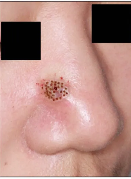
re-Fig. 1. Nonsurgical treatment with a pinhole method with a CO2
laser. duction and dermal collagen remodeling8,9.
In this study, the authors retrospectively reviewed cases of NF occurring on the face that were treated by using surgi-cal and nonsurgisurgi-cal methods such as TA ILI and a pinhole method with a CO2 laser, and evaluated the outcomes of
treatment.
MATERIALS AND METHODS
We performed a retrospective review of 16 patients (6 males, 10 females) with NF on the face clinically and his-topathologically diagnosed from December 2010 to De-cember 2013 at Severance Hospital in the Yonsei University Health System. Each patient’s details from the medical re-cord, including age, sex, onset location, trauma history, clinical photographs, histopathologic findings, treatment methods, clinical progress, and complication profile, were fully reviewed. The study protocol was approved by the institutional review board of Yonsei University Severance Hospital (IRB No. 4-2015-0928) and was conducted ac-cording to the Declaration of Helsinki Principles.
Histopathologic findings
To evaluate the possibility of nonsurgical treatments, we reexamined the histopathologic features. For this review, two dermatologists individually reexamined the H&E-stained sections, and evaluated the following features in a blind manner: depth of the lesion, type of cellular composition, and the presence of cellular atypia. Additionally, the pres-ence of a loose myxoid pattern, which can provide a po-tential space for injectable agents, was also evaluated. If there were atypical cells in the H&E stained section, im-munohistochemical staining for smooth muscle actin, des-min, pan-cytokeratin, CD34, S100 protein, and other mark-ers, was performed to exclude malignancy.
Nonsurgical treatments
The nonsurgical approach was indicated when the diag-nosis of NF is confirmed and the potential for malignancy is excluded with immunohistochemistry in the presence of atypical spindle cells. When recurrence occurred after the surgical treatment, TA ILI was used after the reevaluation of the excised specimen. The pinhole method with CO2
laser and TA ILI were repeatedly performed simultaneously every 4 weeks. Before the treatment, a topical eutectic mixture of 2.5% lidocaine and 2.5% prilocaine (EMLA cream; AstraZeneca AB, Södertälje, Sweden) was applied for local anesthesia. Treatment was performed with an output power of 1 to 4 W to make multiple small holes approximately 1 to 2 mm in diameter and spaced 2 to 5 mm apart (Fig. 1). Posttreatment pain was tolerable, and
erythema spontaneously resolved. Immediately after the pinhole treatment, TA ILI was applied to the deep and firm part of the NF lesion.
Evaluation of the clinical outcomes
For the evaluation of the clinical outcomes, all photo-graphic images of NF patients taken 3 months after the fi-nal treatment were individually evaluated by two derma-tologists by using a visual analogue scale (VAS). The VAS is a 10-cm linear scale in which the left of the scale (score of 0) reflects the worst outcome and the right of the scale (score of 10) reflects the best outcome. At the end of the treatment, patients’ satisfaction was evaluated on a 4-point grading scale (0=unsatisfied, 1=slightly satisfied, 2=sat-isfied, and 3=very satisfied).
Statistical analysis
To compare the outcomes of treatments between the sur-gical and nonsursur-gical methods, we performed statistical analysis by using the nonparametric Mann-Whitney U test, where p-values ≤0.05 were considered statistically significant. All data were analyzed by using SPSS 12.0 (SPSS Inc., Chicago, IL, USA).
RESULTS
The mean age of the 16 patients who had NF on the face was 36.6 years (range, 15∼61 years). The average disease duration was 3.94 months (range, 3 weeks∼12 months). In nine cases (56.3%), patients had histories of physical stimulation or trauma, such as facial massage, at least once
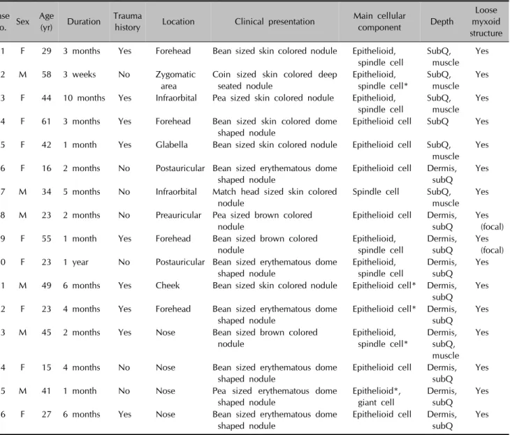
Table 1. Clinical characteristics and histopathologic features of 16 patients with nodular fasciitis Case no. Sex Age (yr) Duration Trauma
history Location Clinical presentation
Main cellular
component Depth
Loose myxoid structure 1 F 29 3 months Yes Forehead Bean sized skin colored nodule Epithelioid,
spindle cell SubQ, muscle Yes 2 M 58 3 weeks No Zygomatic area
Coin sized skin colored deep seated nodule Epithelioid, spindle cell* SubQ, muscle Yes 3 F 44 10 months Yes Infraorbital Pea sized skin colored nodule Epithelioid,
spindle cell
SubQ, muscle
Yes 4 F 61 3 months Yes Forehead Bean sized skin colored dome
shaped nodule
Epithelioid cell SubQ Yes 5 F 42 1 month Yes Glabella Bean sized skin colored nodule Epithelioid cell SubQ,
muscle Yes 6 F 16 2 months No Postauricular Bean sized erythematous dome
shaped nodule
Epithelioid cell Dermis, subQ
Yes 7 M 34 5 months No Infraorbital Match head sized skin colored
nodule
Spindle cell SubQ, muscle
Yes 8 M 23 2 months No Preauricular Pea sized brown colored
nodule
Epithelioid cell Dermis, subQ
Yes (focal) 9 F 55 1 month Yes Forehead Bean sized brown colored
nodule Epithelioid, spindle cell Dermis, subQ Yes (focal) 10 F 23 1 year No Postauricular Bean sized erythematous dome
shaped nodule Epithelioid, spindle cell Dermis, subQ Yes 11 M 49 6 months Yes Cheek Bean sized skin colored nodule Epithelioid cell* Dermis,
subQ
Yes 12 F 23 4 months Yes Forehead Bean sized erythematous dome
shaped nodule
Epithelioid cell* Dermis, subQ
Yes
13 M 45 2 months Yes Nose Bean sized brown colored
nodule Epithelioid, spindle cell* Dermis, subQ, muscle Yes
14 F 15 4 months No Nose Bean sized erythematous dome
shaped nodule
Epithelioid cell Dermis, subQ
Yes
15 M 41 1 month No Nose Pea sized erythematous dome
shaped nodule Epithelioid*, giant cell Dermis, subQ Yes
16 F 27 6 months Yes Nose Bean sized erythematous dome
shaped nodule
Epithelioid cell Dermis, subQ
Yes F: female, M: male, SubQ: subcutaneous fat. Atypical cells were all negative for desmin, pan-cytokeratin, CD34, S100 protein, MART-1, and positive for smooth muscle actin in immunohistochemical stains.
before the development of NF lesions (Table 1).
Histopathologic examination showed that NF consisted mainly of epithelioid and spindle cells. A loose myxoid structure was observed in all cases (Fig. 2). In five cases, a few atypical cells were noted in the H&E stain; however, they were all negative for desmin, pan-cytokeratin, CD34, S100 protein, or MART-1, and positive for smooth muscle actin in immunohistochemical stains. All lesions involved subcutaneous tissues, whereas seven cases (43.8%) showed the involvement of muscle layers, and in 10 cases (62.5%) dermal involvement was observed. Among the 16 cases, surgical excision was performed in 9 and recurrence de-veloped in 7 of these 9 cases (77.8%), which were all his-topathologically confirmed as NF from the excised
spe-cimens. However, the recurred lesions regressed after re-petitive TA ILI (Fig. 3). In case no. 2, Mohs micrographic surgery was performed for the complete removal of the le-sion because of some atypical cells with mitotic figures in the coin-sized lesion and the relatively older age of the pa-tient (Fig. 4). The pinhole method with a CO2 laser and TA
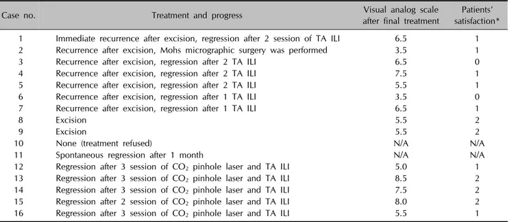
ILI, two to three times every 4 weeks, resulted in re-gression of the five cases of intradermal-type NF (Fig. 5). One of the two patients who did not receive any treatment showed spontaneous regression after 1 month (Table 2). The final outcomes were evaluated on the basis of the VAS. The mean score of cases no. 12 to 16 who received pinhole method treatments (6.90±1.56) was slightly high-er than that of cases no. 1 to 9 who received surgical
ex-Fig. 3. Flow chart of the treatment outcome. TA ILI: triamcinolone in-tralesional injection.
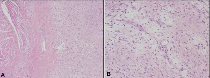
Fig. 2. (A) Benign fibroblastic proliferation in subcutaneous areas around facial muscle, which are mostly composed of epithelioid and spindle cells (H&E stain, ×40). (B) Loose myxoid structure observed on higher magnification (H&E stain, ×100).
cision (5.61±1.36); however, the difference was not stat-istically significant (p=0.163). Concerning patients’ sat-isfaction, the group that received pinhole treatments scor-ed 1 to 2; however, the group with recurrence after the surgical excision scored relatively lower (0∼1) (Table 2). Neither the surgically nor the nonsurgically treated group reported adverse events such as severe pain or secondary infection.
DISCUSSION
Because malignant transformation seldom occurs in be-nign skin lesions, treatment is not always a requirement as long as patients do not mind the cosmetic problems result-ing from the skin lesions. In cases of NF on the face, pro-active efforts are needed to perform histopathologic evalu-ation and treatment because NF tends to grow rapidly on the face of adults between the age of 20 and 40 years (i.e.,
persons who are involved in many social activities)10. Although wide excision is recommended for the treatment of NF on the extremities or trunk, it should be reconsid-ered in facial lesions as it presents several problems in-cluding scarring. First, surgical excision may not remove enough of the area surrounding the lesion because it is dif-ficult to clearly determine the scope of the lesion because inflammation extends to the adjacent muscles and sub-cutaneous tissues. Particularly, the risk of insufficient ex-cision can be higher in the face because surgeons try to re-duce scarring as much as possible. Second, surgical proce-dures may act as another precipitating factor as NF is a myofibroblast proliferation that can be initiated by local injury. In this retrospective study, seven of nine patients (77.8%) who received surgical excision experienced local recurrence, which was a higher rate than those reported for other areas of the body10,11. We assume that this result
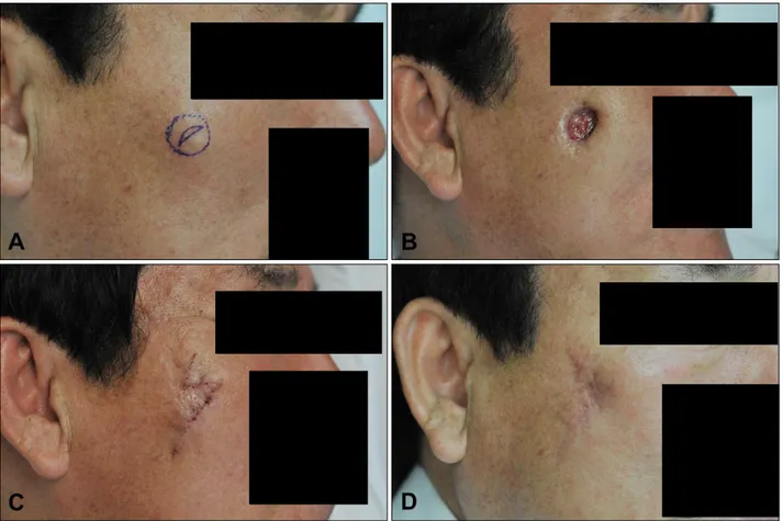
surgi-Fig. 4. Serial photographs of a representative patient (case no. 2) treated with the surgical approach. (A) A coin-sized, skin-colored, deep-seated nodule that recurred on the right zygomatic area. (B) The recurrent lesion was completely removed with Mohs micrographic surgery. (C) Reconstruction was done by using a rhombic flap. (D) Surgical treatment eventually resulted in scarring after 3 months. cal excision in facial lesions or the increased trauma caused
by surgery. Therefore, when only considering scarring and the higher recurrence rate, surgical wide excision is not recommended for the first treatment of NF on the face. Five patients who underwent nonsurgical treatments, such as a repeated pinhole treatments and TA ILI, in this study showed lesion regression and satisfaction. The prerequisite for nonsurgical treatment is a histopathologic confirmation to rule out sarcoma. It was reported that fibrosarcoma typi-cally occurs in patients older than 50 years and in lesions >3 cm12. Although marked cellular atypia is rare in NF le-sions, cells of fibrosarcoma show more nuclear pleo-morphism with dense collagenous stroma and without in-flammatory components. In immunohistochemical stain-ing, lesional cells of NF can be reactive to vimentin, smooth muscle actin, muscle-specific actin, and KP-111.
For the differential diagnosis, desmin, which is rarely ex-pressed in NF, may be used to distinguish NF from leio-myoma and leiomyosarcoma13. In addition, it is possible
to use CD34 to differentiate NF from dermatofibrosarcoma protuberans. However, the Ki67 index, which has been
documented as useful for the diagnosis and prognosis of soft tissue sarcoma14, is not suitable for the assessment of
NF because of the variation in NF lesions12.
Recently, along with the vast development of laser devices, various nonsurgical approaches have been applied in the treatment of benign skin tumors. For dermatofibroma (benign fibrous histiocytoma), one of the most common benign soft tissue tumors, a pulsed dye laser15, fractional laser
with topical corticosteroid16, or traditional cryotherapy17
have been applied. As the cells of newly developed NF are embedded in a loose myxoid stroma6, intralesional de-livery of medication can be done successfully. In our study, loose myxoid patterns were found in all cases, and TA ILI treatment of the lesions resulted in regression. However, the effects of intralesional injection may de-crease for old lesions with inde-creased cellular, fibrous com-ponents and decreased loose parts, because intralesional injection into fibrous and hard lesions is difficult to perform.
From our results, a pinhole method with a CO2 laser was
Fig. 5. Serial photographs of repre-sentative patients treated with the nonsurgical approach. (A) A bean- sized, brown nodule on the nose (case no. 13). (B) Three months later, after three sessions of pinhole laser treatment and triamcinolone intra-lesional injection (TA ILI). (C) A bean-sized, erythematous, dome-sha-ped nodule on the nose (case no. 14). (D) Three months later, after three sessions of pinhole laser trea-tment and TA ILI.
those on anatomic locations that may involve significant aesthetic concern. CO2 laser treatment may induce tissue
shrinkage and decrease in the size of NF lesion. The pin-hole laser treatment has been reported to be an effective method in scar management by inducing tissue shrinkage and scar remodeling18,19. When it is applied to myxoid
le-sions, its effects may become intensified because the CO2
laser wavelength is strongly absorbed by water. In addi-tion, when the pinhole technique is combined with TA ILI, inhibition of fibroblast proliferation would also be achieved. As the scarring left by this procedure is much smaller than the size of the original lesion, scar revision can easily be performed, if necessary, after nonsurgical treatments. In this retrospective study, the superior cosmetic outcomes of the pinhole method resulted in a relatively higher patient satisfaction.
Nine of the 16 patients in this study (56.3%) had a history
of receiving physical stimulation or trauma such as facial massages. Although the relation between NF occurrence and trauma has not been clearly defined, the possible oc-currence of NF on the face should be closely monitored as persons become more interested in skin care that may give physical stimuli to skin.
Despite the limitation of being a retrospective study with a relatively small sample size, we experienced successful outcomes with a nonsurgical approach to NF on the face. However, potential malignancy should not be overlooked, and before any nonsurgical approach, histologic con-firmation with a sufficiently deep skin biopsy including skin and facial muscle parts should be done first. For le-sions showing recurrence or regrowth with different clin-ical features from the original lesion, additional skin biop-sy would be required for further histologic analysis. In conclusion, considering the potential aesthetic
disfigure-Table 2. Treatment outcomes and overall progress of 16 patients with nodular fasciitis
Case no. Treatment and progress after final treatmentVisual analog scale satisfaction*Patients’ 1 Immediate recurrence after excision, regression after 2 session of TA ILI 6.5 1
2 Recurrence after excision, Mohs micrographic surgery was performed 3.5 1
3 Recurrence after excision, regression after 2 TA ILI 6.5 0
4 Recurrence after excision, regression after 2 TA ILI 7.5 1
5 Recurrence after excision, regression after 2 TA ILI 5.5 1
6 Recurrence after excision, regression after 1 TA ILI 3.5 0
7 Recurrence after excision, regression after 1 TA ILI 6.5 1
8 Excision 5.5 2
9 Excision 5.5 2
10 None (treatment refused) N/A N/A
11 Spontaneous regression after 1 month N/A N/A
12 Regression after 3 session of CO2 pinhole laser and TA ILI 5.0 1
13 Regression after 3 session of CO2 pinhole laser and TA ILI 8.5 2
14 Regression after 3 session of CO2 pinhole laser and TA ILI 7.5 2
15 Regression after 2 session of CO2 pinhole laser and TA ILI 8.0 2
16 Regression after 3 session of CO2 pinhole laser and TA ILI 5.5 1
TA ILI: triamcinolone intralesional injection, N/A: not applied. *4-point grading scale: 0=unsatisfied, 1=slightly satisfied, 2=satisfied, 3=very satisfied.
ment and recurrence after conventional wide excision, re-petitive TA ILI and CO2 pinhole laser can be
recom-mended as a less invasive treatment option.
ACKNOWLEDGMENT
This research was supported by the Basic Science Re-search Program through the National ReRe-search Foundation of Korea (NRF) funded by the Ministry of Science, ICT and Future Planning (no. 2013R1A2A2A04015894).
REFERENCES
1. Yanagisawa A, Okada H. Nodular fasciitis with degeneration and regression. J Craniofac Surg 2008;19:1167-1170. 2. Bernstein KE, Lattes R. Nodular (pseudosarcomatous) fasciitis,
a nonrecurrent lesion: clinicopathologic study of 134 cases. Cancer 1982;49:1668-1678.
3. Kang SK, Kim HH, Ahn SJ, Choi JH, Sung KJ, Moon KC, et al. Intradermal nodular fasciitis of the face. J Dermatol 2002;29:310-314.
4. Ko CJ. Dermal hypertrophies and benign fibroblastic/ myofibroblastic tumors. In: Goldsmith LA, Fitzpatrick TB, editors. Fitzpatrick's dermatology in general medicine. 8th ed. New York: McGraw-Hill, 2012:715-716.
5. Graham BS, Barrett TL, Goltz RW. Nodular fasciitis: response to intralesional corticosteroids. J Am Acad Dermatol 1999; 40:490-492.
6. Shimizu S, Hashimoto H, Enjoji M. Nodular fasciitis: an analysis of 250 patients. Pathology 1984;16:161-166. 7. de Feraudy S, Fletcher CD. Intradermal nodular fasciitis: a
rare lesion analyzed in a series of 24 cases. Am J Surg Pathol 2010;34:1377-1381.
8. Whang SW, Lee KY, Cho SB, Lee SJ, Kang JM, Kim YK, et al. Burn scars treated by pinhole method using a carbon dioxide laser. J Dermatol 2006;33:869-872.
9. Lee SJ, Yeo IK, Kang JM, Chung WS, Kim YK, Kim BJ, et al. Treatment of hypertrophic burn scars by combination laser- cision and pinhole method using a carbon dioxide laser. Lasers Surg Med 2014;46:380-384.
10. Spinelli N, Khorassani N. Nodular fasciitis: an uncommon disease with common medical management challenges at a remote Naval Hospital. Mil Med 2013;178:e1051-e1054. 11. Thompson LD, Fanburg-Smith JC, Wenig BM. Nodular
fasciitis of the external ear region: a clinicopathologic study of 50 cases. Ann Diagn Pathol 2001;5:191-198.
12. Lin XY, Wang L, Zhang Y, Dai SD, Wang EH. Variable Ki67 proliferative index in 65 cases of nodular fasciitis, compared with fibrosarcoma and fibromatosis. Diagn Pathol 2013;8:50. 13. Souza e Souza Id, Rochael MC, Farias RE, Vieira RB, Vieira
JS, Schimidt NC. Nodular fasciitis on the zygomatic region: a rare presentation. An Bras Dermatol 2013;88(6 Suppl 1):89-92.
14. Engellau J, Persson A, Bendahl PO, Akerman M, Domanski HA, Bjerkehagen B, et al. Expression profiling using tissue microarray in 211 malignant fibrous histiocytomas confirms the prognostic value of Ki-67. Virchows Arch 2004;445: 224-230.
15. Alonso-Castro L, Boixeda P, Segura-Palacios JM, de Daniel- Rodríguez C, Jiménez-Gómez N, Ballester-Martínez A. Dermatofibromas treated with pulsed dye laser: Clinical and dermoscopic outcomes. J Cosmet Laser Ther 2012;14:98- 101.
a symptomatic dermatofibroma with fractionated carbon dioxide laser and topical corticosteroids. J Drugs Dermatol 2013;12:1483-1484.
17. Lanigan SW, Robinson TW. Cryotherapy for dermatofibro-mas. Clin Exp Dermatol 1987;12:121-123.
18. Cho SB, Lee SJ, Kang JM, Kim YK, Kim TY, Kim DH. The treatment of burn scar-induced contracture with the pinhole
method and collagen induction therapy: a case report. J Eur Acad Dermatol Venereol 2008;22:513-514.
19. Chung BY, Han SS, Kim BW, Chang SE, Lee MW. Effective treatment of congenital melanocytic nevus and nevus se-baceous using the pinhole method with the erbium-doped yttrium aluminium garnet laser. Ann Dermatol 2014;26: 651-653.