Efficient Induction of Oligodendrocytes from Human Embryonic
Stem Cells
S
ANG-M
OONK
ANG,
a,bM
YUNGS
OOC
HO,
cH
YEMYUNGS
EO,
dC
HULJ
ONGY
OON,
eS
UNK
YUNGO
H,
f,gY
OUNGM
INC
HOI,
f,gD
ONG-W
OOKK
IMa,b,ha
Department of Physiology,
bBrain Korea 21st Project for Medical Science,
hCenter for Cell Therapy, Yonsei
University College of Medicine, Seoul, Korea;
cR&D Center, Jeil Pharmaceutical Company, Yongin, Korea;
d
Division of Molecular & Life Sciences, College of Science & Technology, Hanyang University, Ansan, Korea;
e
Laboratory of Electron Microscope, Seoul National University Hospital, Seoul, Korea;
fDepartment of Obstetrics
and Gynecology,
gInstitute of Reproductive Medicine and Population, Medical Research Center, College of
Medicine, Seoul National University, Seoul, Korea
Key Words. Embryonic stem cells • Differentiation • Oligodendrocytes • Myelination • Central nervous system disorders
A
BSTRACTOligodendrocytes form myelin sheaths around axons to support rapid nerve conduction in the central nervous system (CNS). Damage to myelin can cause severe CNS disorders. In this study, we attempted to devise a protocol for the induction of oligodendrocytes from human embry-onic stem (ES) cells to treat demyelinated axons. Four days after embryoid body formation, human ES cells were differentiated into neural precursors through selection and expansion procedures. Neural precursors were then grown in the presence of epidermal growth factor and
then platelet-derived growth factor to generate oligoden-drocyte precursor cells. After withdrawal of the growth factors, the cells were treated with thyroid hormone to induce differentiation into oligodendrocytes. This method resulted in ⬃81%–91% oligodendrocyte precursor cells and⬃81% oligodendrocytes among total cells. The ability of the oligodendrocyte precursors to myelinate axons has been verified by coculturing with rat hippocampal neu-rons, confirming their biological functionality. STEM
CELLS 2007;25:419 – 424
INTRODUCTION
Embryonic stem (ES) cells derived from the inner cell mass of blastocyst-stage embryos can proliferate indefinitely in vitro and differentiate into any desired cell type under specific conditions [1, 2]. These abilities make ES cells the promising source for cell replacement therapies. Additionally, these cells are very useful in the study of developmental biology and for drug/toxin screening. Although mouse ES cells have been studied for more than 2 decades [1, 2], human ES cells have only recently been established [3]. The study of mouse ES cells has provided insight into human ES cell research despite some differences in properties between two types of cells.
Oligodendrocytes are myelinating cells in the central ner-vous system (CNS) that form the myelin sheath of axons for rapid nerve conduction [4]. In CNS disorders, such as stroke, multiple sclerosis, and spinal cord injury, the demyelination of axons contributes to functional deficits. Studies have demon-strated that enhanced remyelination of damaged CNS axons by cell transplantation can restore functions that were lost due to demyelination [5–7]. However, this approach is limited by the availability of a rich and effective source of oligodendrocytes for myelin regeneration. ES cells can be a good cell source for oligodendrocytes because they are pluripotent and are capable of self-renewal.
Recently, Nistor et al. [8] were the first to report the deri-vation of oligodendrocytes from human ES cells through yellow sphere formation. Here, we introduce our newly developed protocol by which oligodendrocyte precursors and mature oli-godendrocytes can be efficiently induced.
MATERIALS AND
METHODS
Human ES Cell Culture and Differentiation
into Oligodendrocytes
The undifferentiated human ES cell line, SNUhES1, was main-tained as previously described [9]. For differentiation, human ES cell colonies were liberated from their culture dishes by incubation in 2 mg/ml collagenase type IV (Invitrogen, Carlsbad, CA, http:// www.invitrogen.com) for 40 minutes at 37°C. The detached human ES cell colonies were transferred into bacterial culture dishes and incubated in EB medium (ES cell culture medium without basic fibroblast growth factor [bFGF]) to induce EB formation. After 4 days, the EBs were transferred onto matrigel (BD, Franklin Lakes, NJ, http://www.bd.com)-coated culture dishes or coverslips. At-tached EBs were cultured for 5 days in a ITSFn medium (Dulbec-co’s modified Eagle’s medium [DMEM]/F12 supplemented with 1.0 mg/ml insulin, 0.55 mg/ml transferrin, 0.5g selenium chloride, and 5.0g/ml Fibronectin) for the selection of neural progenitors and were then continuously cultured for another 5 days in expansion medium (DMEM/F12 including 20 ng/ml bFGF [Invitrogen] and 1⫻ N2 supplements [R&D Systems, Minneapolis, http://www. Correspondence: Dong-Wook Kim, Ph.D., Yonsei University College of Medicine, 134 Shinchon-dong, Seodaemun-gu, Seoul 120-752, Korea, Telephone: 82-2-2228-1703; Fax: 82-2-393-0203; e-mail: dwkim2@yumc.yonsei.ac.kr Received September 30, 2005; accepted for publication October 11, 2006; first published online in STEM CELLSEXPRESSOctober 19, 2006. ©AlphaMed Press 1066-5099/2007/ $30.00/0 doi: 10.1634/stemcells.2005-0482
rndsystems.com]) for the expansion of neural precursors [10, 11]. The neural rosettes and neural tube-like structures observed during neural expansion culture [12] were mechanically isolated and re-plated onto matrigel-coated culture dishes for oligodendroglial dif-ferentiation in a medium (DMEM/F12 supplemented with N2 [R&D Systems], bFGF [Invitrogen; 10 ng/ml] and epidermal growth factor [EGF] [Peprotech, Rocky Hill, NJ, http://www. peprotech.com; 20 ng/ml] for 4 days, and then with N2, bFGF [Invitrogen; 10 ng/ml] and platelet-derived growth factor [PDGF-AA] [Peprotech; 10 ng/ml] for an additional 8 days). When the need arose for further expansion of neural precursors, the neural rosettes were cultured on bacterial culture dishes in expansion medium to form neurosphere-like structures (spherical neural masses [SNMs]) that were expandable for a long period of time. Properly fragmented pieces (40 – 80) from these spheres were plated onto matrigel-coated culture dishes for differentiation of oligodendrocyte precursors, as described above. Oligodendrocytes were then induced by growth factor withdrawal and the addition of 3,3⬘5-triido-L-thyronine (T3) (Sigma, St. Louis, http://www.sigmaaldrich.com; 30 ng/ml) (me-dium composition: DMEM/F12 supplemented with N2 and T3) [13, 14]. Oligodendrocyte precursor cells and mature oligodendrocytes were analyzed by immunocytochemistry, reverse transcription-polymerase chain reaction (RT-PCR) and coculture with hippocam-pal neurons from the brains of rat fetuses as described below. The proportion of differentiated cells among the total number of cells was also analyzed by random field counting and flow cytometry.
Immunostaining and Flow Cytometry Analysis
Immunostaining was performed with a slight modification of the previous method [15]. Human ES cells were fixed with 4% form-aldehyde (Electron Microscopy Sciences, Hatfield, PA, http://www. emsdiasum.com) for 30 minutes, rinsed with phosphate-buffered saline (PBS), and then incubated with blocking solution (PBS, 2% bovine serum, 0.05% Triton X-100 in the case of intracellular proteins) for 30 minutes. Cells were incubated overnight at 4°C with primary antibodies diluted in PBS containing 2% bovine serum. The dishes were washed with PBS and then incubated with fluorescent-labeled secondary antibodies in PBS with 2% bovine serums for 30 minutes at room temperature. The dishes were rinsed for 3⫻ 10 minutes in PBS and mounted using Vectashield (Vector Laborato-ries, Burlingame, CA, http://www.vectorlabs.com). The following primary antibodies were used for immunocytochemistry: mouse anti-human PDGF-R IgG (1:300), mouse anti-human A2B5 IgM (1:500), mouse anti-human O4 IgM (1: 500) and mouse anti-human O1 IgM (1:500, all from R&D Systems); rabbit anti-nestin IgG (1:125), rabbit anti-NG2 IgG (1:200), rabbit anti-mouse myelin basic protein (MBP) IgG (1:200) and mouse anti-human nuclei IgG (1:125; all from Chemicon, Temecula, CA, http://www.chemicon-.com). Other CNS cell types were identified using the following antibodies: rabbit anti-bovine glial fibrillary acidic protein (GFAP) IgG (1:500) and mouse anti--III-tubulin IgG (1:300), both from Chemicon. The following secondary antibodies were then used to localize bound primary antibodies: Alexa Fluor 488 donkey anti-mouse and anti-rabbit IgG, Alexa Fluor 594-conjugated donkey anti-mouse and anti-rabbit IgG (1:200, Molecular Probes, Eugene, OR, http://probes.invitrogen.com).
For flow cytometry analysis, cells were dissociated by incuba-tion for 5 minutes in 0.05% trypsin/0.1% EDTA (Invitrogen) at 37°C and then incubated in perforation solution (0.05% Triton X-100) for 5 minutes in case of intracellular proteins. After the treatment of the antibodies (without the treatment of primary anti-bodies in control), cells were analyzed using FACScan and the Cell Quest pro program (BD Bioscience, San Diego, http://www. bdbiosciences.com).
RT-PCR
RT-PCR was performed as previously described [9]. The primer sequences and the lengths of the amplified products are as follows [16, 17]: glyceraldehyde-3-phosphate dehydrogenase (forward [F]: AGCCACATCGCTCAGACACC-3⬘ [20-mer]; reverse [R]: 5⬘-GTACTCAGCGGCCAGCATCG-3⬘ [20-mer], 302 base pair [bp]), PDGF-R (F: 5⬘-GAAGCTGTCAACCTGCATGA-3⬘ [20-mer]; R: 5⬘-CGACTAGGCACGATTCCTTC-3⬘ [20-mer], 167 bp), Nestin
(F: CAGCTGGCGCACCTCAAGATG-3⬘ [21-mer]; R: 5⬘-AGGGAAGTTGGGCTCAGGACTGG-3⬘ [23-mer], 209 bp), pro-teolipid protein (PLP) (F: 5⬘-GGCGACTACAAGACCACCAT-3⬘ [20-mer]; R: 5⬘-GAAGTTGTGGACCTGGTGGA-3⬘ [20-mer], 247 bp), MBP (F: 5⬘-CTGGGCAGCTGTTAGAGTCC-3⬘ [20-mer]; R: 5⬘-TGGAGCAAAGGTTTGGTGTC-3⬘ [20-mer], 275 bp), NG2 (F: 5⬘-ACTGGCTAGGGGTGTCAATG-3⬘ [20-mer]; R: 5 ⬘-TGAGTCGTCCTGGAACTCCT-3⬘ [20-mer], 271 bp), GFAP (F: 5⬘-TCATCGCTCAGGAGGTCCTT-3⬘ [20-mer]; R: 5⬘-CTGTT-GCCAGAGATGGAGGTT-3⬘ [21-mer], 383 bp), and Neurofila-ment (F: 5⬘-GAGCGCAAAGACTACCTGAAGA-3⬘ [22-mer]; R: 5⬘-CAGCGATTTCTATATCCAGAGCC-3⬘ [23-mer], 209 bp).
In Vitro Myelination
The hippocampal neuron cultures we used were previously de-scribed by Dotti et al. [18, 19]. Briefly, hippocampi were dissected from the brains of 18-day-old rat fetuses, treated with 0.25% trypsin for 15 minutes at 37°C, washed in Ca2⫹,Mg2⫹-free Hanks’ Bal-anced Salt Solution (Gibco, Grand Island, NY, http://www.invitro-gen.com), and dissociated by repeated pipetting. One⫻ 105cells
were added to poly-D-lysine-coated 35-mm culture dishes in mini-mal essential medium (MEM) (Gibco) containing 10% FBS (Gibco). After 3– 4 hours at 37°C, when the cells had attached to the substrate, the media were replaced with MEM supplemented with 10 mM HEPES (Sigma; pH 7.3), pyruvate (Sigma; 0.01 mg/ml), and N2. Then, oligodendrocyte precursor cells were added to the neurons at a concentration of 2⫻ 104cells per cm2and cocultured
for 5 weeks. Electron microscopy and immunostaining were per-formed as previously described [9, 15].
RESULTS
Differentiation of Human ES Cells into
Oligodendrocytes and Their Characterization
We used an EB-based method for in vitro differentiation of oligodendrocytes and their progenitors from human ES cells. Human ES cell colonies were detached by treatment with col-lagenase type IV. After being transferred onto bacterial culture dishes, detached human ES cell colonies were incubated in EB medium for 4 days to form EBs. The EBs were attached on culture dishes and cultured in the ITSFn medium for 5 days for the selection of neural precursors. For the expansion of neural precursors, the EBs were continuously cultured in the presence of bFGF and N2 supplements for an additional 5 days. After differentiation into neural precursors, structures with neural rosettes (Fig. 1, arrows in 1A and 1B) were formed [20, 21]. These neural-specific structures that expressed neural markers such as nestin (Fig. 2Aa) [11] were mechanically isolated and attached on matrigel-coated dishes for differentiation into oli-godendroglial progenitors. To further expand the neural precur-sors, the neural rosettes were cultured in suspension with N2 and bFGF supplements in bacterial dishes. The SNMs that resemble so-called “neurospheres” were formed during the incubation of these neural structures (Fig. 1C). The spheres were mechani-cally dissected and expanded every 5–10 days, depending on size. During this expansion, portions with non-neural morphol-ogies were eliminated by mechanical cutting, which increased the purity of the SNMs. Pure SNMs that were generated after three or four passages could be expanded for a long period of time, thus, continuously increasing the total mass. This means that we could supply neural precursors indefinitely for any experiment that requires mass production.Attached neural precursors (from neural rosettes or SNMs) on matrigel-coated culture dishes were incubated in the presence of EGF for 4 days and, after adding PDGF, for an additional 8 days. EGF stimulates the proliferation of neural precursor cells in serum-free cultures [22], and PDGF
regulates the timing of oligodendrocyte differentiation by inducing the proliferation of early oligodendrocyte progeni-tor cells for several divisions, thereby preventing premature differentiation [23]. The combination of EGF and PDGF was known to promote the proliferation of glial precursor cells [10, 24 –26]. These conditions yielded an isomorphous pop-ulation of round to bipolar cells characteristic of immature cells (Fig. 1D, 1E). These cells expressed oligodendrocyte precursor markers such as PDGF-R, A2B5, and NG2 (Fig. 2Ab–2Ad). PDGF-R, A2B5, and NG2 are membrane epitopes typically expressed in oligodendrocyte precursor cells [27, 28]. We also used neurotrophin (NT)3 with PDGF or NT3 alone, but no differences in effect were found compared with PDGF alone (data not shown).
For further differentiation into mature oligodendrocytes, the growth factors (EGF, PDGF, and bFGF) were removed, and T3 was added. Thyroid hormone is required for oligodendrocyte survival and activates the effector component of the timer in oligodendrocyte precursor cells to initiate differentiation into mature oligodendrocytes [29]. Upon growth factor withdrawal and the addition of thyroid hormone, the cells differentiated into mature oligodendrocytes. Ten days after growth factor with-drawal, many of the cells showed a multipolar morphology characteristic of immature oligodendrocytes (Fig. 1F, 1G). Pro-longed growth factor withdrawal for more than 20 days pro-moted further oligodendroglial differentiation (Fig. 1H, 1I), and these cells expressed oligodendrocyte markers including oligo-dendrocyte surface protein O4 (Fig. 2Ae), O1 (Fig. 2Af), and MBP (Fig. 2Ag). O4 is an antigen on the surface of immature oligodendrocytes to oligodendrocytes and O1 is a protein
ex-pressed on the surface of oligodendrocytes [30]. MBP is local-ized to the major dense lines of myelin and confined to the interior of oligodendrocytes [31]. After differentiation, only a small percentage (less than 5%) of neurons (Fig. 2Ah) and astrocytes (Fig. 2Ai; inset, positive control for astrocytes) were detected.
The expression of several markers was also analyzed by RT-PCR through the differentiation procedure (Fig. 2B). Nestin, a marker of neural precursors, was detected in neural precursor expansion culture (stage III). The reduction in nestin expression was associated with the increased expression of oligodendrocyte
Figure 1. Differentiation of human embryonic stem (ES) cells into oligodendrocytes. Phase contrast microscopic morphology of ES cell-derived neural precursor cells (neural rosettes [A] and [B], spherical neural masses [SNMs] [C]), oligodendrocyte precursor cells (D, E), oligodendrocytes (F–I) following in vitro differentiation of human ES cells. (A, B): Neural rosettes (arrows) were generated from attached embryoid bodies. The neural rosettes were mechanically dissociated and replated on matrigel-coated dishes for differentiation or cultured in bacterial dishes to form SNMs (C) for further expansion of neural precursors. (D, E): Cells show the typical bipolar morphology after⬃10 days of differentiation from neural rosettes (D) or SNMs (E). (F, G): Dendrites were spread in several directions. Ten days after basic fibro-blast growth factor and platelet-derived growth factor withdrawal. (H,
I): Terminal differentiation evidenced by highly branched morphology
(arrows). Scale bar, 50m. Figure 2. Expression of specific markers for neural precursors, oligo-dendrocyte precursors, and oligooligo-dendrocytes. (A): Immunocytochemi-cal staining of specific markers for neural precursors ([a], nestin), oligodendrocyte precursors ([b], PDGF-R; [c], A2B5; [d], NG2), and oligodendrocytes ([e], O4; [f], O1; [g], MBP). Anti--III-tubulin and GFAP antibodies were used for the detection of neurons (h) and astro-cytes ([i], inset; positive control-human normal astroastro-cytes [Cambrex, Walkersville, MD, http://www.cambrex.com]). 4 ⬘,6-Diamidino-2-phe-nylindole staining was also shown in blue in b, d, g, h, and i. Scale bar, 50 m. (B): Semiquantitative reverse transcription-polymerase chain reaction analysis for various markers during in vitro differentiation of human embryonic stem (ES) cells. The expression levels of each gene were normalized to that of GAPDH. Stage I (undifferentiated ES cells); stage II (EB); stage III (neural precursors); stage IV (oligodendrocyte precursor cells); stage V (oligodendrocytes). Nestin (neural marker); PDGF-R and NG2 (oligodendrocyte precursor markers); MBP and PLP (oligodendrocyte markers); GFAP (astrocyte marker); Neurofilament (neuronal marker). Abbreviations: GAPDH, glyceraldehyde-3-phos-phate dehydrogenase; GFAP, glial fibrillary acidic protein; MBP, my-elin basic protein; PDGF-R, platelet-derived growth factor-recpetor; PLP, proteolipid protein.
precursor markers following differentiation. The expression of PDGF-R and NG2 (oligodendrocyte precursor markers) was strongly upregulated during the induction of oligodendrocyte precursors (stage IV) and was downregulated thereafter. After final differentiation (stage V), the expression of oligodendro-cyte-specific genes, such as MBP and PLP, was enhanced. The marker of astrocytes, GFAP, and neuronal marker neurofilament were not nearly detectable after differentiation, showing a good correlation with immunostaining results. This result indicates that the stage-specific marker genes are well expressed follow-ing the differentiation procedure and there is enrichment of the oligodendrocyte lineage relative to neurons or astrocytes.
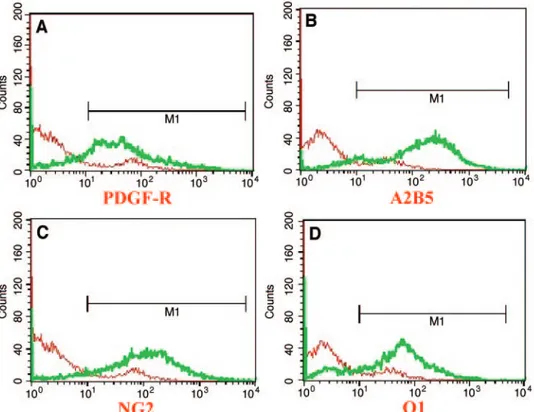
After differentiation, we counted oligodendrocyte precursor cells (stage IV) and oligodendrocytes (stage V) at the cellular level. The majority of the cells were PDGF-R (82%) or A2B5-positive (94%) at stage IV, and O1-A2B5-positive (89%) at stage V. Flow cytometry analysis (Fig. 3) revealed that most of the cells expressed PDGF-R (81%), A2B5 (90.4%), NG2 (91.3%), and O1 (81%), which is similar to the results of random field counting. These results suggest that our in vitro protocol results in efficient induction of oligodendrocytes and their progenitors from human ES cells.
To determine whether these cells were biologically func-tional in terms of myelination activity, the oligodendrocyte precursor cells were cocultured with hippocampal neurons (Fig. 4). The hippocampal neurons were dissected from the brains of 18-day-old rat fetuses, treated with trypsin, and dissociated by pipetting. After the neurons had attached on culture dishes, oligodendrocyte precursors at a concentration of 2⫻ 104 per
cm2were added and incubated for 5 weeks to induce
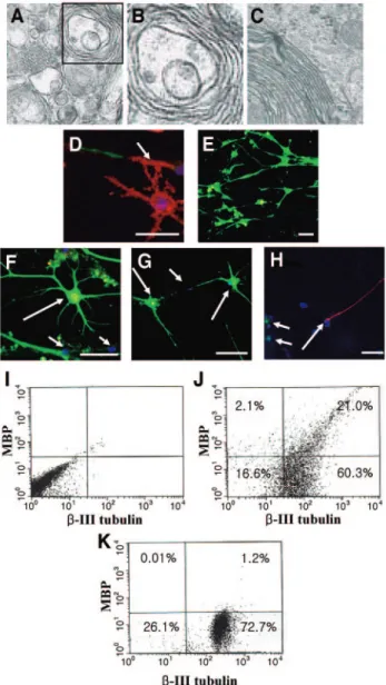
myelina-tion of the axons. The electron micrograph in Figure 4A– 4C shows a transverse section of a representative myelinated axon by coculture. As shown in Figure 4B and 4C, the myelin sheets
compactly enclosed the axon of a neuron. Myelination of neu-rons (red: arrow) were also confirmed by immunostaining using anti-MBP (red) and-III-tubulin (green) antibodies (Fig. 4D). MBP was hardly detected in cultures of hippocampal neurons alone (Fig. 4E), indicating that myelination of neurons was due to oligodendrocytes differentiated from human ES cells, but not endogenous oligodendrocytes. This fact was confirmed using antibody against human nuclei (Fig. 4F– 4H). MBP-positive oligodendrocytes (Fig. 4F, 4G, green) were human nuclei-pos-itive (red), yielding yellow color (big arrows). In contrast, hippocampal neurons (Fig. 4H, big arrow; Fig. 4F and 4G, small arrows) were human nuclei-negative (blue). Figure 4J and 4K showed that⬃25.8% of the hippocampal neurons in the cocul-ture were myelinated MBP-positive cells, whereas⬃1.6% of the neurons were MBP-positive in the culture of hippocampal neu-rons alone.
DISCUSSION
Oligodendrocytes produce myelin, which is wrapped around axons in the CNS. The myelin sheath is lipid-rich and contains several proteins such as MBP and PLP [31]. MBP and PLP are important structural components of myelin that are involved in the compaction of the myelin sheath. The myelination of axons is essential for rapid conduction of nerve impulses. Thus, dam-age to myelin can cause the loss of function that occurs in many CNS disorders such as multiple sclerosis and spinal cord injury. Oligodendrocyte-based cell therapy through remyelination may be a good approach for inducing functional recovery in CNS disorders [32–34] and ES cells are a good source for obtaining oligodendrocytes and their progenitors [10, 35]. Several studies have reported that mouse ES cells can differentiate into
oligo-Figure 3. Flow cytometry analysis of oligodendrocytes and their progenitors. Antibodies against PDGF-R (A), A2B5 (B), NG2 (C) (oligodendrocyte
precursor markers), and O1 (D) (oligodendrocyte marker) were used for the detection of oligodendrocytes and their progenitors. Stained cells were analyzed using a FACScan. This analysis revealed that 81%–91% of total cells were oligodendrocyte progenitors and⬃81% of total cells were oligodendrocytes. Abbreviation: PDGF-R, platelet-derived growth factor receptor.
dendrocytes [10, 35, 36]. A previous study showed that when ES cell-derived glial precursors were transplanted in a rat model of a human myelin disease, the precursors efficiently myelinated axons in brain and spinal cord [10]. Liu et al. [35] reported achieving enriched cultures of oligodendrocytes through oligo-sphere formation. When these cells were transplanted into the spinal cord of shiverer mutant mice in which myelin was defi-cient due to mutation in a MBP gene, the cells produced myelin and myelinated host axons [34, 35].
More recently, Keirstead et al. [8, 34] introduced a method for generating oligodendrocytes from human ES cells through yellow sphere formation and showed their functionality after transplantation into spinal cord injury model. They made spheres by culturing detached human ES colonies on low bind-ing plates and exposed the spheres to glial restriction media (GRM) supplemented with growth factors with/without retinoic acid throughout differentiation. Oligodendrocyte-lineage cells within the cultures were enriched by a mechanical selection procedure to select for yellow spheres based on preferential adhesion.
In contrast, in our protocol, we dissected each stage (undif-ferentiated ES cells-EB stage-neural precursor stage-oligoden-drocyte precursor stage-mature oligodenstage-oligoden-drocyte) through differ-entiation. Undifferentiated human ES cells were maintained on an STO feeder layer and detached with the treatment of colla-genase type IV. Detached human ES cell colonies were trans-ferred onto bacterial culture dishes to induce EB formation. Four days after EB formation, the EBs were transferred onto matri-gel-coated culture dishes. We attempted to create neural rosettes (neural precursors; Fig. 1A, 1B, arrows) from EBs through neural selection (5 days in the ITSFn medium) and expansion procedures (5 days in the presence of bFGF and N2 supple-ments). These neural structures were then directly differentiated into oligodendrocyte progenitor cells on matrigel-coated dishes or used to create SNMs (Fig. 1C) in bacterial culture dishes for further expansion of the neural precursors. One of the features of our protocol is the creation of pure SNMs (pure neural precur-sors) after undergoing mechanical selection upon the initial expansion of the SNMs. After pure SNMs are created, they are expandable for a long period of time and, thus, can provide ample amounts of neural progenitors. These pure SNMs or purely detached neural rosettes gave rise to oligodendrocytes and their progenitors in high purity following differentiation. Neural precursor cells (from neural rosettes or SNMs) are treated with EGF for 4 days, followed by PDGF, for an addi-tional 8 days to induce the formation of oligodendrocyte pre-cursors. Using this method, our results yielded oligodendrocyte progenitors as 81%–91% of the total cells (Fig. 3A–3C). These precursors were bipolar (Fig. 1D, 1E) and positive for markers such as PDGF-R, A2B5, and NG2 (Fig. 2Ab–2Ad). After with-drawal of growth factors, bFGF, EGF, and PDGF, and the addition of T3, the cells differentiated into mature oligodendro-cytes for 14 –21 days. With the passage of time, the cells were morphologically changed into more mature oligodendrocyte cells (Fig. 1F–1I), and the majority of the cells at final stage expressed oligodendrocyte markers such as oligodendrocyte surface proteins O4 (Fig. 2Ae) and O1 (Fig. 2Af), and MBP (Fig. 2Ag). Neurons (Fig. 2Ah) or astrocytes (Fig. 2Ai) were less than 5% of total cells. RT-PCR results showed that each stage of differentiation expressed the representative markers well (Fig. 2B), indicating that our in vitro protocol is sequen-tially well dissected for the induction of neural precursors, oligodendrocyte precursors, and mature oligodendrocytes, re-spectively. When we analyzed the functionality of the differen-tiated cells, we found that they formed multilayered compact myelin sheaths in cocultures with hippocampal neurons from the brains of rat fetuses (Fig. 4A– 4C). Myelination of neurons was
Figure 4. Human embryonic stem (ES) cell-derived cells myelinate axons in cocultures with fetus hippocampal neurons. Oligodendrocyte precursor cells were cocultured with hippocampal neurons from the brains of 18-day-old rat fetuses for 5 weeks. (A–C): The transmission electron micrograph illustrates that the axons of hippocampal neurons are surrounded by multilayered compact myelin sheets ([A],⫻70,000;
[B],⫻140,000; [C], ⫻720,000). (D): Double immunostaining of MBP
(red) and-III-tubulin (green) after coculturing for 5 weeks. Myelina-tion of axons (arrow, red) is shown. (E): Hippocampal neurons cultured without human ES cell-derived oligodendrocytes were stained with anti--III-tubulin (green) and anti-MBP antibodies (red), but MBP-positive cells (red) as shown in (D), are hardly detectable. (F, G): Double immunostaining of MBP (green) and human nuclei (red) after coculture shows that the myelinating MBP-positive cells are human cells (big arrows, yellow). Hippocampal neurons are shown as small arrows. [H]: Costaining of-III-tubulin (red) and human nuclei (green) after coculture.-III-tubulin-positive cells (hippocampal neurons) were not labeled with anti-human nuclei antibody (big arrow). Human cells are shown as small arrows (green). 4⬘,6-Diamidino-2-phenylindole staining (blue) is also shown in (D–H). Scale bar, 50m. (I–K): Flow cytometry analysis using antibodies against-III-tubulin and MBP. Of total cells,⬃21% (⬃25.8% of the neurons) were MBP-positive cells in the coculture (J), whereas⬃1.2% of total cells (⬃1.6% of the neurons) were MBP-positive cells in the culture of hippocampal neurons alone
(K). Cells stained only with secondary antibodies were used as the
control (I). Abbreviation: MBP, myelin basic protein.
also confirmed by immunostaining and flow cytometry after cocultures with hippocampal neurons. Axons were stained with anti-MBP antibody (Fig. 4D, red), and⬃25.8% of the neurons were found to be positive (Fig. 4J). MBP-positive cells were barely detectable in cultures of hippocam-pus neurons alone (Fig. 4E, 4K). The MBP-positive oligo-dendrocytes (green) were labeled with human-specific antibody, anti-human nuclei antibody (red). (Fig. 4F, 4G, merged yellow in big arrows). These results suggest that myelination of hippocampal neurons is formed by oligoden-drocytes differentiated from human ES cells, but not endog-enous oligodendrocytes.
In conclusion, the in vitro protocol that we have developed provides a high level of functional oligodendrocytes and their progenitors from human ES cells. Our study may pave the way
for human ES cell-derived oligodendrocyte therapy in various CNS myelin disorders.
ACKNOWLEDGMENTS
This research was supported by grants (codes: SC1020, SC2140, and SC2160) from the Stem Cell Research Center of the 21st Century Frontier Research Program funded by the Ministry of Science and Technology, Republic of Korea.
DISCLOSURES
The authors indicate no potential conflicts of interest.
REFERENCES
1 Evans MJ, Kaufman MH. Establishment in culture of pluripotential cells from mouse embryos. Nature 1981;292:154 –156.
2 Martin GR. Isolation of a pluripotent cell line from early mouse embryos cultured in medium conditioned by teratocarcinoma stem cells. Proc Natl Acad Sci U S A 1981;78:7634 –7638.
3 Thomson JA, Itskovitz-elder J, Shapiro SS et al. Embryonic stem cell lines derived from human blastocysts. Science 1998;282:1145–1147 4 Miller RH. Oligodendrocyte origins. Trends Neurosci 1996;19:92–96. 5 Keirstead HS, Blakemore WF. The role of oligodendrocytes and
oligo-dendrocyte progenitors in CNS remyelination. Adv Exp Med Biol 1999; 468:183–197.
6 McDonald JW, Becker D, Holekamp TF et al. Repair of the injured spinal cord and the potential of embryonic stem cell transplantation. J Neurotrauma 2004;21:383–393.
7 Zhang SC, Duncan ID. Remyelination and restoration of axonal function by glial cell transplantation. Prog Brain Res 2000;127:515–533. 8 Nistor GI, Totoiu MO, Haque N et al. Human embryonic stem cells
differentiate into oligodendrocytes in high purity and myelinate after spinal cord transplantation. Glia 2005;49:385–396.
9 Oh SK, Kim HS, Ahn HJ et al. Derivation and characterization of new human embryonic stem cell lines, SNUhES1, SNUhES2 and SNUhES3. STEMCELLS2005;23:211–219.
10 Brustle O, Jones KN, Learish RD et al. Embryonic Stem Cell-Derived Glial Precursors: A Source of Myelinating Transplants. Science 1999; 285:754 –755.
11 Okabe S, Forsberg-Nilsson K, Spiro AC et al. Development of neuronal precursor cells and functional postmitotic neurons from embryonic stem cells in vitro. Mech Dev 1996;59:89 –102.
12 Zhang SC, Wering M, Duncan ID et al. In vitro differentiation of transplantable neural precursors from human embryonic stem cells. Nat Biotechnol 2001;19:1117–1118.
13 Barberi T, Klibenyi P, Calingasan NY et al. Neural subtype specification of fertilization and nuclear transfer embryonic stem cells and application in parkinsonian mice. Nat Biotechnol 2003;21:1200 –1207.
14 Billon N, Jolicoeur C, Ying QL et al. Normal timing of oligodendrocyte development from genetically engineered, lineage-selectable mouse ES cells. J Cel Sci 2002;21:6452– 6450.
15 Kim DW, Chung SM, Hwang MK et al. Stromal cell-derived inducing activity, Nurr 1, and signaling molecules synergistically induce dopami-nergic neurons from mouse embryonic stem cells. STEMCELLS2006;
24:557–567.
16 Ruffini F, Arobour N, Blain M et al. Distinctive properties of human adult brain-derived myelin progenitor cells. Am J Pathol 2004;165:2167–2175. 17 Itsokovitz-Eldor J, Schuldiner M, Karsenti D et al. Differentiation of
Human embryonic stem cells into embryoid bodies compromising the three germ layers. Mol Med 2000;6:88 –95.
18 Dotti CG, Sullivan CA, Banker GA. The establishment of polarity by hippocampal neurons in culture. J Neurosci 1988;8:1454 –1468. 19 Rubio N, Rodriguez R, Arevalo MA. In vitro myelination by
oligoden-drocyte precursor cells transfected with the Neurotrophin-3 gene. Glia 2004;47:78 – 87.
20 Yan Y, Yang D, Zarnowska ED et al. Directed differentiation of dopa-minergic neuronal subtypes from human embryonic stem cells. STEM
CELLS2005;23:781–790.
21 Park CH, Min YK, Lee JY et al. In vitro and in vivo analyses of human embryonic stem cell-derived dopamine neurons. J Neurochem 2005;92: 1265–1276.
22 Nakafuku M, Nakamura S. Establishment and characterization of a multi-potential neural cell line that can conditionally generate neurons, astrocytes, and oligodendrocytes in vitro. J Neurosci Res 1995;41:153–168. 23 Baron W, Metz B, Bansal R et al. PDGF and FGF-2 signaling in
oligodendrocyte progenitor cells: regulation of proliferation and differ-entiation by multiple intracellular signaling pathways. Mol Cell Neurosci 2000;15:314 –329.
24 Woodruff RH, Franklin RJ. Growth factors and remyelination in the CNS. Histol Histopathol 1997;12:459 – 466.
25 McMorris FA, McKinnon RD. Regulation of oligodendrocyte develop-ment and CNS myelination by growth factors: prospects for therapy of demyelinating disease. Brain Pathol 1996;6:313–329.
26 Raff MC, Miller RH, Noble M. A glial progenitor cell that develops in vitro into an astrocyte or an oligodendrocyte depending on culture medium. Nature 1983;303:390 –396.
27 Zhou Q, Wang S, Anderson DJ. Identification of a novel family of oligodendrocyte lineage-specific basic helix-loop-helix transcription fac-tors. Neuron 2000;25:331–343.
28 Reubinoff BE, Itsykson P, Turetsky T et al. Neural progenitors from human embryonic stem cells. Nature 2001;19:1134 –1140.
29 Durand B, Raff M. A cell-intrinsic timer that operates during oligoden-drocyte development. Bioessays 2000;22:64 –71.
30 Cai Z, Pang Y, Xiao F et al. Chronic ischemia preferentially causes white matter injury in the neonatal rat brain. Brain Res 2001;898:126 –135. 31 Zhang SC. Defining glial cells during CNS development. Nat Rev
Neurosci 2001;2:840 – 843.
32 Martin S, Hans-Peter H. Remyelinating strategies for the treatment of multiple sclerosis. Prog Neurobiol 2002;68:361–376.
33 Martin ES. Repairing the injured spinal cord. Science 2002;295:1029 –1031. 34 Keirstead HS, Nistor G, Bernal G et al. Human embryonic stem cell-derived oligodendrocyte progenitor cell transplants remyelinate and restore locomo-tion after spinal cord injury. J Neurosci 2005;25:4694 – 4705.
35 Liu S, Qu Y, Stewart TJ et al. Embryonic stem cells differentiate into oligodendrocytes and myelinate in culture and after spinal cord trans-plantation. Proc Natl Acad Sci USA 2000;97:6126 – 6131.
36 McDonald JW, Liu XZ, Qu Y et al. Transplanted embryonic stem cells survive, differentiate and promote recovery in injured rat spinal cord. Nat Med 1999;5:1410 –1412.
![Figure 1. Differentiation of human embryonic stem (ES) cells into oligodendrocytes. Phase contrast microscopic morphology of ES cell-derived neural precursor cells (neural rosettes [A] and [B], spherical neural masses [SNMs] [C]), oligodendrocyte precurso](https://thumb-ap.123doks.com/thumbv2/123dokinfo/5073612.72276/3.877.455.800.114.639/differentiation-embryonic-oligodendrocytes-microscopic-morphology-precursor-spherical-oligodendrocyte.webp)