이
이
이학
학
학 석
석
석사
사
사학
학
학위
위
위 논
논
논문
문
문
Mesenchymal Stem Cells Therapy
in Progressive Rat Model of
Parkinson's Disease
아
아
아 주
주
주 대
대
대 학
학
학 교
교
교 대
대
대 학
학
학 원
원
원
의
의
의 학
학
학 과
과
과
박
박
박 현
현
현 정
정
정
Mesenchymal stem cells therapy in
progressive rat model of Parkinson's Disease
지
지
지도
도
도교
교
교수
수
수 이
이
이 필
필
필 휴
휴
휴
이
이
이 논
논
논문
문
문을
을
을 이
이학
이
학
학 석
석
석사
사
사학
학
학위
위
위 논
논
논문
문
문으
으
으로
로 제
로
제
제출
출
출함
함
함.
.
.
2
2
20
0
00
0
07
7
7년
년
년 2
2
2월
월
월
아
아
아 주
주
주 대
대
대 학
학
학 교
교
교 대
대
대 학
학
학 원
원
원
의
의
의 학
학
학 과
과
과
박
박
박 현
현
현 정
정
정
박
박
박현
현
현정
정
정의
의 이
의
이
이학
학
학 석
석
석사
사
사학
학
학위
위
위 논
논
논문
문
문을
을
을 인
인
인준
준
준함
함
함.
.
.
심
심
심사
사
사위
위
위원
원
원장
장
장
이
이
이 필
필
필 휴
휴
휴 인
인
인
심
심
심사
사
사위
위
위원
원
원
안
안
안 영
영
영 환
환
환 인
인
인
심
심
심사
사
사위
위
위원
원
원
이
이
이
광
광
광 인
인
인
아
아
아 주
주
주 대
대
대 학
학 교
학
교
교 대
대
대 학
학
학 원
원
원
2
2
20
0
00
0
06
6
6년
년
년 1
1
12
2
2월
월
월 2
2
22
2
2일
일
일
M
M
Me
e
es
s
se
e
en
n
nc
ch
c
h
hy
y
ym
m
ma
a
al
l
lS
S
St
t
te
e
em
m
mC
C
Ce
e
el
l
ll
l
ls
s
sT
T
Th
he
h
e
er
r
ra
a
ap
p
py
yi
y
i
in
n
nP
P
Pr
r
ro
o
og
g
gr
r
re
e
es
s
ss
s
si
iv
i
v
ve
e
eR
R
Ra
a
at
t
t
M
M
Mo
o
od
d
de
e
el
l
lo
o
of
f
fP
P
Pa
a
ar
r
rk
k
ki
i
in
ns
n
s
so
o
on
n
n'
'
's
s
sD
D
Di
i
is
s
se
e
ea
a
as
s
se
e
e
by
Hyun Jung PARK
A Dissertation Submitted to The Graduate School of Ajou University
in Partial Fulfillment of the Requirements for the Degree of
MASTER OF SCIENCE
Supervised by
Phil Hyu Lee, M.D., Ph.D.
Department
Department
Department
Department of
of
of
of Medical
Medical
Medical
Medical Sciences
Sciences
Sciences
Sciences
The
The
The
ACKNOWLEDGEMENTS
우선 학위과정 내내 지도해 주시고 항상 염려해주신 든든하고 존경스러운 저희 이필휴 선생님을 비롯하여 작은 데이터 하나하나 꼼꼼히 체크해주신 안영환 선생님, 방오영 선생님, 이광 선생님, 남효석 선생님, 김세혁 선생님, 백만정 선생님, 박창석 선생님께 고개 숙여 감사를 표합니다. 그 동안의 학위과정 동안 옆에서 힘이 되어준 실험실 식구들...우영이, 아르미, 화정언니, 유정이, 선처리, 지여니 언니, 소유니, 창미, 민서니, 윤정이, 문옥언니, 경아……그리고 특히나 내 옆에서 땡깡 다 받아주며 힘들 때마다 손 꼭 잡아주면서 함께 나가준 진영이…. 귀찮게 이것저것 물어도 항상 기분좋게 웃으면서 가르쳐준 멀리 있는 다용언니와 가까이 있는 근우오빠….정말 정말 고맙습니다… 뭔지도 잘 모르면서 항상 잘한다…힘내라.. 해준 내 똘마니 친구들… 미경, 지현, 해명… 그리고, 여기까지 올 수 있게끔 항상 기도해주시고 믿어주시고 응원해주신 울 엄마… 아빠.... 언니랑… 형부… 외할머니, 꼬모 그리고 정진, 반야, 바라, 밀타.. 많이 많이 감사하고 사랑합니다. 세상 중심에서 꽃을 피울 수 있는 그 날까지 더 열심히 노력하는 현정이가 되겠습니다.. 모두모두 다시 한번 감사드립니다.. - 이 논문을 위해 죽어간 쥐들에게도 감사하고 미안한 마음을…-- ABSTRACT -
Mesenchymal Stem Cells Therapy in Progressive Rat Model of
Parkinson’s Disease
Parkinson’s disease (PD) is a common progressive neurodegenerative disorder caused by the loss of dopaminergic neurons in the substantia nigra (SN). In present study, we investigated the effect of human mesenchymal stem cells (hMSCs) therapy in vitro and progressive SD rat model of PD using MG-132, proteasome inhibitor. Immunocytochemical analyses in primary mesencephalic culture revealed that hMSC treatment increased dopaminergic neuronal survival compared to dopaminergic neuronal death in MG-132 treatment. Additionally, hMSCs treatment showed that there was a significant reduction of increased caspase 3 activity following MG-132 treatment. In Rats received systemic injection of MG-132, quantification of the extent of neuronal loss assessed by stereological analysis revealed that TH+ positive cell loss was 23% ± 7.2% ( p <0.05, n=5 per group) at 8 weeks, 70 ± 9.5 at 10 weeks (p <0.01, n=5 per group), and 92 ± 7.2 at 13 weeks (p <0.01, n=5 per group). hMSCs treatment dramatically reduced a decline in the number of TH-immunoreactive cells in the SN of MG-132-treated rats, showing approximately 50% increase of survival in TH-immunoreactive cells in the SN compared to only 132-treated group (p<0.05). Additionally, hMSC treatment in MG-132-treated rats revealed that increased OX-42 immunoreactivity induced by MG-132 was markedly decreased. Histological analysis showed that the number of
NuMA-positive cells was 1.7% of total injected hMSCs and the number of cell double-stained with NuMA and TH was 35.7 % of NuMA-positive cells. In addition, these cells seem to have a functional characteristic of neurons, demonstrating triple-stained with NuMA, TH, and synaptophysin. Behavioral analysis revealed that hMSC treatment in MG-132-treated rats had a tendency of increase in mean stay time on the rod at a 25 rpm (p<0.1).
In conclusion, we have shown that hMSC treatment have a protective effect of dopaminergic neuronal cell death induced by MG-132 in vitro and in vivo. Complex mechanisms mediated by trophic effect of hMSCs and differentiation of hMSCs into functional TH-positive neurons may work in the neuroprotective process of hMSCs. In addition, neuroprotective strategies of hMSCs in this study may be clinically applicable.
Key words : Parkinson’s disease (PD), MG-132, proteasome inhibitor, human
TABLE OF CONTENTS
ACKNOWLEDGEMENTS ··· ⅰ ABSTRACT ··· ⅱ TABLE OF CONTENTS ··· ⅲ LIST OF FIGURES ··· ⅴ . Ⅰ INTRODUCTION ··· 1 . Ⅱ MATERIALS AND METHODS ··· 3A. MATERIALS ··· 3
1. Antibodies ··· 3
B. METHODS ··· 4
1. Isolation of human bone marrow MSCs ··· 4
2. Administration of proteasome inhibitors and hMSCs ··· 4
3. Primary culture of Mesencepharic tissue and proteasome inhibitor treatment · 5 4. Tissue preparation ··· 6
5. Immunocytochemistry and Immunohistochemistry ··· 7
6. Immunofluorescence double-labeling ··· 7
7. Gas chromatography-mass spectrometry··· 8
8. Western blot analysis ··· 8
9. Rotarod test··· 9
10. Stereological cell counts ··· 10
.
Ⅲ RESULTS ··· 12
1. Characterization of hMSCs··· 12 2. Effect of hMSC on dopaminergic neurons in vitro··· 12 3. Progressive decline in the number of TH-immunoreactive cells in
MG-132-treated rats ··· 14 4. Effect of cell therapy with hMSC on animals treated with MG-132··· 15 5. Histological analysis of transplanted hMSCs in animal model of PD induced
by MG-132 ··· 17 6. Behavioral assessment ··· 19 . Ⅳ DISCUSSION ··· 20 . Ⅴ CONCLUSION ··· 25 REFERENCES ··· 26 국문요약 ··· 28
LIST OF FIGURES
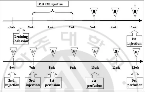
Fig. 1. Timeline and summary of treatment regimens and analyses ··· 4
Fig. 2. Characterization of hMSCs··· 12
Fig. 3. Effect of hMSC on dopaminergic neurons in vitro··· 13
Fig. 4. Progressive decline in the number of TH-immunoreactive cells in MG-132-treated rats··· 14
Fig. 5. Effect of cell therapy with hMSC on animals treated with MG-132··· 16
Fig. 6. Histological analysis of transplanted hMSCs in animal model of PD induced by
MG-132 Characterization of hMSCs··· 18
Fig. 7.Behavioral assessment of transplanted hMSCs in animal model of PD induced by
I. INTRODUCTION
Parkinson’s disease (PD) is a chronic neurodegenerative disease characterized by a selective loss of dopaminergic neurons and the presence of Lewy bodies, proteinaceous inclusions that contain alpha-synuclein, synphilin-1, components of the ubiquitin proteasomal pathway and parkin in substantia nigra (SN) (Moore. et al., 2005). Recently, various genes responsible for familial PD have been identified and these have led to an increased understanding of its molecular pathogenesis (Healy. et al., 2004, Morris. et al., 2005). Of those, mutations in the parkin gene are relatively common in familial PD, with mutations found in 50% of familial early-onset cases compatible with recessive inheritance (Healy et al., 2004, Schrag. et al., 2006). Parkin has E3 ligase activity which is an important part of the cellular machinery that covalently tags target proteins with ubiquitin through the ubiquitin-proteasome system (Hattori. et al., 2004). Therefore, loss of function of parkin from mutations is thought to result in the improper targeting of its substrates for proteasomal degradation leading to inadequate protein degradation and consequently unfolded protein accumulation.
Recently, McNaught and colleague reported that systemic injection of proteasome inhibitor induces a chronic progressive neurodegenerative disorder in rats that closely recapitulates the features of PD in pathological, neurochemical and behavioral aspects (Reaney. et al., 2006). Furthermore, they suggested that this model could be a useful tool for testing putative neuroprotective therapies for PD. There are a variety of proteasome inhibitors, lactacystin, PSI, epoxomicin and MG-132. These proteasome inhibitors consistently caused a significant loss of viability of dopaminergic neurons, although selective vulnerability to dopaminergic
neurons was different among these proteasome inhibitors with the least selectivity in PSI ( McNaught. et al., 2004 ).
Mesenchymal stem cells (MSCs) are present in adult bone marrow and represent <0.01% of all nucleated bone marrow cells. MSCs are themselves capable of multipotency, with differentiation under appropriate conditions into chondrocytes, skeletal myocytes, and neurons (Pittenger. et al., 1999, Woodbury. et al., 2000). The application of MSCs in neurodegenerative diseases is seldom studied. Jin and colleague (Jin. et al., 2002) reported that intracerebral transplantation of MSCs had significant effects on the progression of neurological deficits and life span in a knockout mouse model of Niemann-Pick disease, a lysosomal storage disorder showing progressive ataxia and Purkinje cell loss. Mazzini and colleague (Mazzini. et al., 2003) in a study of MSC transplantation into spinal cord of patients with amyotrophic lateral sclerosis reported that most patients showed a slowing down of the linear decline or increasing muscle strength. Li and colleague ( Li. et al., 2001) reported that using lesional model of PD by 6-hydroxydopmaine, MSCs injected intrastriatally survive, express dopaminergic protein tyrosine hydroxylase (TH) immunoreactivity, and promote functional recovery. However, there were no reports of hMSCs application in similar clinical setting of PD with the viewpoint of neuroprotective strategies. The aim of this study was to investigate whether cell therapy with MSCs has a protective effect on progressive dopaminergic neuronal loss in vitro and in vivo system using MG-132. In addition, we also evaluated whether transplanted MSCs have the capacity of differentiation to TH-positive neuron and functional recovery.
Ⅱ
Ⅱ
Ⅱ
Ⅱ. MATERIALS AND METHODS
A. MATERIALS
1. Antibodies
Antibodies and dilutions used in this study include: mouse anti-TH (1:2000 dilution for brain tissue ; 1:7500 dilution for cell culture, Pel-freez, USA), rabbit anti-TH (1:2000 dilution for brain tissue; 1:7500 dilution for cell culture, Chemicon), mouse anti-Nuclear Matrix (NuMA, 1:100 dilution, Calbiochem ), rabbit anti-Synaptophysin (Syn, 1:200 dilution, invitrogen ), rabbit anti-α-synuclein (α-syn, 1:200 dilution, SIGMA ), mouse anti-ubiquitin (Ub, 1:200 dilution, SANTA CRUZ), Ox-42 ( 1:200, Seratec, Raleight, NC, USA), D105/CD34 for immunochemistry, immunofluorescence, and mouse anti-ubiquitin (Ub, 1:1000 dilution, SANTA CRUZ ) for Western Blotting.
The different primary antibodies were codetected by immunofluorescence, using goat anti-mouse IgG Alexa Fluor-488 (green) and goat anti-rabbit IgG Alexa Fluor-594 (red) (1:200, Molecular probes).
B. METHODS
1. Isolation of human bone marrow MSCs (hMSCs)
Bone marrow aspirates (10ml) were obtained from iliac crests of human donors. The aspirates were with 10ml of PBS. The mononuclear cell layer was isolated by Ficoll-Hypaque, washed in PBS, and plated in polystyrene plastic 100mm culture dishesand were cultivated in low-glucose Dulbecco modified eagles’ medium (Gibco-BRL, Grand Island, NY) containing 10% fetal bovine serum (Hyclone, Irvine, CA) and 1% penicillin/streptomycin (Sigma, St. Louis, MO) in a humidified incubator at 37°C under 5% CO2. Nonadherent cells were removed after 24hr. When these primary cultures of MSCs
reached 80% confluence, the cells were harvested using 0.25% trypsin and subcultured. At passage 6 of hMSCs, cells were injected by tail vain.
2. Administration of Proteasome Inhibitors and hMSCs
Male Sprague–Dawley (SD) rats (200-220g) were used for the study. The proteasome inhibitor, MG-132 was freshly dissolved in sterile dimethylsulfoxide (DMSO). Rats were injected subcutaneously with MG-132 on six occasions on alternate days over 2 weeks (Monday, Wednesday, and Friday). Control rats were injected with DMSO using the same administration method.
These animals were divided into 3 groups. Group 1 was a control group of male rats (n=18) treated with subcutaneously DMSO. Group 2 was a group of male rats (n=18) treated with MG-132 (3mg/kg ) subcutaneously dissolved in DMSO for 2weeks. Group 3 was a group of male rats (n=18) injected human Mesenchymal stem cells once (1×10⁶) at 1 week for 3weeks via tail vain, two weeks after last administration of MG-132. The detailed experimental schedule was
illustrated in figure 1.
3. Primary culture of mesencephalic tissue and proteasome inhibitor
treatment
Dopaminergic neurons were cultured from SN of 14-day-old embryonic SD rats (Chung et al., 2001; Cho et al., 2003; Han et al., 2003). The tissues incubated in Ca2+-, Mg2+- free Hanks’ balanced salt solution (CMF-HBSS) for 10min and a 0.01% Trypsin solution in CMF-HBSS for 9 min at 37℃. Cultures were rinsed twice in RF (Dulbecco’s modified
Eagle’s medium supplemented with 10% fetal bovine serum, 6mg/ml glucose, 204µg/ml L-glutamine, 100U/ml penicillin/streptomycin(P/S)) for trypsin inhibition and then dissociated into single cells by trituration. Dissociated cells were plated on 12-mm round aclar plastic coverslips or cultureslides pre-coated with 0.1mg/ml poly-D-lysin and 4µg/ml laminin and seeded in 24-well culture plates at a density of 1.0 × 10⁵ cells/coverslip or slide. Cells were incubator in a humidified incubator at 37℃, 5% CO2 for 24 hr. At two day-old in vitro
cultures (DIV), the culture medium was replaced with chemically defined serum-free medium (DM) composed of Ham’s nutrient mixture (F12-DMEM) and with 1% ITS (insulin, transferring, selenium), glucose, L-glutamine and P/S. At DIV 4, the cultures were pre-treated with MG-132 (10µM). Primary culture of mesencephalic tissue was co-cultured with 3×10⁵ hMSCs/ well (using transwell) 24 hr after MG-132 treatment.
4. Tissue preparation
For immunohistochemistry, the animals were perfused with a saline solution containing 0.5% sodium nitrate and heparin (10 U/ml) and fixed with 4% paraformaldehyde dissolved in 0.1M PB (both at almost 200 ml/rat) 8, 10, 13 weeks after MG-132, respectively. The brain was removed from the skull, post-fixed overnight in buffered 4% paraformaldehyde at 4℃ and stored in a 30% sucrose solution for 1 to 2 days at 4℃ until they sank. They were then sectioned on a sliding microtome to obtain a 30-µm-thick coronal section or after embedded in paraffin, 4µm sections from SN were immunofluorescenced. The 30-µm-thick coronal sections were stored in tissue stock solution (30% glycerol, 30% etholene glycol, 30% 3rd D.W., 10% 0.2M PB) at 4℃ until required. For Western blotting and high-performance liquid chromatographic-mass spectrometer (HPLC), animals were euthanized 13weeks after MG-132 treatment, and the SN area was rapidly removed from the brains and frozen at –70℃ .
5. Immunocytochemisrty and Immunohistochemistry
The 30-µm-thick coronal brain sections and co-cultured cells were rinsed twice in PBS and incubated in 0.2% Triton X-100 for 30 min at room temperature (RT). They were rinsed thrice with 0.5% bovine serum albumin (BSA) in 1× PBS for blocking. After blocking, they were incubated overnight at RT or 4℃ with primary antibodies; the primary antibodies for TH (1:2000 for immunohistochemistry, 1:7500 for immunocytochemistry; Pel-freez, St. Rogers, AR, USA), Ox-42 ( 1:200, Seratec, Raleight, NC, USA). After 24 hr, primary culture of mesencephalic tissue and brain sections were rinsed thrice in 0.5% BSA in 1× PBS (10 min each rinse) and incubated with appropriate biotinylated secondary antibody and avidin–
biotin complex (Elite Kit; Vector Laboratories, Burlingame, CA, USA) for 1 hr at RT. The bound antiserum was visualized by incubating with 0.05% diaminobenzidine–HCL (DAB) and 0.003% hydrogen peroxide in 0.1M PB. The primary mesencephalic culture and brain sections was rinsed with 0.1M PB for DAB inhibition.
The immunostained cells were analyzed under bright-field microscopy.
6. Immunofluorescence double-labeling
The 4µm sections of embedded paraffin tissues and the 30µm-thick coronal brain sections were rinsed twice in PBS and incubated in 0.2% Triton X-100 for 30 min at RT. They were rinsed thrice with 0.5% BSA in 1× PBS for blocking. After blocking, they were incubated overnight at RT or 4℃ with primary antibodies; TH+
NuMA, TH+syn, NuMA+syn, Ub+ α -syn. After 24 hr, the brain sections were rinsed thrice in 0.5% BSA in 1× PBS (10 min each rinse) and incubated with secondary antibody; goat anti-mouse IgG (Alexa Fluor-488, green) and goat anti-rabbit IgG (Alexa Fluor-594, red) for 1 hr at RT. Brain tissues were then washed and mounted using Prolong Antifade Kit (Molecular Probes, USA). Stained tissues were viewed using an Olympus I×71 confocal laser scanning microscope (Olympus, Tokyo, Japan). To analyze the localization of different antigens in double-stained samples, immunofluorescence images were created from the same tissue section and merged using interactive software.
7. Gas chromatography-mass spectrometry (GC-MS, dopamine level
assay )
GC–MS analyses both in scan and Selected Ion Monitorimg (SIM) modes were performed with an Agilent 6890 gas chromatograph, interfaced to an Agilent 5973 mass-selective detector (70 eV, electron impact mode) and installed with an Ultra-2 (5% phenyl–95% methylpolysiloxane bonded phase; 25m×0.20mm i.d.,0.11m film thickness) cross-linked capillary column (Agilent Technologies, Atlanta, GA, USA). The temperatures of injector, interface and ion source were 260, 300 and 230 ℃, respectively. Helium was used as carrier gas at a flow rate of 0.5 ml/min−1 with constant flow mode. Samples were introduced in the split-injection mode (10:1) and the oven temperature was initially at 170℃ for 2 min and programmed at 5℃/min to 205℃ and at 2℃/min to 220℃ then finally to 30℃/min to 300℃ (3min). The mass range scanned was 50-800u at a rate of 0.42scan/s. All the GC– SIM–MS runs were performed in triplicate.
8. Western blot analysis
Brain tissues from the SN were dissected and homogenized in ice-cold lysis buffer containing (in mM) 20 Tris-HCl, pH 7.5, 1 EDTA, 5 MgCl2, 1 dithiothretol, 0.1
phenylmethylsulfonyl fluoride plus protease inhibitor cocktail (Sigma). Tissue homogenates was centrifuged for 20min at 14,000 × g at 4℃ and the supernatant was transferred to a fresh tube. Their proteins were analyzed by Bio-Rad Protein Assay Kit (USA). Equal amounts of protein (50µg) were loaded in each lane with loading buffer containing 0.125M Tris-HCl, pH 6.8, 20% glycerol, 4% SDS, 10% mercaptoethanol, and 0.002% bromophenol blue. Samples were boiled for 5min before gel loading. Protein analyzed on gel electrophoresis was
transferred to polyvinylidiene difluoride membranes (Millipore, Bedford, MA, USA) using an electrophoretic transfer system (Bio-Rad). The membranes were washed with Tris-buffered saline solution and 2.5 mM EDTA (TNE) and then bloked in TNE containing 5% skim milk for 1hr. Membranes were then incubated overnight at 4℃ with specific primary antibodies of Ub and actin (1:2000; Santa Cruz). After washing, the membranes were incubated with secondary antibodies (1:2000; Amersham) for 1hr at RT, and wash again, and the blots were finally developed with the ECL Western blotting detection reagents (Amersham).
9. Rotarod test
For training, the animals were placed on the rotarod at a constant speed of 5 rpm for 10min during 3 consecutive days before the beginning of the experiment. For rotarod testing, the rats were placed on the rotarod and tested at different rotation speeds (for 10min each 5, 15, 25 rpm.). Each rat generally rested for 20-30 min between tests at each speed; this helps to reduce stress and fatigue. The test was performed on 2 consecutive days every week, and the result was averaged to obtain a stay time on the rod for each rat at a given rotation speed. The test commenced at the same time.
10. Stereological cell counts
The unbiased stereological estimation of the total number of the staining cells in the SN was made using the optical fractionator, as previously described in detail with some modifications (Kirik et al., 1998, 2000). This sampling technique is not affected by tissue volume changes and does not require reference volume determinations (West et al., 1991).
The sections used for counting covered the entire SN, from the rostral tip of the pars compacta back to the caudal end of the pars reticulate. This generally yielded 8-9 sections in a series. Sampling was performed using the Olympus C.A.S.T.-Grid system (Olympus Denmark A/S, Denmark), using an Olympus BX51 microscope, connected to the stage and feeding the computer with the distance information in the Z-axis. SN was delineated at 1.25× objective. A counting frame (60%, 35,650µm2) was placed randomly on the first counting area and systemically moved though all counting areas until the entire delineated area was sampled. Actual counting was performed using a 40× oil objective. Guard volume (4µm from the top 4-6µm from the bottom of the section) were excluded from both surfaces to avoid the problem of lost cap, and only the profiles that came into focus within the counting volume (with depth of 10µm) were counted. The total of staining cells was calculated according to the optical fractionator formula (West et al, 1991).
11. Statistical analysis
The Mann-Whitney test and Kruskal-Wallis analysis were used to compare the means in pairs of groups and multiple comparisons, respectively. P values less than 0.05 were considered statistically significant. Statistical analyses were performed using commercially available software (version 10.0; SPSS Inc., Chicago, IL, USA).
Ⅲ
Ⅲ
Ⅲ
Ⅲ. RESULTS
1. Characterization of hMSCs
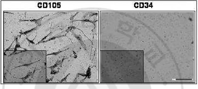
The hMSCs plated on 12-mm round aclar plastic coverslips for characterization by immunocytochemistry. We were identified positive marker of CD 105 and negative marker of CD 34 of the hMSCs (Fig. 2).
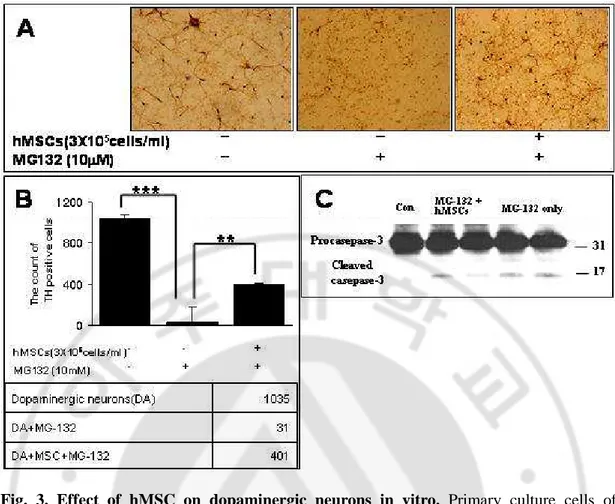
2. Effect of hMSC on dopaminergic neurons in vitro
After 24 hr of treatment by proteasome inhibitor (MG-132, 10 µM), dopaminergic neurons in primary mesencephalic culture were treated hMSCs for 24 hr. Immunocytochemical analyses revealed that hMSC treatment increased dopaminergic neuronal survival compared to dopaminergic neuronal death in MG-132 treatment (Fig. 3A). Quantitative analysis of TH+ cells showed that hMSC treatment significantly decreased dopaminergic neuronal loss induced by MG-132 (p<0.01, Fig. 3B).
In addition, hMSCs treatment showed that there was a significant reduction of increased caspase 3 activity following MG-132 treatment (Fig. 3C).
3. Progressive decline in the number of TH-immunoreactive cells in
MG-132-treated rats
Rats received systemic injection of MG-132 did not exhibit evidence of systemic toxicity and none died. MG-132 had no effect on significant weight changes compared with controls. At 2 and 4 weeks after initiation of treatment, mean body weights were, respectively, 291.8 ± 5.6gm and 381 ± 10gm for controls (n =5) compared with 290.8 ± 5.1 and 386 ± 9.4 for
MG-132-treated rats (n=8).
Immunohistochemical analysis showed that there was a progressive decline in the number of TH-immunoreactive cells in the SN of MG-132-treated animals (Fig. 4A). This TH+ neuronal loss was more prominent in the lateral portion of SN than medial region of SN. Quantification of the extent of neuronal loss assessed by stereological analysis revealed that TH+ positive cell loss was 23% ± 7.2% ( p <0.05, n=5 per group) at 8 weeks, 70 ± 9.5 at 10 weeks (p <0.01, n=5 per group), and 92 ± 7.2 at 13 weeks (p <0.01, n=5 per group; Fig. 4B). In rats treated with MG-132, but not in controls, intracellular protein accumulation was identified, which was immunostained with α-synuclein and ubiquitin (Fig. 4C)
4. Effect of cell therapy with hMSC on animals treated with MG-132
3 weeks after last administration of MG-132, hMSCs were injected via tail vain 3 times for 3weeks. Rats were killed at 10 weeks after the first hMSCs injection for immunohistochemistry, Western Blot, and HPLC. Immunohistochemical analysis showed that hMSCs treatment dramatically reduced a decline in the number of TH-immunoreactive cells in the SN of MG-132-treated rats (Fig. 5A). stereological analysis revealed that the number of TH+ positive cells was significantly higher in hMSC treatment group than in only MG-132-treated group (p<0.05), showing approximately 50% increase of survival in TH-immunoreactive cells in the SN (Fig. 5B). Consistent with the loss of dopaminergic neurons in the SNc, dopamine level in striatum was significantly decreased in MG-132-treated rat compared with controls. However, hMSCs treatment significantly increased dopamine level in the striatum of MG-132-treated rats, which was consistent with increased survival of TH-immunoreactive cells in the SN after hMSCs treatment (Fig. 5C).
Since inhibition of proteasomal enzymatic activity resulted in increased levels of ubiquitinated proteins due to the reduced clearance of proteins by the UPS (Rideout and Stefanis, 2002), we evaluated levels of high molecular weight poly-ubiquitinated proteins (200 and 20 kDa) in MG-132-treated animals. MG-132 treatment resulted in accumulation of poly-ubiquitinated proteins as determined by Western blot, however, hMSCs treatment markedly decreased accumulation of poly-ubiquitinated proteins in MG-132-treated rats (Fig. 5D).
To determine whether hMCS modulate microglial activation, SN was immunostained with OX-42, a marker of microgia. In rats treated with MG-132, there was a marked increase in OX-42 immunoreactivity (a marker of microglia), however, there was little immunoactivity with OX-42 in control rats. hMSC treatment in MG-132-treated rats revealed that increased OX-42 immunoreactivity induced by MG-132 was markedly decreased (Fig. 5E).
In rat treated with MG-132, there was a significant increase of caspase 3 activity compared with controls. However, hMSCs treatment in MG-132-treated rats significantly decreased caspase 3 activity (data not shown).
5. Histological analysis of transplanted hMSCs in animal model of
PD induced by MG-132
To determine if transplanted hMSCs would survive, we identified existence of hMSCs in the SN of MG-132-treated rats using human specific NuMA staining. In controls and only MG-132-treated animals, there were no positive cells (Fig. 6A). In contrast, positive cells were only observed in animals treated with hMSCs. The number of NuMA-positive cells was 51957 ± 35, which was corresponded to 1.7% of total injected hMSCs (Fig.
6B). To determine whether transplanted hMSC possessed phenotype of TH neurons, we co-stained NuMA-positive cells with antibodies to TH (Fig. 6C). The number of cell double-stained with NuMA and TH was 687 ± 35, 35.7 % of NuMA-positive cells (Fig. 6D). Furthermore, we evaluated if the cell merged with MuNA and TH staining harbored immunoreactivity with human specific synaptophysin, a major membrane protein of synaptic vesicle using thin paraffin section (4µm). As shown in Fig. 6E, transplanted hMSC was triple-stained with NuMA, TH, and synaptophysin, indicating that transplanted hMSC harbored phenotype of TH as well as human specific synaptophysin.
6. Behavioral assessment
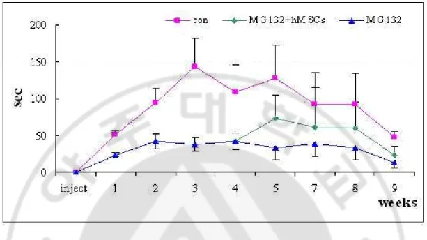
In controls, the mean stay time on the rod at a 25 rpm was increased to 100-150 sec after two weeks after initiation of rotarod test, however, MG-132-treated rat showed that the mean stay time at a 25 rpm was significantly reduced two weeks after MG-132 treatment (p<0.01). hMSC treatment in MG-132-treated rats had a tendency of increase in mean stay time on the rod at a 25 rpm (p<0.1). There was a significant difference of mean stay time on the rod at a 5 and 15 rpm among groups. (Fig. 7)
Fig. 1. Timeline and summary of treatment regimens and analyses. Before injection,
adult Sprague-Dawey rats (SD rat) trained rotarod for 1week. MG132 (3mg/kg, SC) or vehicle (dimethyl sulfoxide;DMSO) as a control were injected six times into adult SD rats (approximately 200g at start) over a period of 2weeks (Mon, Wed, Fri, Mon, Wed, Fri). 2 weeks after injected MG132, rats injected human Mesenchymal stem cells once (1×10⁶) at 1 week for 3weeks via tail vain. 8, 10, 12 weeks after MG-132, rats were killed for immunohistochemistry, western blot, RT-PCR. Behavior test at each week (B:behavior).
Fig. 2. Characterization of hMSCs. Isolation of hMSCs were identified positive marker of
CD 105 and negative marker of CD 34 of the hMSCs. Big box is x200 and small box is x400. (scale bar – 100µM).
Fig. 3. Effect of hMSC on dopaminergic neurons in vitro. Primary culture cells of
mesencephalic tissue were pretreated with MG132 (10 µM) for 2h and then co-cultured hMSCs for 24hr. Imunocytochemistry with TH revealed that hMSCs co-culture group significantly decreased MG132-induced dopaminergic neurons death (A). The count of TH-ip cells in slTH-ip (B). The MSCs group showed reduction of increased caspase 3 activity following MG-132 treatment (Fig. 3C).
Fig. 4. Progressive decline in the number of TH-immunoreactive cells in MG-132-treated rats. SD rats were injected with MG132 (3mg/ml) for 2weeks and then identified
progressive of dopaminergic neurons loss by imunohistochemistry (using TH) in SN (A). The count of TH-ip cells using C.A.S.T grid (B). The tissues identified inclusion by immunofluorescence (using ubiqutin-green, alpha-synuclein-red) (C).
Fig. 5. Effect of cell therapy with hMSC on animals treated with MG-132. Rats were
killed at 10 weeks after the first hMSCs injection or 13weeks after the first MG132 injection for immunohistochemistry. The hMSCs treatment dramatically reduced a decline in the number of TH-immunoreactive cells in the SN of MG-132-treated rats (A). Stereological analysis revealed that the number of TH+ positive cells was significantly higher in hMSC treatment group than in only MG-132-treated group (p<0.05) (B), Dopamine level in striatum was significantly decreased in MG-132-treated rat compared with controls. However, hMSCs treatment significantly increased dopamine level in the striatum of MG-132-treated rats (C), hMSCs treatment markedly decreased accumulation of poly-ubiquitinated proteins in MG-132-treated rats (D). hMSC treatment in MG-132-treated rats revealed that increased OX-42 immunoreactivity induced by MG-132 was markedly decreased (E).
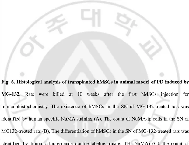
Fig. 6. Histological analysis of transplanted hMSCs in animal model of PD induced by MG-132. Rats were killed at 10 weeks after the first hMSCs injection for
immunohistochemistry. The existence of hMSCs in the SN of MG-132-treated rats was identified by human specific NuMA staining (A), The count of NuMA-ip cells in the SN of MG132-treated rats (B), The differentiation of hMSCs in the SN of MG-132-treated rats was identified by Immunofluorescence double-labeling (using TH, NuMA) (C), the count of TH+NuMA merge cells in the SN of MG-132-treated rats (D). The transplanted hMSC was triple-stained with NuMA, TH, and synaptophysin, indicating that transplanted hMSC harbored phenotype of TH as well as human specific synaptophysin (E).
Fig. 7.Behavioral assessment of transplanted hMSCs in animal model of PD induced by
MG-132 Characterization of hMSCs. MG-132-treated rat showed that the mean stay time
at a 25 rpm was significantly reduced two weeks after MG-132 treatment. The hMSC treatment in MG-132-treated rats had a tendency of increase in mean stay time on the rod at a 25 rpm.
. DISCUSSION
Ⅳ
Ⅳ
Ⅳ
Ⅳ
Although PD is the only chronic neurodegerative disease for which there are effective for symptomatic treatment of dopaminergic regimens, these therapies do not change the progressive nature of PD and moreover, some axial symptoms of gait freezing and postural instability which are more disability than tremor and rigidity do not respond to dopaminergic therapies. In addition, as PD progress, dopaminergic therapies results in disabling drug-induced motor complications such as wearing off and dyskinesia and a variety of nom-motor symptoms such as dementia, autonomic dysfunction, and mood disorders also emerge, which is comparatively disabling as much as motor symptoms. Therefore, neuroprotective strategy that favorably influences the disease process or underlying pathogenesis to produce enduring benefits for PD patients is required.
There were numerous candidates for neuroprotective agents which are tested in animal model of PD.
However, most drug candidates have been tested in acute or subacute neurotoxin-induced animal model and these compounds are administrated before the development of PD model by neurotoxin. To test neuroprotective agents in animal model of PD similar to clinical settings, animal model of PD should have the nature of chronic progressive nature and administration of candidate drugs should start after neuronal loss of SN has started. Like progressive PD model studied by McNaught and colleagues using proteasome inhibitors, chronic administration of MG-132 in this study also showed progressive decline in the number of TH-immunoreactive cells, from approximately 23% TH-positive cell loss at 8 weeks to 92% TH-positive cell loss at 13 weeks, and this decline in TH-positive neurons in
SN accompanied the decreased dopamine level in the striatum as well as mean stay time on the rod compared with controls. In addition, the treatment of hMSCs in this study started 4 weeks after initial administration of MG-132 in animal and 24 hr after MG-132 treatment in primary mesencephalic culture.
Present study demonstrated that hMSC treatment significantly decreased neurodegeneration of dopaminergic neuron resulted from MG-132 treatment, showing approximately 50% increase of survival in TH-immunoreactive cells in the SN. Increased dopamine level in the striatum and a tendency of reduction in behavioral deterioration after hMSCs treatment were well corresponded to the increase in survival of dopaminergic neurons following MSCs treatments in MG-132-treated animals. The mechanism of neuroprotective effects in hMSCs seems to be more complex and pleiotrophic, which may be mediated by trophic mechanisms. First, this study demonstrated that hMSCs treatment markedly decreased accumulation of poly-ubiquitinated proteins and the activity of caspase 3 activity in vitro as well as in vivo following MG-132 treatment. Second, hMSC treatment had an effect of anti-inflammatory effect, showing significantly reduced microglial activation in MG-132-treated animals. Although initially triggering events in the dopaminergic neuronal death in PD are still unknown, apoptosis and alteration of proteasome activity play a pivotal role in the pathogenesis of PD. The presence of DNA fragmentation and chromatin clumping in the dopaminergic neurons coupled with upregulation of signals associated with apoptosis in PD patients supports that dopaminergic neurons would be die by apoptosis in PD (annals). Recent genetic, postmortem, and experimental studies suggest that proteasomal dysfunction might play an important role in the accumulation of toxic proteins and consequently, neurodegeneration in the SN, mediating by imbalance between the degradation and clearance
of abnormal proteins. Evidence has been presented in several studies of in vitro or in vivo disease models that inhibition of the inflammatory response can prevent degeneration of nigrostriatal dopaminergic neurons. For example, sodium salicylate, COX-2 inhibitor, or minocycline have been shown to significantly reduce striatal dopaminergic depletion and dopaminergic neuronal loss induced by MPTP or LPS-models (Aubin et al. 1998; Du et al. 2001; He et al. 2001; Sairam et al. 2003; Teismann et al. 2003). Furthermore, recent epidemiological study has demonstrated that regular use of nonsteroidal anti-inflammatory drugs (NSAID) is associated with a 45% lower risk of PD compared to a nonuser (Chen et al. 2003), supporting the neuroprotective effects of NSAID in the development or progression of PD. It has been known that the MSCs could be mobilized from the marrow and produce a variety of trophic factors that inhibit apoptosis and inflammation (J Cell Biochem. 2006 Aug 1;98(5):1076-84). MSCs have a characteristic of migration towards damaged tissues in animal model of ischemia as well as PD model by 6-hydroxydopamine. In this study, the number of survival hMSCs in SN (a key structure damaged from MG-132 treatment) 5 weeks after last hMSCs administration was approximately 1.7% of total injected hMSCs. We speculate that these migrated cells may contribute to the production of numerous trophic factors and inhibit microenvironmental cascade of the neurodegenerative process in nigral dopaminergic neurons.
Recent studies indicate that mouse, rat, and human MSCs can be induced to differentiate into neuron-like cells. Moreover, Blondheim and colleagues demonstrated that MSCs had an expression of several specific neuronal markers and transcriptional factors, of which a large proportion of the genes was participating in the neuro-dopaminergic system, and suggested that the expression of neural gene as well as gene associated with dopaminergic system is a
widespread phenomenon of MSCs. However, there were few reports regarding that MSCs can differentiate into TH-immnuoreactive neurons in vivo study and their results were contradictive. Li and colleagues and Blondheim and colleagues reported that using 1-Methyl-4-Phenyl-1,2,3,6-Tetrahydropyridine and 6-hydroxydopamine -PD model, respectively, MSCs injected intrastriatally express the phenotype of dopaminergic neurons. In a recent study by Ye and colleague, they did not find BrdU and TH-positive cell in the striatum and suggested that functional recovery in MSCs-treated rat may not be associated with differentiation of MSCs into TH-positive neurons. Our data demonstrated that approximately 35.7% of survival hMSCs in SN had an expression of TH-positive immunoreactivity and furthermore, TH-positive cells were immunostained with human specific synaptophysin, suggesting that TH-positive cells may have a dopaminergic function. Taken with trophic effects of anti-apoptosis, anti-inflammation, and decreased poly-ubiquitinated proteins, differentiation of hMSCs into functional TH-positive neurons may in part contribute to functional recovery and decreased neurodegeneration in MG-132 treated animals.
Besides molecular and cellular benefits of hMSCs, cell therapy with hMSCs has an advantage in clinical application. hMSCs can be easily harvested from self bone marrow, culture in vitro, and administrated to patients through various roots including intravenous, intraarterial, intrathecal, or intralesional infusion. In contrast with therapy with embryonic stem cell, there is no immunological rejection, and cell therapy with hMSCs is free from ethical problems. Importantly, regarding the safety in hMSCs application, our group recently documented that cell therapy with hMSCs in patients with ischemic stroke is feasible and quite safe. This is the first report of hMSCs application in similar clinical setting of PD with
the viewpoint of neuroprotective strategies, and we believe it is an approach with direct clinical application.
. CONCLUSION
Ⅴ
Ⅴ
Ⅴ
Ⅴ
In conclusion, we have shown that hMSC treatment have a protective effect of dopaminergic neuronal cell death induced by MG-132 in vitro and in vivo. Complex mechanisms mediated by trophic effect of hMSCs and differentiation of hMSCs into functional TH-positive neurons may work in the neuroprotective process of hMSCs. In addition, neuroprotective strategies of hMSCs in this study may be clinically applicable.
REFERENCES
1. Adler CH: Nonmotor Complications in Parkinson’s Disease. Mov Disord 20 Suppl 11, S23-29, 2005.
2. Aubin N, Curet O, Deffois A, Carter C: Aspirin and Salicylate Protect Against MPTP-Induced Dopamine Depletion in Mice. Journal of neurochemistry 71, 1635-1642, 1998.
3. Bang OY, Lee JS, Lee PH, Lee G.: Autologous Mesenchymal Stem Cell Transplantation in Stroke Patients. Annals of neurology 57, 874-882, 2005.
4. Blondheim NR, Levy YS, Ben-Zur T, Burshtein A, Cherlow T, Kan I, Barzilai R, Bahat-Stromza M, Barhum Y, Bulvik S et al: Human Mesenchymal Stem Cells Express Neural Genes. Stem cells and development 15, 141-164, 2006.
5. Caplan AI, Dennis JE: Mesenchymal Stem Cells as Trophic Mediators. Journal of cellular biochemistry 98, 1076-1084, 2006.
6. Chen H, Zhang SM, Hernan MA, Schwarzschild MA, Willett WC, Colditz GA, Speizer FE, Ascherio A: Nonsteroidal anti-inflammatory drugs and the risk of Parkinson disease. Archives of neurology 60, 1059-1064, 2003.
7. Chen J, Li Y, Katakowski M, Chen X, Wang L, Lu D, Lu M, Gautam SC, Chopp M: Intravenous Bone Marrow Stromal Cell Therapy Reduces Apoptosis and Promotes Endogenous Cell Proliferation After Stroke in Female Rat. Journal of neuroscience research 73, 778-786, 2003.
8. Cho BP, Song DY, Sugama S, Shin DH, Shimizu Y, Kim SS et al: Pathological dynamics of activated microglia following medial forebrain bundle transaction. Glia 53:92-102, 2006.
9. Chung ES, Joe EH, Ryu JK, Kim J, Lee YB, Cho KG et al: GT1b ganglioside induces death of dopaminergic neurons in rat mesencephalic cultures. Neuroreport 12: 611-614, 2003.
10. Du Y, Ma Z, Lin S, Dodel RC, Gao F, Bales KR, Triarhou LC, Chernet E, Perry KW, Nelson DL et al: Minocycline prevents nigrostriatal dopaminergic neurodegeneration in the MPTP model of Parkinson’s disease. Proceedings of the National Academy of Sciences of the United States of America 98, 14669-14674, 2001.
11. Emborg ME: Evaluation of animal models of Parkinson’s disease for neuroprotective strategies. Journal of neuroscience methods 139, 121-143, 2004.
12. Han BS, Hong HS, Choi WS, Markelnios GJ, Oh YJ: Caspase-dependent and – independent cell death pathways in primary cultures of mesencephalic dopaminergic neurons after neurotoxin treatment. J Neurosci 23: 5068-5078, 2003.
13. Hattori N, Mizuno Y: Pathogenetic mechanisms of parkin in Parkinson’s disease. Lancet 364, 722-724, 2004.
14. He Y, Appel S, Le W: Minocycline inhibits microglial activation and protects nigral cells after 6-hydroxydopamine injection into mouse striatum. Brain research 909, 187-193, 2001.
15. Healy DG., Abou-Sleiman PM, Wood NW: PINK, PANK, or PARK? A clinicians’ guide to familial parkinsonism. Lancet neurology 3, 652-662, 2004.
16. Hellmann MA, Panet H, Barhum Y, Melamed E, Offen D: Increased survival and migration of engrafted mesenchymal bone marrow stem cells in 6-hydroxydopamine-lesioned rodents. Neuroscience letters 395, 124-128, 2006.
17. Jin HK, Carter JE, Huntley GW, Schuchman EH: Intracerebral transplantation of mesenchymal stem cells into acid sphingomyelinase–deficient mice delays the onset of neurological abnormalities and extends their life span. J Clin Invest 109, 1183-1191, 2002.
18. Kirik D, Rosenbald C, Björklund A: Characterization of behavioral and neurodegenerative changes following partial lesions of the nigrostriatal dopamine system induced by intrastriatal 6-hydroxydopamine in the rat. Exp Neurol 152:259-277, 1998.
19. Li Y, Chen J, Wang L, Zhang L, Lu M, Chopp M: Intracerebral transplantation of bone marrow stromal cells in a 1-methyl-4-phenyl-1,2,3,6-tetrahydropyridine mouse model of Parkinson’s disease. Neurosci Lett 316, 67-70, 2001.
20. Makino S, Fukuda K, Miyoshi S, Konishi F, Kodama H, Pan J, Sano M, Takahashi T, Hori S, Abe H et al: Cardiomyocytes can be generated from marrow stromal cells in vitro. J Clin Invest 103, 697-705, 1999.
21. Mareschi K, Novara M, Rustichelli D, Ferrero I, Guido D, Carbone E, Medico E, Madon E, Vercelli A, Fagioli F: Neural differentiation of human mesenchymal stem cells:
evidence for expression of neural markers and eag Kþ channel types. Experimental hematology 34, 1563-1572, 2006.
22. Mazzini L, Fagioli F, Boccaletti R, Mareschi K, Oliveri G., Olivieri C, Pastore I, Marasso R, Madon E: Stem cell therapy in amyotrophic lateral sclerosis: a methodological approach in humans. Amyotroph Lateral Scler Other Motor Neuron Disord 4, 158-161, 2000.
23. McNaught KS, Perl DP, Brownell AL, Olanow CW: Systemic Exposure to Proteasome Inhibitors Causes a Progressive Model of Parkinson’s Disease. Annals of neurology 56, 149-162, 2004.
24. Moore DJ, West AB, Dawson VL, Dawson TM: Molecular Pathophysiology of Parkinson’s Disease. Annual review of neuroscience 28, 57-87, 2005.
25. Morris HR: Genetics of Parkinson's disease. Annals of medicine 37, 86-96, 2005.
26. Olanow CW, McNaught KS: Ubiquitin–Proteasome System and Parkinson’s Disease. Mov Disord 21, 1806-1823, 2006.
27. Pittenger MF, Mackay AM, Beck SC, Jaiswal RK, Douglas R, Mosca JD, Moorman MA, Simonetti DW, Craig S, Marshak DR: Multilineage Potential of Adult Human Mesenchymal stem cells. Science 284, 143-147, 1999.
28. Reaney SH, Johnston LC, Langston WJ, Di Monte DA: Comparison of the neurotoxic effects of proteasomal inhibitors in primary mesencephalic cultures. Exp Neurol 202, 434–440, 2006.
29. Sairam K, Saravanan KS, Banerjee R., Mohanakumar KP: Non-steroidal anti-inflammatory drug sodium salicylate, but not diclofenac or celecoxib, protects against 1-methyl-4-phenyl pyridinium-induced dopaminergic neurotoxicity in rats. Brain research 966, 245-252, 2003.
30. Schrag A, Schott JM: Epidemiological, clinical, and genetic characteristics of early-onset parkinsonism. Lancet neurology 5, 355-363, 2006.
31. Tatton WG., Chalmers-Redman R, Brown D, Tatton N: Apoptosis in Parkinson’s Disease: Signals for Neuronal Degradation. Annals of neurology 53 Suppl 3, S61-70; discussion S70-62, 2003.
32. Teismann P, Tieu K, Choi DK, Wu DC, Naini A, Hunot S, Vila M, Jackson-Lewis V, Przedborski S: Cyclooxygenase-2 is instrumental in Parkinson’s disease neurodegeneration. Proceedings of the National Academy of Sciences of the United States of America 100, 5473-5478, 2003.
33. West MJ, Slomianka L, Gundersen HJ: Unbiased stereological estimation of the total number of neurons in thesubdivisions of the rat hippocampus using the optical fractionator. Anat Rec 231: 482-497, 1991.
34. Woodbury D, Schwarz EJ, Prockop DJ, Black IB: Adult Rat and Human Bone Marrow Stromal Cells Differentiate Into Neurons. J Neurosci Res 61, 364-370, 2000.
35. Yang MS, Park EJ, Sohn S, Kwon HJ, Shin WH, Pyo HK et al: Interleukin-13 and -4 induce death of activated microglia. Glia 38: 273-280, 2002.
36. Ye M, Wang XJ, Zhang YH, Lu G.Q, Liang L, Xu JY, Sheng-Di C: Therapeutic effects of differentiated bone marrow stromal cell transplantation on rat models of Parkinson’s disease. Parkinsonism Relat Disord, 2006.
- 국문요약 -
SD rat
SD rat
SD rat
SD rat 의
의
의
의 PD model
PD model
PD model 에서
PD model
에서
에서
에서 Human Mesenchymal S
Human Mesenchymal Stem C
Human Mesenchymal S
Human Mesenchymal S
tem C
tem C
tem Cell
ell
ell
ell 의
의
의
의
효과
효과
효과
효과
박 현 정
아주대학교 대학원
의학과
(
지도교수 : 이 필 휴)
파킨슨 씨 병은 흑색질 안에 도파민 세포의 감소가 원인이 되는 진행성 신경 퇴화병이다. 현재 우리의 실험에선, 단백질 분해 억제제인 MG 132 를 이용하여 파킨슨 씨 병의 진행성 신경퇴화 랫트 동물 모델과 세포 모델을 만든 후 human mesenchymal stem cells (hMSCs) 치료제의 효과를 확인하였다. 중뇌 신경 세포의 면역화학염색법을 이용하여 MG-132 만을 첨가한 그룹과 hMSCs 를 함께 첨가한 그룹을 비교하였을 때 hMSCs 를 첨가한 그룹의 세포 사멸이 감소한 것을 확인하였다. 또한 MG-132 만을 첨가한 그룹에서는 caspase-3 의 활성이 증가하였는데 hMSCs 를 첨가한 그룹에서는 caspase-3 의 활성이 감소하였다.MG-132 만를 투여한 동물 모델들의 신경 세포는 8 주 때 23% ± 7.2%, 10 주 때 70 ± 9.5, 13 주 때 92 ± 7.2 로 진행성 있게 감소하였다. hMSCs 를 반복적으로 정맥에 투여 후에 흑색질의 도파민 세포의 감소는 MG 132 만 투여한 그룹에 비해 50% 정도 줄어들었고 운동성은 증가하였다. hMSCs 를 투여한 그룹에서 염증반응의 활성화 감소를 확인하기 위해 OX-42 항체를 이용한 면역화학염색결과 피질의 마이크로글리아의 활성화가 매우 감소되는 것을 관찰할 수 있었다. 또한 면역염색법을 통해 흑색질 안에 hMSCs 마커인 NuMA positive 세포가 hMSCs 총량의 1.7%가 확인됨으로써 hMSCs 의 존재를 확인하였고, 도파민 세포 마커인 tyrosine hydroxylase 와 NuMA 의 merge 되는 세포가 NuMA-positive 세포 총량의 35.7%가 확인됨으로써 hMSCs 는 흑색질에서 도파민 세포로 분화 된다는 것을 보여주였다. 추가로 이 세포들의
기능적인 면도 TH, NuMA, synaptophysin 의 merge 를 보여주는
면역염색실험과 행동분석실험을 통해 확인하였다. 결론적으로, hMSCs 의 trophic effect 와 기능성이 있는 도파민 세포로 분화되는 것은 MG-132 로 인한 도파민 세포의 사멸을 보호하는 역할일 것이고, 추가로, 우리가 밝힌 hMSCs 의 이 세포 보호 작용은 임상 적용 치료제로 가능할 것을 시사한다. 핵심어 : 파킨슨 씨 병, MG-132, 단백질 분해 억제제, human mesenchymal stem cell