「쿄짙「교
rnat 01Medicineand Ufe Science L ←→~← S ←←←←마우스의
중추신경계에서
osteopontin의 발현
박준우’,인 미 정2 l제주대학교 수의과대학 수의왜부학교션 쩨주대학교 의학전문대학왼 해부학교섣--~----~
Vol. 7. No. 1. 2민갇」r
二t二二二二
---
AbstractExpression of osteopontin in the central nervous system of BALB/c mice
.Junwoo Park'
,
Meejung Ahn'IDepa끼menl 01Velerinary Anatomy.Jeju Nali
。때
I Universily c이ege 01Veterinary Medicine,1Departmenl01Anatomy,Jeju Nalional Universily SChool01 Medicine,Jeju. Korea
Osteopontin (OPN) is an integrin and calcium-binding phosphoprotein Ihal is produced by mineralized tissue cells. many epithelial cells. and activaled immune system cells The aim 01 Ihis sludy was to examine the expression and cellular localization
。
1 OPN in the central nervous syslem. including the cerebrum,cerebellum and s미nal cord. Female BALBfc mice (25g,8 weeks。
Id) tissues were collected and used lor Western blot anatysis and immuoohistochemistry. Weslern blol analysis showed that OPN was delecled 55- (upper) and 29-kDa (lower) in" Ihe central nervous system. OPN immunoreaclivily was low in the cerebrum: moderate in Ihe cerebetlum; and high in Ihe s미nal cord. lmmunohislochemical study showed Ihal lhe expression of OPN was immunoslained neuron and astrocytes in cenlral nervous system. In the 이aclory bulb. OPN immunoreaclivily wasfound in milral cell layer and glomerular tayer,but not in external plexiform layer,In the hippocampus. OPN expression was
。
bserved in the peria1erial area bul nol CA regions’
n 1he cerebellum,OPN immunoreaclivity was found in purkinje cell layerand molecular layer,bul not in granule cell layer. In spinal cord ,OPN immunoreaclivity was weakly found in neuron and some astrocyte. The presen1 study is the firsl to rep。이 that OPN is variably expressed in CNS tissue,This linding suggests that OPN
may play an impor1anl r,이e in neurons and aslrocytes in lhe normal central nervous syslem. Further s1udy is needed 10 elucidate the functional role 01 OPN in central nervous system. (J Med Ufe 501 2010;7:117-121)
Key Words : Osteoponlin. immunohistochem넌lry. Balb/c mouse. central nervous system
Address 1α corres
∞
ndence : MeejungAhnDepartmenl01Analomy,Jeju National Universily SChool01 Medicine.66 Jejudaehakno.690-756. Jeju,Korea E-mai1:heallhy@jejunu.ac.kr Osteopontin(OPN)은 고유한 Arg-Gly-Asp fRGD) 세포부착 서염을 포함하고 있는 인단액질 (phosphoprotein) 로,무기블화된 소의 뼈에서 처음으흑 발견되었고I)‘그 후 사랍의 뼈",생쥐의 신장 둥에서 확인되었다 ~I. OPN은 뼈모세포와 뼈따괴세포에서 분비되어 일종의 가쿄 역할을 하여 세포틀을 기질에 부착시키는 역할을 한다4l 그러나 많은 연구지듣은 OPN이 뼈 외에도 신장 을 비롯한 속귀,동백의 명환근욕, 태반 내이 중앙조직 그리고 소회관, 태반,혈관 풍플 포함하는 다양한 상피표연에서도 발견 되었고4,5). 그 기능 또한 매우 다촬 것이라는 의견을 제시하고 있다-6. 7) 뼈에서 따팔세포는 자신의 세포악에 있는 정착분자인 인테그 서
론
린을 이용해 골 매트릭스에 존재히는。
PN 등의 RGD 배열윤 인식함으로 골 홉수 기능플 한다~I 이 외에도。
PN은 RGD뜸 배깨로 몇몇 인태그린과 상호 작용하여 다른 종류의 세포에 부확 항 수 있는 부착인자로 보고되여, RGD틀 매개하지 않더악도 CD44,뼈β1‘뻐'Pl 인태그린과 상호작용하여 다른 종류의 세포에 부착한 수 있는 것으로 알려져 있다., 이와 갇은 OPN의 따팔세포에서의 기능이외에도 신징계에서의 염증세포 및 신경세포에서 딸현하며 다른 기능음 나타낸다는 보고들이 있다 OPN은 발달충인 생쥐 뇌에서 신경세포의 분화와 성숙에 관여하고 10),pro-imflammaiory mediator.로 알려져 있으 며,척수손상 '11)‘끼1eiler 바이러스로 유도원 뇌척수염1"
,"r:
가연 역성 뇌척수염13~운 포함히는 마양한 중추신경계 질병 상태에서 증가된 발현양상을 나타낸다 이러한 신경계에서pro-imf1anunatory mediator로서의 OPN의 기놓은 자가연역성 뇌적
수영에서는 염아교세포의 환성화시키여 14),류마티스 관절염에서
inducible nitric ox:ide synthase의 억제틀 용해 염증의 본젤적인 억계자로서의 역한을 한다15)
JunwooPark. Mee,iung Ahn 1.실험동울 BALB/c마우스등 중앙실협동결애 1서 구입 후,제주대학쿄 설협 동울 관련 규정에 의거하여 사육하였다 본 설험에서는 25g.8주 령의 B떠13lc 마우스 암컷 5마리틀 사용하였다 정착 단백으로써의 역할과 연역계에서의 세포 정착. 이주 풍에 중요한 역한을 단당하는 것으로 연구원 바가 있으나, 정상 마우 스의 충추 신경계에서의 발현 몇 분포 세포에 관해서는 연구된 바가 거의 없다 따라서 본 연구에서는 정상 마우스의 중추 신경 계에서 OPN의 발현을 면역조직화학기법옴 이용하여 확인하고자 한다
「
← →→→--- --재료
및
멍법
←←」
간 반응시컸다 비득이적 반웅을 방지하기 위해 10% normalgoat semm으로 1시간 반웅시컸다‘ 1차 항체로 mouse anti-osteopontin (1:200) (BD Biosciences. San jose. CA. U. S. A)
을 설온에서 1시간 반웅시킨 후 biotinylated goat anti-mouse IgG (] :200)(Vector Laborat<lries‘8urlingame. CA)(Veclor
Laboralories. 8urlingame. CA)로 45분간 반응시컸다 이어
avidin-biotin peroxidase complex Elite kit (Vector Laboratories. 8urlingame‘CA)로 실온에서 45분간 반용시켰다 각 단계가 끔냐고 PBS (pH 7.4)로 5분간 3회 충분히 세적했으 며, 면역반웅이 끔난 조직정연은 3.3 -diami no be nzidin e tetrahydrochloride (D뼈)(Sigma. St. Louis‘MO. USA)용액으 로 멍
f
색했다 그리고 hematox'ylin용액으로 대조염색을 한 후 에단올과 자일렌으로 닫수와 투영화 과정을 거쳐 봉입하여 광학 현"1
경으로 관찬하였다‘ 결 과5
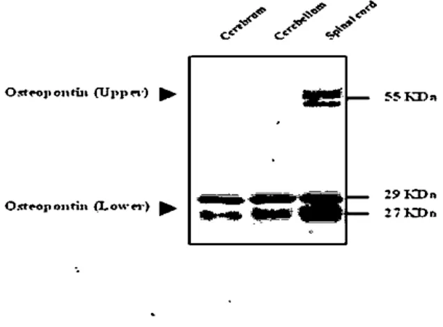
면역반응세포의평가 C어veolin-l의 연역반웅의 결과는 염색의 강도에 따리, 염색이 안된 경우!.(-)약힘~+).보원++) 강함(+++)으로명가하였다Figure 1. Wcstern blot analysis of Osteoponlin in thc central nervous system of mouse. Protein (20ug) from cerebrum. cerebcllum and spinal cord of adult mice were separated by SDS-PAGE: (12%). and then immunoblotted 、꺼th osteopontin antibodies. 810ts arc represent.ativc of dat.a from a scrics of three different animals identical rcsults
1. Osteopontin 의Western blotting 결과
정상 마우스의 중추신경계에서의 OPN의 받현 양상을 얄아보
기 위해 Westem blot analysis룹 봉해서 마우스의 대뇌 소뇌 척수의
。
PN단백양의 연화룹 관잔하였다。
PN은 55 kDa과 27 kDa에서 쌀현하였으며 약 60과 30 kDa의 크기로 말현한다는 μledtke둥의 보고와 일치하였다 16) 이'N의 발현을 죠직에 따라 확인한 결과.55kDa과 27 kDa에서 모두 대뇌에서 가장 약하게 말현하였고. 소뇌는 대뇌에 비해 강하게 받현하였으며. 척수에서 가장 강하게 발현하였다{Fig. 1)‘
ø ....•,
P •.••<‘
...•....r _-<!-••".'. ~#~ c.. c.• t:;/<툴툴~
~~KD"。‘
_J'on’
u‘(UI'P".)‘
3. Western blotting 적출한 장기 조직은 leupeptin <0.5 ug/ml)‘PMSF (1 mM). aprotinin (5. ug/mJ)등의 protein. inhibiωr가 포함된 40 mIM Tris-HCI. pH 7.4. 120 mM NaCI. 0.1% Nonidiet P-40 (poiyoxYethylene [9] p-t-octyi phenoi)의 huffe에서 넣은 후‘ 완전히 따쇄한 후‘14α)() g.로 30분간 원심분리하여 상총액융회수한다 이듣을 단백칠 정량하여 변성시킨 후 동량
(20ug/20ui)으로 sodium dodecyl sulfate- polyacrylamide gel
에서 전기영동하고 gei 상의 단백질 밴드릅 다시 mσ
。
cellulos membrane 에 100V에서 2시간 동안 이동시켰다 옮겨진membrane을 5% skim milk로 blocking.한 후. 1차 항체로
mouse anti-osteopontin (1:200) (BD Biosciences. San jose CA. U.S.A)을 실옹에서 1시간 반웅시킨 후 2차 항체로는
horseradish peroxidase-c 。이ugat.ed horse anti-mouse IgG
(Veclor Laboratories. Burlingame. CA)로 실온에서 60분간
반응시킨다 면역반응이 끝난 membrane은 Amersham ECL
reagents (Amersham Life Science. 8uckinghamshire. UK)로
반응시격. X-ray 옐륜에 노출시키고, 그 절과룹 densitometer (M GS-700 Imaging Densit<lmeter‘8io-Rad laboratories‘
Hercules. CA)로 측정하였다
2.
조직표본 준비와 조직검사 싣험동붉은c
。
ι로 마취하여 방현하였고. 대뇌‘ 소뇌 척수뜸 적출하여, 각 조직의 일부분은 단빽칠 발현융 보기위한 웨스턴 생흉로 급속 냉동하여 사용까지 -70t 의 deep freezer에보관하 였다 면역조직화학영색용 죠칙은 4% 따라포픔알데히드 조직고 정액에 고정하고‘ 에탄용과 자일렌으로 탈수와 투영화 과정을 거쳐 따라핀에 포뱃한 후5""
,의 두께로 조직젤연을 만틀어 hematα<;yiin-eosin염색 및 연역염색을 실시하였다 4 연역조직화학 슬라이드 준비된 조직의 따라핀을 제거하고 내재성 peroxidase룹 제거하기 위해 0.3% H,α
가 포함된 어1탄올에 20분。
:n-_poutillιω…
~I:
틀를룰
1
톨흩
29KD •• 2iKDr. 118-2. Osteopontin 의 연억조직화학 결과
신경계에서 O메이 가지는 분포학적, 기능적 의미에 관해 연구
된 바는 많지 않으며, 그 연구대상도 소수의 통결-종에 한갱되어
Figure 2. Immunohistochemical localization of osteopontin in the cerebrum (A,B). olfacl.ory bulb (C)‘hippocampus
(D.E.F.G) of mouse. ML. miσal cell layer‘GL,glomerular layer: EP. extemaJ plexifonn layer‘DG. dantaù:! gyrus.
E-G: high magnification of D. Scale bar in A-C‘25 ωn: D‘
200/ßll: E-G. 25 αn
중추신경계 조직에 따라
。
steopontin의 발현올 연역초직화학영색법올 이용하여 확인한 결과 주로 말현히는 세포는 열아교세
포(astrocy1e)2J-신경원 세포(neurons)였다
대뇌에서는 분자충{molec비와 Iayer파 과립총(granular I잉er)
에서 신경원(neuron)과 신경아세포 (neuroglia)에서 강경}게 발현 하였다{Fig. 2A. 2B). Choroid plexus의 뇌실악세포 (ependymal cells)에서 강경}게발현하였으며. Olfacl.ory bulb 부위에서 OPN
의 발현은 숭모세포충(mitral cell layer)과 사구제총{gjomerular layer)에서 발현하였으냐 바왈그올충앤 xtcrnal plexiform layer)
에서는 발현하지 않았다(Fig. 2C). Hippocampus에서는 CA1. CA2. dent800 gyrus의 신경원에서 발현하였다{Fig. 2D-2Gl. 소 뇌에서
。
PN의 발현은 연질막"(pia maær)에서 강하게 반현하였 고 조콩박신경세포충 {purkinje cell I어er)의 신경세포에서도 강하게 발현하였다 분자충{molecular 1어er)에서 다소 약하게 받현 하였으나. 과립세포충 {granule cell layer)에서는 발현하지 않았다
(Fig. 3A-3D
、
'"스에셔。
PN2I 받현은 뇌설악세포 (ependymal cells)에서 아주 조ι
하게 발현하였고 운동신경원 (motor neurons)과 신경아세포 (neurogli외 cells)에서 강하게 발현하였고 일부 현관내피세포에서도 발현이 확인되었다 {Fig. 4A-4D) 중추신경 계의 조직에 따른
。
steopontin의 발현양상은 Table 1에 요약하 여 정리하였다 ‘ f~험연펠편燮평첼천
:r:
‘•-ι
샤 5
“‘
*:’'
....
_~~
짧
IIf
렐¥
rfτT
션까
γ'....
‘
ι“;‘-
,
~‘
,'. ‘r、’},I、
1 1:'‘↑
f“-.-
,
t.‘"
‘i;η~~O
‘、“?;
i
댐쐐
.#;
i:.
〔
1
뇨~
Figure 4. lmmunohisωchemical localization of osteopontin in the spinai cord of mouse. WM:.white matter: GM. grey matter. Scale bar in A‘100 αn‘B and C. 50/ßll: D. 25 ωn
,
A
「꽤-「펴
찰고
.
{ I)G ’ ( ι‘
l C.\3:
’
F(
.\-
CAI‘,
-.
--Junwoo Park. Meejung Ahn 차이를보인다 OPN은 뇌에서 항성된다는 보고 후에18),사람의 중추신경계 및말초신경에서 OPN의 세포에 따른 말현 및분포에 대해 보고 되었다19). 0얀J은 대뇌, 소뇌, 척수 및 여러 신경조직에 분포하 였고, 신경원에서 강한 발현을 보였고, 신경아교세포에서도 다양 하게 발현하였다 19) 본 연구에서도 이미 보고된 사람의 신경계에 서의 발현과 유사한 양상을 확인하였다 발달증인 랫트에서는 신겸계통이 분화하는 동안 초기 척삭과 후뇌에서 OPN의 발현이 관잘되었디는 보고가 있고20),마우스에 서 OPN의 발현은 태아기에 관잘되고‘ 플생 후 점차 증가하다가 60일령에는 감소하는 경항을 나타냈고, 뇌에서는 카잘간길핵
(interstitia1 necleus C셰aI)과 흑잭질 그물부「분핵의 신경원에서
OPN이 벨현하였고, 그 외 디
"J'
한 대뇌부위의 신경원에서 OPN 이 발현하였다 10)발생과정중의 OPN의 발현은 마우스의 뇌에서 。PN이 특정한 신경원 집단의 분화와 성숙을 포합ðf-
는 받달과정 에 기여하고 10),정상 조직의 중추신경계서의 발현은 발생 과정 중에 발현히는 것과 마찬가지로 성숙된 신경세포에서 세포 이주 및 세포 접착 분자로서 중요하다고 사료된다 이와 더불어 신경퇴보와 관련된 질병 중 알츠하이머병에서의 。메의 말현은 신경원에서 그 발현이 증가되었고있 J ,파킨슨명에 서는 신경아세포에서 발현이 증가되었다 -22)신경계에서 OPN이 가지고 있는 생물학적 의미는 생태 병려학적 상태에서 정확하게 설명되어야 하고, 앞으로 연구하여야 할 중요한 부분이다 이상의 결과를 종합해 볼 때,본 연구에서는 마우스 중추신경 계의 다양한 조직에서 OPN의 말현을 처음으로 확인하였고, 0맨은 신경원 및 신경아교세포에서 중요한 역할을 할 것으로 사료되며, 마우스 중추 신경계에서의 OPN의 기능적인 역할에 대한 추가적인 연구가 이루어져한 것이다 침고문헌 Table 1. Immunohisíochemical localization of osteopontinÎn the central nervous system of mouse
8t..ained sections were scored for the densîty of positive cells per field. - negative ‘+,weak: ++,moderate; 十十t
intense Tiss니e Cerebrum Choroid plexus
。
lfactory bulb Hippocampus Cerebellum Spina! cordCells α layer lype Osleoponlin
Pia mater ++
Molecular layer
Granular layer ++
얘Tamidal cells. cytoplasm ++
Pyramidal celh익,neucleus
Neuroglîal cells ++
Epitheliwn
Ependymal c허!s ++
Mitral cell layer ++
Extemal plexifonn layer
Glomerular layer ++ Neurons of CAl + Neurons of CA2 + Dentate gyrus + Pia mater ++ Molecular layer + Purkinje cells ++ Granular layer Ependymal cel1s +++ Motor neurons ++ Neuroglial cells ++
Vascular endothelial cells +
120
1) Franzeo A,Heinegard D. Isolation and characterization of two sialoproteins present only in bone calcified matrix Biochem J 1985:232: 715-24
2) Fisher LW,Hawkins GR,Tuross N,Termine JD Purification and partial characterization of smal1 proteoglycans 1 and 11,bone sialoproteins 1 and 11,and osteonectin from the mineral compartment of developing human bone. J Biol Chem 1987:262:9702-8
3) Xie Y,Sakatsume M,Nisru S,Narita 1,Ar와mwa M,Gejy
。
F. Expression ,roles ,receptors ,and regulation of osteopon 디n in the kidney. Kidney Int 2001:60:1645-57 4) Reinholt FP,Hultenby K,Oldberg A,Heinegård DOsteopontin-a possible anchor of osteoclasts ío bone Proc Natl Acad Sci U S A 1990:87:4473-5
5) Davis RL,Lopez CA,Mou K. Expression of osteopontin in the inner ear. Ann N Y Acad Sci 1995:760:279-95
6) Brown LF,Berse B,Van de Water L,Papadopoulos 8ergiou A,Perruzzi CA. Manseau EJ,et a1.,Expression and distribution of osteopontin in human tissues widespread association with luminal epithelial swfaces Mol Biol CeU 1992:3:1169-80
7) Denhardt DT,Guo X. Osteopontin: a protein with diversE functions. FASEB J 1993η 1475-82
8) Nakamura 1,Rodan GA,Duong le T. Regulatory mechanism of osteoclast activation. J Electron Microsc 2003:52:527-33
9) Chellaiah MA,Hruska KA. The integrin 머pha(v)beta(3) and CD44 regulate the actions of osteopontin 00
。
sæoclast motility. Calcif T\ssue Int 2003:72:197-205 10) Kim GB. Hwang IS,Moon C,Shin T,Son H,Jee YHExpression of osteopontin in developing mouse brain Korean J Vet Res 2004:44: 335-41
-11) Hashimoto _M,.Koda~M,_Ino H,Murakami M,Y밍naz하d
M,Moriya H. Upre밍lation of osteopontin expression in rat spinal cord microglia after traumatic injury. J Neurotrauma 2003:20:287-96
12) Shin T, Koh CS. Immunohistochemical detection of osteopontin in the spinal cords of mice with Thei1er's murine encephalomyelitis virus-induced demyelinating disease. Neurosci Lett 2004:356:72-4
13) Kim rvtD,Cho HJ ,Shin T. Expression of osteopontin and l않 ligand,CD44,in the spinal cords of Lewis rats with
experimental autoimmune encephalomyelitis. J Neuroimmunol 2004:151:78-84
14) Chabas D,Baranzini SE,Mitehell D,Bernard CC,
Rittling SR,Denhardt DT,et 31.,The influence of the proinflammatorγ cytokine,osæopontin ,on autoimmune demyelinating disease. Science 2001;294:1731-5
15) Attur MG,Dave "MJ\f,Stuchîn S,Kowalski AJ,Steiner G,
Abramson SB,et a1.,Osteopontin 없1 intrinsic inhibitor
of inflammation in eartilage. Arthritis Rheum 2001:44:578-845
16) Luedtke CC,McKee MD,Cyr DG,Gregory M,Ka앙tinen
MT,Mui J,et a1.,Osteopontin expression 없d regl니lation
in the testis ,efferent duets,and epididymis of rats during postnatal development 야lTough to adulthood. Biol Reprod 2002:66:1437-48
17) Jeon YS,Kim IB,Lee EJ,Moon SH. Lim YG,Chun MH
‘ .
Expression of osteopon
“
n in the central nervous system of BALB/cmice Ch밍1ges in Osteopontin Expression in the Rat Ltunb81Spinal Cord Following the Avulsion of Ltunbar Nerve Roots. The Korean J Anat 2004:37:89-101
18) But1er WT. The nature and signifieanee of ost8opontin Connect Tissue Res 1989:23:123-36
19) Kunii Y,Niwa S,Hagiwara Y,Maeda M,Seitoh T,
Suzuki T. The ÎInInrmorustoehemieal expression profile of osteopontin in normal htunan tissues using two sit8 speeific antibodies reveals a wide distribution of positive eells and extensive expression in the central ahd peripheral nervous systems. Med Mol Morphol 2009:42:155-61
20) Thayer JM,Schoenwolf GC. Early expression of Osteopontin in the cmck is restricted to rhombomeres 5
킹ld 6 and to a subpopulation of nemal erest cells that
arise from these segments. Anat Rec 1998:250:199-209 21) Wung JK,Peπγ G,Kowalski A,Harris PL,Bishop GM
Trivedi MA,et a1. Increased expression of the rernodeling- and turnorigenicassoeiated faet01 osteopon이n in pyramidal neurons of the Alzheimer’↑S
disease braÎIl. Curr Alzheimer Res 2007;4:67-72
22) Maetzler W,Berg D‘Sehalamberidze N,Melms A,Sehott K,Mueller JC,et a1.,Osteopontin is elevated in P,때nson' ’s disease and its absenee leads to reduced neurodegeneration in the. MPTP rnodel. Neurobiol Dis 2007:25:473-82