Comparing volumetric and biological
aspects of 3D-printed interim restorations
under various post-curing modes
Gun Song1, Ji-Won Son2,Ji-Hyun Jang2, Sung-Hyeon Choi2, Woo-Hyung Jang3, Bin-Na Lee2*, ChanPark3*
1Department of Conservative Dentistry, School of Dentistry, Chonnam National University, Gwangju, Republic of Korea 2Department of Conservative Dentistry, School of Dentistry, Dental Science Research Institute, Chonnam National University, Gwangju, Republic of Korea
3Department of Prosthodontics, School of Dentistry, Chonnam National University, Gwangju, Republic of Korea
PURPOSE. This study aims to compare the volumetric change, degree of conversion (DOC), and cytotoxicity of 3D-printed restorations post-cured under three different conditions. MATERIALS AND METHODS. 3D-printed interim restorations were post-cured under three different conditions and systems: 5 min, 30 min, and 24 h. Three-unit and six-unit fixed dental prostheses (n = 30 for each case) were printed; ten specimens from each group were post-cured and then scanned to compare their volumetric changes. Root-mean-squared (RMS) values of the data were acquired by superimposing the scanned files with original files. Thirty disk-shaped specimens were printed to evaluate the DOC ratio. Fourier transform infrared spectroscopy was used to compare the DOCs of 10 specimens from each group. Human gingival fibroblasts were used to measure the cell viability of every specimen (n = 7). The data from this experiment were employed for one-way analysis of variance and Tukey’s post-hoc comparisons. RESULTS.
Differences between the three-unit restorations were statistically insignificant, regardless of the post-curing conditions. However, for the six-unit restorations, a high RMS value was acquired when the post-curing duration was 30 min. The average DOC was approximately 56 - 62%; the difference between each group was statistically insignificant. All the groups exhibited cell viability greater than 70%, rendering them clinically acceptable. CONCLUSION. The post-curing conditions influenced the volume when the length of the restoration was increased. However, this deviation was found to be clinically acceptable. Additionally, post-curing did not significantly influence the DOC and cytotoxicity of the restorations. [J Adv Prosthodont 2021;13:71-8]
KEYWORDS
3D printing; Interim restoration; Post-curing ORCID Gun Song https://orcid.org/0000-0003-1070-0310 Ji-Won Son https://orcid.org/0000-0003-2874-5576 Ji-Hyun Jang https://orcid.org/0000-0002-4672-3381 Sung-Hyeon Choi https://orcid.org/0000-0002-4324-6567 Woo-Hyung Jang https://orcid.org/0000-0001-8077-6877 Bin-Na Lee https://orcid.org/0000-0001-8017-1835 Chan Park https://orcid.org/0000-0001-5729-5127 Corresponding author Chan Park*, Bin-Na Lee** *Department of Prosthodontics, **Department of Conservative Dentistry, School of Dentistry, Chonnam National University 33 Yongbong-ro, Buk-gu, Gwangju, 61186, Republic of Korea *Tel +82625305896 *E-mail upgradepc@jnu.ac.kr **Tel +82625305868 **E-mail bnlee13@jnu.ac.kr Received November 2, 2020 / Last Revision March 2, 2021 / Accepted April 5, 2021 *Chan Park and Bin-Na Lee contributed equally to this work. This study was supported by the National Research Foundation of Korea (NRF) grant funded by the Korean government (No. 2019R1F1A1060935, 2019R1A5A2027521, and 2021R1C1C1008165), a grant (BCRI20030 and BCRI21030) of Chonnam National University Hospital Biomedical Research Institute, and the Technology development Program (S2975345 and S2850798) funded by the Ministry of SMEs and Startups (MSS, Korea).
© 2021 The Korean Academy of Prosthodontics
ccThis is an Open Access article distributed under the terms of the Creative Commons Attribution Non-Commercial License
(http://creativecommons.org/licenses/by-nc/4.0) which permits unrestricted non-commercial use, distribution, and reproduction in any medium, provided the original work is properly cited.
INTRODUCTION
Three-dimensional (3D) printing technology is widely used in clinical dentistry.1 This technology facilitates
printing at any time by using the saved patient infor-mation. Additionally, simulated implant surgery on dental computer-aided design (CAD) software can be actualized using a 3D printed surgical guide.2 Interim
restorations can be manufactured faster and are more aesthetic when created using 3D printing technology, as compared to conventional methods.3 Moreover,
the development of biocompatible 3D printing mate-rials for permanent use is expected to accelerate the application of 3D printing technology in dentistry.4
Various mechanisms are involved in 3D printing.1
The stereolithography apparatus (SLA), which uses photopolymerizing material, is widely adopted in clinical dentistry. For high efficiency, vat polymeriza-tion with digital light processing (DLP) or liquid crys-tal display is utilized in SLA methods.5,6 As multiple
restorations can be printed simultaneously using this technique, the printing is economical.
The process of 3D printing begins with CAD or scan-ning.7 The acquired data are exported to a standard
triangulated language (STL) file and then printed us-ing the appropriate materials and conditions. The printed resin is rinsed with isopropyl alcohol; then, the support structure is removed. The remnant mono-mer of the resin is eliminated during the post-curing process. Previous studies have focused on the accu-racy and speed of printing, as well as the mechanical properties of 3D-printed dental prostheses.8-12
How-ever, apart from the printing process itself, saving time in the other stages is the key to achieving a more efficient workflow. Among the multiple printing stag-es, this study focuses on post-curing, which is the last stage of printing.
It is well known that the remnant monomer in a dental resin causes several issues.13-16 In particular,
irritation of the oral mucosa and a decrease in the solidity of the monomer are significant concerns. Therefore, the remnant monomer must be removed through a process called post-curing. In general, den-tal clinicians consider long post-curing durations to be safe. However, innovative solutions and tech-niques can potentially reduce the post-curing
dura-tion. Thus far, there have been no evaluations of the currently used post-curing systems that simultane-ously consider the volumetric changes and biological effect of the remnant monomer after post-curing.
Therefore, this study aims to evaluate the volumet-ric changes, degrees of conversion (DOCs), and cyto-toxicity of 3D-printed interim restorations developed under various post-curing durations. The null hypoth-esis of this study is that there would be no significant differences in the volumes and biological traits of the interim restorations due to the different post-curing conditions.
MATERIALS AND METHODS
Fig. 1 depicts the experimental protocol of the study. Because this study aimed to evaluate the influence of post-curing alone on the 3D-printing workflow, a sin-gle type of 3D printer and material were used for the experiment. Moreover, three- and six-unit fixed dental prostheses for the posterior molar area and anterior teeth, respectively, were selected as the final designs for printing. After extracting the first molar on a stan-dard dentiform, a three-unit fixed dental prosthesis was prepared. Then, the dentiform was scanned and designed using a standard CAD process. For the six-unit fixed dental prosthesis model, both the lateral incisors were removed, and both canines and central incisors were prepared. The following CAD process was undertaken, using the same method to prepare both the three- and six-unit fixed dental prostheses: the group with a post-curing duration of 30 min (group name: 30N) was considered as the control group for this study, as this is the most common post-curing du-ration. 17-19 The group with a short post-curing time (5
min), wherein a higher intensity light source was em-ployed (group name: 5F), and that with a post-curing duration of 24 h, wherein natural light was employed without a post-curing machine (group name: 24H), were considered the experimental groups. The print-ed specimens were cleanprint-ed with isopropyl alcohol for 20 min; subsequently, post-curing was performed un-der the three different conditions. The materials and equipment used in the experiment are summarized in Table 1, and the experimental groups are listed in Ta-ble 2.
The following experiment was performed on the three-unit (n = 30) and six-unit fixed dental prosthe-ses (n = 30) to examine the influence of post-curing on volumetric changes. A statistical power analysis was conducted to estimate the sample size required from each group. With an effect size of 0.22, an alpha of 0.05, and a power of 0.80, the calculations revealed that eight specimens per group would be needed to detect the postulated effect size. For this study, ten specimens from each group were post-cured
un-der the abovementioned conditions. The post-cured specimens were scanned using a model scanner (DOF Freedom; DOF), and the STL files were acquired. The original and scanned STL files were superimposed us-ing the best-fit algorithm to determine the influence of post-curing conditions on volumetric changes us-ing a 3D data analysis program (Geomagic control X, 3D systems, Santa Ana, CA, USA).20 The volumetric
changes were calculated using the root mean square (RMS) formula.21
Fig. 1. Experimental protocol.
Post-polymerization Test
Cell viability (n = 21)
Volumetric change (n = 60) Degree of conversion (n = 30)
Group 3-unit (n = 30)
6-unit (n = 30)
5F 30N 24H 5F 30N 24H
5F 30N 24H 5F 30N 24H
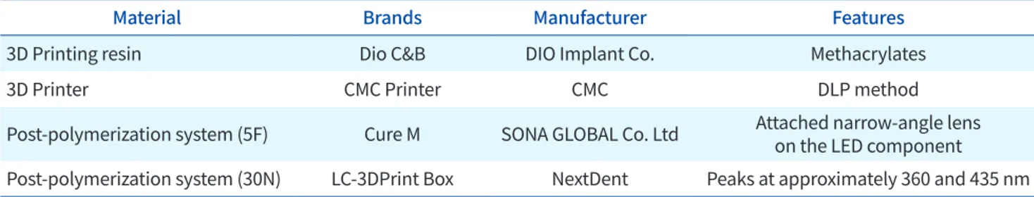
Table 1. Summary of materials, as well as composition, manufacturers, and features of equipment used
Material Brands Manufacturer Features
3D Printing resin Dio C&B DIO Implant Co. Methacrylates
3D Printer CMC Printer CMC DLP method
Post-polymerization system (5F) Cure M SONA GLOBAL Co. Ltd Attached narrow-angle lens on the LED component Post-polymerization system (30N) LC-3DPrint Box NextDent Peaks at approximately 360 and 435 nm
Table 2. Experimental groups
Group Post-polymerization system Time Condition
5F Cure M 5 min Fast curing
30N LC-3DPrint Box 30 min Normal curing
Next, to measure the remnant monomer after post-curing under the various conditions, the DOCs were calculated. Disk-shaped specimens (with a di-ameter of 5 cm and thickness of 2 cm) were print-ed and post-curprint-ed using the same methods as em-ployed for the fixed dental prosthesis specimens. A spectrometer (Fourier-transform infrared, FTIR, spec-trometer, Perkin–Elmer Corporation, Norwalk, CT, USA) was used to examine ten specimens from each post-curing group. Then, the FTIR equation was used to calculate the DOCs by referencing the results ob-tained before post-curing.22
Human gingival fibroblasts (hGF, ATCC CRL-2014, American Type Culture Collection, Manassas, VA, USA) were used for the cell viability tests. 3D-print-ed specimens were design3D-print-ed to mimic three-unit fixed dental prostheses, and the specimens for all the three groups were prepared using the same process as in the previous experiment. An additional group of specimens that were only printed and rinsed without post-curing was included to further validate the re-search. The specimens were transferred into 50 cm3
tubes and incubated in 40 cm3 ofDulbecco’s
modi-fied Eagle’s medium (DMEM, Invitrogen, Carlsbad, CA, USA) for 24 h, which was supplemented with 10% fe-tal bovine serum (FBS, Invitrogen, Carlsbad, CA, USA) and 1% antibiotics (100 U/cm3 of penicillin and 100 μ
g/cm3 of streptomycin, Invitrogen, Carlsbad, USA) at
37°C in a humid atmosphere containing 5% CO2.
Ex-tracts were collected and thlen filtered using 0.20 μ -m filters (Minisart, Sartorius Stedim Biotech, Goet-tingen, Germany). The experimental groups were treated with their respective material extracts. In this test, the control group was set to media only. The hGF were seeded into 96-well culture plates at a density of 1 × 10⁴ cells per well and incubated in the DMEM,
containing 10% FBS and 1% antibiotics, for 24 h to promote adhesion. Then, the medium was replaced with 100 mm3 of each material extract and
incubat-ed for another 24 h. The hGF with the mincubat-edium were used as controls. The cell viability was examined us-ing 4-[3-(4-Idophenyl)-2-(4-nitrophenyl)-2H-5-tetrazo-lio]-1,3-benzene disulphonate (WST-1 assay) (EZ-Cy-tox, Daeil Lab Service, Seoul, Korea), according to the manufacturer’s instructions. Next, 10 mm3 of Ez-Cytox
(tetrazolium salts) was added to the culture medium
and incubated with the cells at 37°C for 2 h. The ab-sorbance of each well was measured at a wavelength of 450 nm using a microplate spectrophotometer (Multiskan GO Microplate Spectrophotometer, Ther-mo Scientific, Vantaa, Finland) and SkanIt Software 4.1 (Thermo Scientific Multiskan GO™, Waltham, MA, USA). All procedures of this experiment were based on those in previous studies.23 The schematics of all
experiments are summarized in Fig. 1.
Statistical analyses were performed using statistical software (IBM SPSS Statistics, version 22.0, IBM Corp, Chicago, IL, USA). The data from the experiment were employed for a one-way analysis of variance, and Tukey’s post-hoc test was used to determine any sta-tistically significant differences. A P-value of < .05 was considered statistically significant.
RESULTS
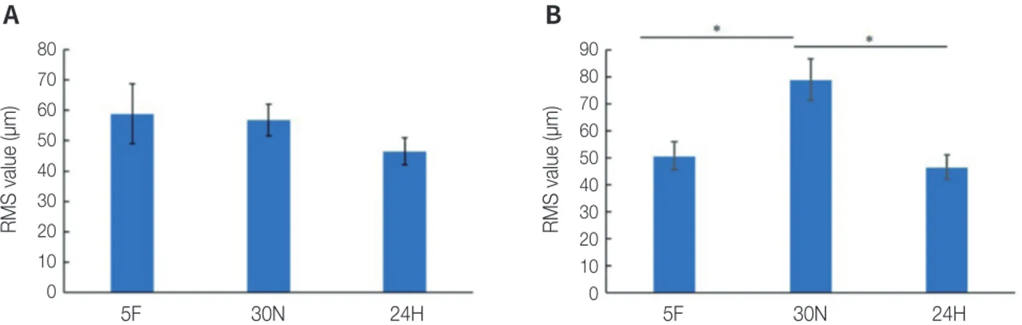
The comparison of the volumetric changes under the three different post-curing conditions is illustrated in Fig. 2. The RMS values of the three-unit fixed dental prostheses were 58.82 ± 9.81 μm for the 5F group, 56.86 ± 5.16 μm for the 30N group, and 46.47 ± 4.38 μm for the 24H group. These outcomes were not sta-tistically significant. In contrast, the RMS values for the six-unit fixed dental prostheses were 46.58 ± 5.02, 79.00 ± 7.59, and 50.90 ± 4.60 μm for the 5F, 30N, and 24H groups, respectively. The specimens of the 30N group exhibited the highest RMS values. The results of Tukey’s post-hoc test revealed statisti-cally significant differences between the 5F and 30N groups and between the 30N and 24H groups. How-ever, no significant difference was observed between the specimens of the 5F and 24H groups. A P-value of < .05 was considered statistically significant.
The DOC results are depicted in Fig. 3. In summary, the DOCs were 56% for the 5F group, 62% for the 30N group, and 56% for the 24H group. These results were not statistically significant.
The cell viability results are depicted in Fig. 4. The specimens that did not undergo post-curing exhibited relative cell viabilities of 0.839 ± 0.05, 0.976 ± 0.05, and 0.866 ± 0.02 for the 5F, 30N, and 24H groups, re-spectively. The results show that cell viability was sta-tistically lower after 30 min of post-curing using the
RMS value (µm) 80 70 60 50 40 30 20 10 0 5F 30N 24H RMS value (µm) 90 80 70 60 50 40 30 20 10 0 5F 30N 24H Fig. 2. Volumetric change corresponding to three different post-curing conditions by RMS value. (A) RMS values for three-unit fixed dental prosthesis, and (B) RMS values for six-unit fixed dental prosthesis. The columns connected by the black lines showed statistically signifi-cant differences (*P < .05).
A
B
DOC (%) 70 60 50 40 30 20 10 0 5F 30N 24H Fig. 3. Degree of conversion corresponding to three different post-curing conditions. All values were not statistically significant.Relative cell viability (%)
120 100 80 60 40 20 0 5F 30N 24H
Fig. 4. Relative cell viability of each group. The columns connect-ed by the black lines showconnect-ed statistically significant differences (*P < .05). **ISO10993-5:2009 describes test methods to assess the in vitro cytotoxicity of medical devices. These methods are designed to determine the biological response of mammalian cells in vitro using appropriate biological parameters.
ISO 70%
standard method. However, each group surpassed the cut-off level (70%), which is certified by ISO 10993-5.
DISCUSSION
In this study, we compared the volumetric and bi-ological changes of 3D printed interim restorations prepared following various post-curing procedures. Among the interim restorations, the volumetric changes for the six-unit fixed dental prosthesis spec-imens were statistically significant. In contrast, there was no statistically significant difference in the DOCs between groups. However, a significant difference was observed in cell viability. Therefore, the null hy-pothesis of this study was partially rejected.
The polymerization process begins with the radia-tion of a photopolymer under light at a wavelength at which the polymerizing initiators of the photo-polymer are sensitive.24 The wavelengths for the
po-lymerization of conventional composite resins and 3D-printing resins are known to be different. Dental products for 3D printing are polymerized in the pres-ence of light with wavelengths of approximately 405 nm.25-26 Consequently, their polymerization itself may
differ from conventional composite resins; there-fore, additional experiments for post-curing under multiple conditions are required. 3D-printed spec-imens that exhibit a significant volumetric change after post-curing cannot be used in clinical practice.
Hence, it is crucial to develop a means to minimize post-curing contraction.17,27 Gradual and sufficient
post-curing is the key to minimizing contraction and the remnant monomer; however, high production ef-ficiencies cannot be achieved if the post-curing dura-tion is extremely long. Thus, determining the shortest post-curing duration that results in clinically accept-able shrinkage and remnant monomer is important. If the polymerization is performed in a short duration with minimal contraction and remnant monomer, not only can practitioners save time, but patients can also be treated more comfortably. To this end, this study focused on reducing the post-curing duration to max-imize comfort in clinical practice.
Various ideas have been developed and adopted to cure resins rapidly. In this experiment, for the 5F post-curing group, a low-angle lens was attached to the ultraviolet LED lamp of the fast post-curing ma-chine. Consequently, the machine could emit light at ~60 mW/cm2, which was twice its usual radiation
in-tensity. Thus, the post-curing duration could theoret-ically be reduced from 30 min to 5 min, while main-taining adequate mechanical properties, as regulated by ISO 10477. Reduction in the post-curing duration to maximize comfort in clinical practice can only be achieved when the outcomes are accurate. Howev-er, because post-curing with intense light induces contraction28, the printed specimens that undergo
post-curing using this method need to be clinically evaluated.
In this experiment, three- and six-unit fixed dental prostheses for the posterior teeth and anterior teeth, respectively, were selected. Because the amount of resin used is directly proportional to the amount of contraction, no problem should occur with gener-al crown restorations if there are no flaws with these long fixed dental prosthesis units. The volumetric change results indicate that for the three- and six-unit fixed dental prosthesis interim specimens, the RMS values were 45 - 55 µm and 45 - 80 µm, respectively. The DLP 3D printer used in this experiment is known to print with deviations of up to 50 µm; a deviation of approximately 100 µm is considered to be clinical-ly acceptable. Although the deviation of the six-unit fixed dental prosthesis that was post-cured for 30 min was greater than that of the other specimens, it was
still within the clinically acceptable range.
The average DOC of the dental composite resin’s C=C is 50 - 70%,20 which also applies to dental resins
for 3D printing. The DOC is affected by the filler con-tent and monomer constitution. Higher DOCs can be achieved by the refraction and scattering of light, which increases the depth of curing. An increase in the DOC implies that the polymers in the interim res-torations are stable, resulting in enhanced mechan-ical properties. Previous studies have demonstrated the correlation of hardness and tensile strength with the DOC.8 Therefore, evaluating the DOCs of various
specimens with diverse post-curing conditions can help in indirectly evaluating the mechanical proper-ties of 3D printed interim restorations. In this exper-iment, even the post-curing group with the shortest duration (group 5F) exhibited a DOC of 56%, with no statistically significant differences between the groups. Furthermore, this DOC value was similar to the expected value provided by the manufacturer. Therefore, in terms of DOC, every post-curing tech-nique and duration set for this experiment was found to be clinically acceptable.
Without appropriate post-curing, monomers may remain in the printed dental resin. Remnant mono-mers can be harmful in the oral environment and re-duce the biocompatibility of the printed resin.15,16
hGF were used to evaluate the cell viability of print-ed interim restorations. Without proper post-curing, the cytotoxicity of the printed specimens is likely to be high. In this experiment, every group exhibited an acceptable cell viability, as certified by ISO10993. Ad-ditionally, a group of specimens without post-curing was evaluated to further validate our findings. How-ever, even specimens without post-curing exhibited cytotoxicity levels that complied with the ISO stan-dards. This may be attributed to the sufficient light exposure during the original printing process in all the groups. Therefore, the techniques used in the post-cured groups and the control group can be used in clinical practice.
However, this study has several limitations. For ex-ample, various post-curing durations were consid-ered. A more controlled experiment could be realized if a single type of post-curing machine was used for the experiment, while varying the post-curing
du-ration alone. Moreover, a scientifically controlled, shorter post-curing duration was not considered in this study. Nevertheless, as more data are accumulat-ed, the 3D printing of dental prosthetics is expected to provide increased comfort to patients and exhibit higher production efficiency.
CONCLUSION
In this study, various experiments were conducted to assess the influence of volumetric change, DOC, and cytotoxicity on multiple groups of specimens. The results revealed some differences relative to the post-curing time; however, these differences were sta-tistically insignificant.
In conclusion, 3D-printed interim restorations that underwent post-curing for a duration of 5 min were found to exhibit clinically acceptable volumetric changes. The post-curing duration was found to have an insignificant influence on the DOC and cell via-bility. Therefore, based on the results of the current study, a post-curing duration of 5 min for 3D-printed interim restorations is considered to be clinically ac-ceptable and can thus be applied in clinical practice.
REFERENCES
1. Dawood A, Marti Marti B, Sauret-Jackson V, Darwood A. 3D printing in dentistry. Br Dent J 2015;219:521-9.
2. Abduo J, Lyons K, Bennamoun M. Trends in comput-er-aided manufacturing in prosthodontics: a review of the available streams. Int J Dent 2014;2014:783948.
3. Barazanchi A, Li KC, Al-Amleh B, Lyons K, Waddell JN. Additive technology: update on current materials and applications in dentistry. J Prosthodont 2017;26: 156-63.
4. Quan H, Zhang T, Xu H, Luo S, Nie J, Zhu X. Photo-cur-ing 3D printPhoto-cur-ing technique and its challenges. Bioact Mater 2020;5:110-5.
5. Seime L, Hardeberg JY. Characterisation of LCD and DLP projection displays. Color Imaging Conf Final Progr Proc 2002:277-82.
6. van Noort R. The future of dental devices is digital. Dent Mater 2012;28:3-12.
7. McGurk M, Amis AA, Potamianos P, Goodger NM. Rap-id prototyping techniques for anatomical modelling
in medicine. Ann R Coll Surg Engl 1997;79:169-74.
8. Tahayeri A, Morgan M, Fugolin AP, Bompolaki D, Athi-rasala A, Pfeifer CS, Ferracane JL, Bertassoni LE. 3D printed versus conventionally cured provisional crown and bridge dental materials. Dent Mater 2018; 34:192-200.
9. Taft RM, Kondor S, Grant GT. Accuracy of rapid proto-type models for head and neck reconstruction. J Pros-thet Dent 2011;106:399-408.
10. Goodacre BJ, Goodacre CJ, Baba NZ, Kattadiyil MT. Comparison of denture base adaptation between CAD-CAM and conventional fabrication techniques. J Prosthet Dent 2016;116:249-56.
11. Steinmassl O, Dumfahrt H, Grunert I, Steinmassl PA. CAD/CAM produces dentures with improved fit. Clin Oral Investig 2018;22:2829-2835.
12. Favero CS, English JD, Cozad BE, Wirthlin JO, Short MM, Kasper FK. Effect of print layer height and printer type on the accuracy of 3-dimensional printed ortho-dontic models. Am J Orthod Dentofacial Orthop 2017; 152:557-65.
13. Park SH, Lee CS. The difference in degree of conver-sion between light-cured and additional heat-cured composites. Oper Dent 1996;21:213-7.
14. Susila AV, Balasubramanian V. Correlation of elution and sensitivity of cell lines to dental composites. Dent Mater 2016;32:e63-72.
15. Samanidou VF, Kerezoudi C, Tolika E, Palaghias G. A simple isocratic HPLC method for the simultane-ous determination of the five most common residual monomers released from resin-based dental restor-ative materials. J Liq Chromatogr Relat Technol 2015; 38:740-9.
16. Bourbia M, Ma D, Cvitkovitch DG, Santerre JP, Finer Y. Cariogenic bacteria degrade dental resin composites and adhesives. J Dent Res 2013;92:989-94.
17. Reymus M, Lümkemann N, Stawarczyk B. 3D-printed material for temporary restorations: impact of print layer thickness and post-curing method on degree of conversion. Int J Comput Dent 2019;22:231-7.
18. Reymus M, Stawarczyk B. In vitro study on the influ-ence of postpolymerization and aging on the Martens parameters of 3D-printed occlusal devices. J Prosthet Dent 2020;19:S0022-3913(20)30077-9.
19. Kalberer N, Mehl A, Schimmel M, Müller F, Srini-vasan M. CAD-CAM milled versus rapidly prototyped
(3D-printed) complete dentures: an in vitro evalua-tion of trueness. J Prosthet Dent 2019;121:637-43.
20. Kang SY, Park JH, Kim JH, Kim WC. Accuracy of pro-visional crowns made using stereolithography appa-ratus and subtractive technique. J Adv Prosthodont. 2018;10:354-60.
21. Schaefer O, Watts DC, Sigusch BW, Kuepper H, Guentsch A. Marginal and internal fit of pressed lith-ium disilicate partial crowns in vitro: a three-dimen-sional analysis of accuracy and reproducibility. Dent Mater 2012;28:320-6.
22. Leloup G, Holvoet PE, Bebelman S, Devaux J. Ra-man scattering determination of the depth of cure of light-activated composites: influence of different clini-cally relevant parameters. J Oral Rehabil 2002;29:510-5.
23. Lee BN, Hong JU, Kim SM, Jang JH, Chang HS, Hwang YC, Hwang IN, Oh WM. Anti-inflammatory and osteo-genic effects of calcium silicate-based root canal seal-ers. J Endod 2019;45:73-8.
24. Fuh J, Lu L, Tan C, Shen Z, Chew S. Processing and characterising photo-sensitive polymer in the rapid prototyping process. J Mater Process Technol 1999;89: 211-7.
25. Piedra-Cascón W, Sadeghpour M, Att W, Revilla-León M. A vat-polymerized 3-dimensionally printed du-al-material occlusal device: a dental technique. J Prosthet Dent 2020;S0022-3913(20)30438-8.
26. Lin CH, Lin YM, Lai YL, Lee SY. Mechanical proper-ties, accuracy, and cytotoxicity of UV-polymerized 3D printing resins composed of Bis-EMA, UDMA, and TEGDMA. J Prosthet Dent 2020;123:349-54.
27. Reymus M, Fabritius R, Keßler A, Hickel R, Edelhoff D, Stawarczyk B. Fracture load of 3D-printed fixed dental prostheses compared with milled and conventional-ly fabricated ones: the impact of resin material, build direction, post-curing, and artificial aging-an in vitro study. Clin Oral Investig 2020;24:701-10.
28. Par M, Marovic D, Attin T, Tarle Z, Tauböck TT. The ef-fect of rapid high-intensity light-curing on microme-chanical properties of bulk-fill and conventional resin composites. Sci Rep 2020;10:10560.