The Fidgetin-like1 gene regulates
proliferation and differentiation of
osteoblasts and is inhibited by
basic fibroblast growth factor
Thesis by
Su Jin Kim
Department of Medical Science
The Fidgetin-like1 gene regulates
proliferation and differentiation of
osteoblasts and is inhibited by
basic fibroblast growth factor
Directed by Professor Sung-Kil Lim
The Master’s Thesis
submitted to the Department of Medical Science
the Graduate School of Yonsei University
in partial fulfillment of the requirements for the
degree of Master of Medical Science
Su Jin Kim
This certifies that the Master’s Thesis of
Su Jin Kim is approved.
---
[Thesis Supervisor : Sung-Kil Lim]
---
[Thesis Committee Member]
---
[Thesis Committee Member]
The Graduate School
Yonsei University
Acknowledgments
지식과 경험이 부족한 저에게 지난 2년간의 대학원 생활은 많은 것 을 느끼고 배우게 하였습니다. 힘들고 지칠 때도 많았던 저에게 그럴 때마다 힘과 용기를 주신 고마운 분 들께 감사의 말씀을 전하고 싶습 니다. 먼저 세심한 지도와 격려를 아끼지 않으셨던 지도 교수님인 임승 길 교수님께 진심으로 감사드리며, 바쁘신 중에도 논문 내용을 세심하 게 살펴주시고, 각별한 조언을 아끼지 않으신 김명희 교수님, 용태순 교수님께도 감사드립니다. 한 실험실에서 공부하고 서로 의지하면서 슬픔과 기쁨을 함께 나눈 박수진 선생님과 변지현 선생님, 한동하 선생님께 감사드리며, 항상 밝 은 미소의 내분비내과 이유미 선생님과 김세화 선생님께도 감사를 드 립니다. 가족이란 이름으로 편히 연구에만 전념할 수 있도록 물심양면으로 도와주시고, 저의 부족한 면들을 따뜻한 믿음으로 보살펴 주신 아버지 와 어머니, 동생에게 감사드립니다. 아울러 힘들 때마다 잦은 짜증과 트집에도 항상 아낌 없는 격려와 용기를 준 평생지기 친구들에도 감사 를 전하면서 이 논문을 마칩니다. 저자 씀Contents
ABSTRACT---1
І. INTRODUCTION---3
П. MATERIALS AND METHODS---6
1. Plasmid constructions---6
2. bFGF dose response and WST-1 assay---6
3. Cell culture and transfection---6
4. bFGF dose response and RT-PCR reaction---7
5. Cell proliferation and differentiation---8
6. Trypan blue staining---10
7. LDH assay---10
8. Flow cytometric analysis---10
9. Synthesis and transfection of small interfering RNA---11
10. Green fluorescent protein analyses---12
11. Statistical analysis---12
Ш. RESULTS---14
1. Effect of bFGF treatment on proliferation of MC3T3-E1 cells and Fignl1 expression---14
3. The effect of suppressed Fignl1 on osteoblastogenesis---20
4. Nuclear translocation of Fignl1 by bFGF treatment---22
. Ⅳ DISCUSSION---25
V. CONCLUSION---28
REFERENCES---30
LIST OF FIGURES
Figure 1. Effect of bFGF exposure on the proliferation of MC3T3-E1 cells-15
Figure 2. The effect of short-term exposure of bFGF on Fignl1 mRNA expression in MC3T3-E1 cells---16
Figure 3. Effects of Fignl1 on proliferation and differentiation of MC3T3-E1 cells---18
Figure 4. Correlation of PI staining and LDH assay for estimating cell death19
Figure 5. The effects of siFignl1 on Fignl1 mRNA expression in MC3T3-E1 cells---21
Figure 6. Effects of siFginl1 on proliferation and differentiation of MC3T3-E1 cells---21
ABSTRACT
The Fidgetin-like1 gene regulates proliferation and
differentiation of osteoblasts and is inhibited
by basic fibroblast growth factor
Su Jin Kim
Department of Medical Science
The Graduate School, Yonsei University
(Directed by Professor Sung-Kil Lim)
The Fidgetin-like1 (Fignl1) gene encodes a new subfamily member of ATPases associated with diverse cellular activities (AAA proteins). Though the Fignl1 protein localizes to both the nucleus and cytoplasm, the function of Fignl1 remains unknown. Using a microarray, we identified several genes that mediate the anabolic effects of bFGF on bone. Fignl1 was one of the genes
down-regulated greater than twofold in MC3T3-E1 cells after treatment with bFGF. Reduced expression of Fignl1 by bFGF treatment was verified by RT-PCR analysis. Overexpression of Fignl1 caused up to 20% fewer MC3T3-E1 cells (P < 0.05), and WST-1 analysis also revealed a 20% reduction, compared to the mock transfected control cells. In contrast, siRNA treatment against Fignl1 increased cell number (P < 0.05) and viability (P < 0.05), and overexpression of Fignl1 does not appear to alter apoptosis in osteoblasts. Meanwhile, Fignl1 enhanced mRNA expression of alkaline phosphatase (ALP) and osteocalcin (OC), both of which are genes expressed during the late stage of osteoblast differentiation. In contrast, siRNA treatment against Fignl1 decreased the expression of ALP and OC. A green fluorescent protein-Fignl1 fusion protein (pEGFP-Fignl1), transfected into MC3T3-E1 cells, had an initially ubiquitous distribution that rapidly changed to a nuclear distribution one hour after bFGF treatment. From the above results, we proposed that Fignl1, a subfamily member of the AAA family of proteins, might play some regulatory role in osteoblast proliferation and differentiation. Further analyses of Fignl1 will be required to better delineate the mechanisms contributing to the inhibition of proliferation and stimulation of osteoblast differentiation.
The Fidgetin-like1 gene regulates proliferation and
differentiation of osteoblasts and is inhibited
by basic fibroblast growth factor
Su Jin Kim
Department of Medical Science
The Graduate School, Yonsei University
(Directed by Professor Sung-Kil Lim)
І. INTRODUCTION
Osteoporosis is a multifactorial disease characterized by reduced bone mass, microarchitectural deterioration of bone, increased bone fragility, and increased risk of fracture1,2. Anti-resorptive agents stabilize the bone remodeling by reducing the number and activity of osteoclasts, thereby reducing the risk of fracture without actually increasing true bone mass3. Thus, although the prevalent treatment for osteoporosis consists primarily of anti-resporptive agents, these agents have the significant limitation that they cannot reverse decreased bone
strength. Thus, the identification of agents that actually stimulate bone formation would be a great advance in the treatment of osteoporosis. The only anabolic agent presently approved to treat osteoporosis is the parathyroid hormone [PTH(1-34)], but, unfortunately, the use of this agent has been limited because it may cause bone tumors4. The development of new anabolic agents is, thus, an important research objective 4-6.
Basic fibroblast growth factor (bFGF) is one of the 23 known FGFs, and it is involved in the regulation of various cell types7. bFGF is expressed abundantly in osteoblasts8, and in vitro treatment of bFGF increases replication of calvarial cells and stimulates cell growth and matrix mineralization of bone marrow precursor cells11. bFGF stimulates bone formation in vitro by stimulating proliferation while inhibiting differentiation9-12. In vivo treatment of bFGF increases bone mass and formation of new trabeculae by increasing connectivity or bridging of the trabeculae11,13. bFGF treatment also increases recruitment of osteoblast precursor cells to the endo-steal bone14,15. Conditioned FGFR2 knock out mice revealed defects in proliferation of pre-osteoblast cells and osteogenesis16,17. bFGF is also a strong stimulator of osteoclastogenesis18-20. The effects of bFGF on osteoblasts are quite complex and variable. Although key transcription factors, matrix proteins, and growth factors have all been associated with bFGF activity, we wished to use microarray analysis to identify bFGF-associated genes that play
key roles in osteoblastogensis.
Fidgetin-like 1 gene (Fignl1) was identified as one of the genes down-regulated greater than two-fold after treatment of MC3T3-E1 cells with bFGF. The protein encoded by the fidgetin gene is a member of the subfamily of the AAA family proteins and plays a regulatory role in signaling pathways that regulate development of the eye, inner ear, and some skeletal bones. Despite having some role in these processes, however, the function of Fignl1 in bone remains unclear. Therefore, the goal of this study was to better elucidate the function of Fignl1 in osteoblasts.
П. MATERIALS AND METHODS
1. Plasmid constructions
The mammalian expression vectors pcDNA3.1 and pEGFP-C1 were used to drive the expression of all fusion proteins. The Fignl1 insert was first generated by PCR. For pcDNA3.1-Fignl1, the PCR product and vector were digested with Xba1 and BamH1. For pEGFP-Fignl1, PCR product and vector were digested with Sal1 and BamH1. The digested gene fragments were then inserted into pcDNA3.1 and pEGFP-C1, respectively, to generate the expression constructs pcDNA3.1-Fignl1 and pEGFP-Fignl1.
2. bFGF dose response and WST-1 assay
At 50% cell confluence, the medium was switched to 0.1% FBS for one day before addition of 1-20 ng/ml bFGF. Cells were maintained for 1-3 days, and a cell proliferation assay was performed, using the cell proliferation reagent WST-1(RocheApplied Science, Mannheim, Germany), following the manufacturer’s recommendation.
The mouse osteoblastic cell line, MC3T3-E1, was cultured in α–MEM
(minimal essential medium), containing 10% (v/v) fetal bovine serum (FBS) and 1% penicillin/streptomycin. During the culture period, cells were incubated at 37 °C in a humidified atmosphere of 5% CO2 and 95% air, and
the medium was changed daily. The concentration of DNA was transfected into each well using LipofectAMINE Plus Reagent (Invitrogen, Carlsbad, CA, USA), according to the manufacturer’s recommendation. After 24-72 hr, we performed the cell proliferation and differentiation assays.
4. bFGF dose response and RT-PCR reaction
For RNA preparation, MC3T3-E1 cells were plated into 100 mm Petri dishes
in α–MEM containing 10% (v/v) FBS, 1% penicillin/streptomycin. During the culture period, cells were incubated at 37 °C in a humidified atmosphere of 5% CO2 and 95% air and switched to α–MEM containing 0.1% (v/v) FBS at
50% confluence. Cells were then treated with various concentrations (10-10, 10-8, 10-6M) of bFGF for 6 hr at 70% confluence. After 6 hr, total RNA from MC3T3-E1 cells was extracted by standard protocol, using the Trizol reagent (Invitrogen, Carlsbad, CA, USA). The RNA pellet was dissolved in DEPC-treated H20, extracted with chloroform/isopropanol, and precipitated with
ratio of absorbances of samples at 260 nm. The quality of the RNA samples was assessed, using agarose gel electrophoresis to directly visualize the integrity of the 18S and 28S ribosomal RNA bands. 2 µg of total RNA was reverse transcribed, using 2 U of MMLV reverse transcriptase (Promega, WI, USA), 1 µl of Oligo dT (Roche, Mannheim, Germany), 2.5 mM dNTP (Promega, WI, USA) and 10 U RNAsin (Promega, WI, USA) in a final volume of 25 µl. PCR amplification was performed with 1µl of RT products, 10 mM dNTP and 1.5 U Taq polymerase (Promega, WI, USA) in a final volume of 20 µl. PCR amplification of Fignl1 was performed with 27 cycles as follows: 94°C for 40 sec, 53 °C for 40 sec and 72 °C for 1 min. The primer sequences used for Fignl1 amplification were 5’-TCGGCTGCCTAAG GAAGGAAACTA-3’ in the forward direction and 5’-ATGCTGCTCACTTC CGTCTT-3’ in the reverse direction. The PCR products were separated by electrophoresis on a 1.5% agarose gel and visualized by ethidium bromide staining.
5. Cell proliferation and differentiation assay
One thousand MC3T3-E1 cells were plated in a 96-well plate for the proliferation assay and were transfected at 70% confluence, using Lipofectamine. Within 24-72 hr, cell proliferation assays were performed,
using the WST-1 reagent. For the analysis of cell differentiation, MC3T3-E1 cells were transfected at 70% confluence with 1 µg pcDNA3.1(vector) and Fignl1, using Lipofectamine. After cell transfection, total RNA was extracted by standard protocol, using the Trizol reagent, and then reverse transcribed into cDNA. 1 µl of RT product was then used as a template for PCR amplication of type 1 collagen (COL1), alkaline phosphatase (ALP), osteocalcin (OC), and β-actin, using the following cycles: COL 1, 94 °C for 30 sec, 58 °C for 45 sec, and 72 °C 1 min for 27 cycles; ALP and OC, 94 °C for 30 sec, 58 °C for 45 sec, and 72 °C for 1 min for 35 cycles; β-actin, 94 °C for 30 sec, 53 °C for 40 sec, and 72 °C for 30 min for 20 cycles. Primer pairs were as follows: COL1, sense primer 5’- GAGGCATAAAGGGTCGTGG-3’ and antisense primer 5’-CATTAGGCGCAGGAAGGTCAGC-3’; ALP, sense primer GGGACTGGTACTCGGATAACG-3’ and antisense primer 5’-CTGATATGCGATGTCCTTGCA-3’; OC, sense primer 5’-CGGCCCTGAG TCTGACAAA-3’ and antisense primer 5’-GCCGGAGTCTGTTCCTCCTT-3’; β-actin, sense primer 5’-TTCAACACCCCAGCCATGT-3’ and antisense primer 5’-TGTGGTACGACCAGAGGCATAC-3.’ The PCR products were separated by electrophoresis on a 1.5% agarose gel and visualized by ethidium bromide staining.
6. Trypan blue staining
Fewer than 2x10⁵ cells were seeded into six-well plates. After transfection with the indicated plasmid, cells were recovered in 10% FBS α–MEM medium. After 48 hr, cells were collected by trypsin – EDTA and suspended in 10% FBS α–MEM medium. Cell viability was then determined by direct counting using a hemocytometer in the presence of 0.5% trypan blue.
7. LDH assay
5x10³ cells per well in a 96-well plate were cultured for 24 hr at 37 °C in a humidified atmosphere of 5% CO2 and 95% air. Forty-eight hours after
transfection with 0.1 µg pcDNA3.1 (vector) and Fignl1, cells were harvested for LDH analysis, and cytotoxicity was determined according to the manufacturer’s recommendation. The LDH released into the extracellular medium was quantified using the Promega Cytotox 96 non-radioactive cytotoxicity assay (Promega, WI, USA). The resulting absorbance at 490 nm was measured within 1 hr, using a plate reader, and the relative cell viabilities were determined.
To quantify the level of apoptosis, we chose a flow cytometric propidium iodide (PI) based staining method to detect nuclear DNA fragmentation, a late apoptotic event. The Fignl1 treated MC3T-E1 cells were harvested and washed twice with 1x cold PBS and permeabilized with 80% ethanol. The cells were centrifuged at 2,000 g for 10 min and resuspended in 500 µl TE buffer, containing 40 µg/ml PI and 40 µg/ml RNase A, and incubated at 37 °C for 30 min. The PI stained cells at different fluorescence intensities, and this was measured with an FACScan flow cytometer (Coulter, Fullerton, CA, USA)
9. Synthesis and transfection of small interfering RNA
The 21-nucleotide small interfering (siRNA) sequences, specifically targeting mRNA, were designed and synthesized, using the Silencer siRNA Construction kit (Ambion, Austin, TX, USA), following the manufacturer’s recommendation. The siRNAs procured from Ambion were evaluated for their effectiveness by measuring the extent of suppression of Fignl1 mRNA. The optimal oligonucleotide siRNA sequences were 5’-AAAGCAGAAGGC GATGCATACCTGTCTC-3’ and 5’-AATATGCATCGGCCTTCTGCTCCT GTCTC-3,’ which were synthesized by in vitro translation with a siRNA Construction kit, following the manufacturer’s recommendation. MC3T3-E1
cells were plated onto six-well plates for RNA preparation, and they were transfected at 70% confluence with 50 nM control siRNA or Fignl1 siRNA, using Lipofectamine. After 48 hr, the effect of the siRNA on Fignl1 was
measured by quantitative RT-PCR gel analysis.
10. Green fluorescent protein analyses
To determine the localization of Fignl1 in osteoblast cells, MC3T3-E1 cells were grown on glass coverslips in 10% FBS α–MEM, 1% penicillin/streptomycin. These cells were then transiently transfected with the pEGFP-Fignl1 plasmid, using Lipofectamine. After treatment with 10-6 M bFGF for 1 hr, pEGFP-Fignl1 transfected cells were washed twice with PBS and fixed in 3% formaldehyde at room temperature for 20 min and then rinsed thoroughly with PBS. For nuclear staining, Hoescht stock solution (0.5 µg/ml, Sigma, Dorset, UK) was added and incubated for 15 min at room temperature. Fluorescence images were acquired, using a CCD camera.
All of the results were expressed as the means and standard deviation. Statistical significance was determined, using the Student’s t-test. A value of p < 0.05 was considered to be statistically significant.
Ш. RESULTS
Effect of bFGF treatment on proliferation of MC3T3-E1 cells and Fignl1 expression
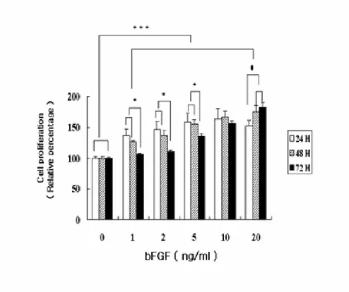
To confirm the effect of bFGF on the proliferation of osteoblasts, MC3T3-E1 cells were stimulated with bFGF in various concentrations (0-20 ng/ml). Cell viability was measured at 24, 48, and 72 hr after bFGF treatment. Cell proliferation increased in a dose-dependent manner, using dosages of 1-10 ng/ml (p < 0.05), and the proliferation was highest after treatment for 24 hr ( p < 0.05, Fig. 1).
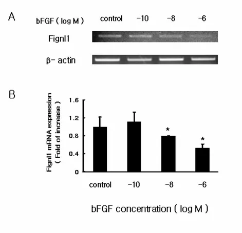
To confirm the effect of bFGF on the expression of Fignl1 at the mRNA level, RT-PCR was performed, using RNA obtained from MC3T3-E1 cells that were either untreated or treated with various concentrations (10-10, 10-8, 10-6M) of bFGF for 6 hr. Fig.2A shows that various concentrations of bFGF suppressed Fignl1 mRNA expression. The effect of bFGF of Fignl1 mRNA levels was dose-dependent. All Fignl1 mRNA levels were normalized to the level of β-actin mRNA, and this analysis revealed that the level of Fignl1 reduction was significant (p < 0.05, Fig.2B).
Fig. 1 . Effect of bFGF exposure on the proliferation of MC3T3-E1 cells. The
effect of bFGF on MC3T3-E1 cells was determined after 24, 48, and 72 hr of treatment. After the incubation periods, the absorbance was determined by an enzyme-linked immunosorbent assay reader. Each value represents the means ± S.D. of triplicate determinations. *, p < 0.05 vs 72 hr ; #, p < 0.05 vs 24 hr ; ***, p < 0.005 bFGF vs control.
Fig. 2. The effect of short-term exposure of bFGF on Fignl1 mRNA expression in MC3T3-E1 cells . The short-term effect of bFGF on MC3T3-E1 cells was determined
after 6 hr of treatment in various concentrations (10-10, 10-8, 10-6M). The results of gel analysis of RT-PCR are shown. Each bar represents the mean and standards of deviation for three separate experiments of Fignl1 densitometry values normalized to β-actin. Subscripts (control and Fignl1) showed significantly different values. Fignl1 mRNA expression was significantly decreased by Fignl1. * p < 0.05 vs control.
The effects of overexpressed Fignl1 on osteoblastogenesis
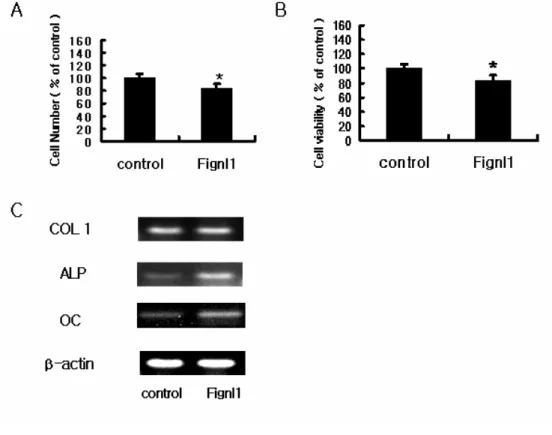
To investigate the impact of Fignl1 overexpression on osteoblastic function, MC3T3-E1 cells were transiently transfected with Fignl1. After 48 hr of transfection, the actual cell count of Fignl1 transfected cells was significantly decreased (Fig. 3A). Cell viability also decreased by 20% in Fignl1 overexpressing cells, compared to a mock transfected group (p < 0.05, Fig 3B).
Overexpession of Fignl1 also affected the expression of osteoblast differentiation marker genes. COL1 mRNA showed no significant change after Fignl1 introduction. However the osteoblast differentiation markers, ALP and OC, were remarkably increased by overexpression of Fignl1 (Fig. 3C).
To determine whether Fignl1 overexpression caused cells to undergo apoptosis, we performed LDH assays and FACS analysis with PI staining. After 48 hr of transfection, there was no change in the proportion of apoptotic cells nor LDH enzymatic activity between mock transfected and Fignl1 overexpressing cells (Fig. 4A & B).
Fig. 3. Effects of Fignl1 on proliferation and differentiation of MC3T3-E1 cells.
(A) The cell count of MC3T3-E1 cells transfected with Fignl1 decreased, compared with mock transfected control cells. (B) MC3T3-E1 cells were transfected with Fignl1, and the proliferation level was determined, using the WST-1 reagent. Proliferation of MC3T3-E1 cells was significantly decreased at 48 hr. Fignl1 appeared to suppress cell proliferation. The results are expressed as means ± S.D from triplicate individual experiments. * p < 0.05 vs control (C) ALP and OC mRNA expression levels significantly increased by overexpression of Fignl1, compared to cells
transfected with vector alone. The amplification of β-actin was used as an internal control for cDNA quality and PCR efficiency in each lane. Data are shown from one experiment representative of three or four similar experiments.
Fig. 4. Correlation of PI staining and LDH assay for estimating cell death. (A)
Flow cytometry analysis. PI staining of apoptotic cells revealed that there was no significant difference between cells transfected with vector alone and cells transfected with Fignl1. Data are shown from one experiment representative of three or four similar experiments. (B) The effects of Fignl1 and agonist on LDH activity in MC3T3-E1 cells. The LDH activity of the control ( transfected vector alone cells ) group was taken as 100%. There were no significant differences between control and Fignl1 groups. The data represent the means of the triplicate determination.*, p < 0.05 vs control (Student’s t-test).
The effect of suppressed Fignl1 on osteoblastogenesis
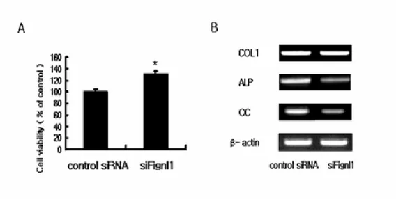
To further examine the role of Fignl1 on MC3T3-E1 cells, Fignl1 expression was knocked down, using RNA interference against Fignl1 (siFignl1). RT-PCR analysis revealed significant inhibition of Fignl1 mRNA expression after siFignl1 treatment, while no change was observed with a control siRNA. The expression of Fignl1 mRNA was suppressed by 70%, compared with the control (Fig. 5). Proliferation of MC3T3-E1 cells was significantly increased by siFignl1 introduction for 48 hr (p < 0.05, Fig 6A). There was no change in COL1, but we observed a significant decrease in ALP and OC levels (Fig. 6B).
Fig. 5.. The effects of siFignl1 on Fignl1 mRNA expression in MC3T3-E1 cells.
Gel analysis results of RT-PCR, showing specific inhibition of Fignl1 expression by siRNA. Each bar represents the mean and standards of deviation for three separate experiments, with Fignl1 densitometry values normalized to those of β-actin. *, p < 0.05 vs control
Fig. 6. Effects of siFignl1 on proliferation and differentiation in MC3T3-E1 cells.
(A) The effects of siFignl1 on proliferation of MC3T3-E1 cells transfected with siFignl1. The proliferation levels were determined, using the WST-1 reagent. Proliferation of MC3T3-E1 cells significantly increased at 48 hr. The results are expressed as means ± S.D from triplicate individual experiments. * p < 0.05 vs control. (Student’s t-test). (B) The effect of siFignl1 transfection on mRNA expression of osteoblast differentiation markers. COL1 mRNA expression level was not significantly different between siFignl1 transfected cells and vector-only transfected
cells. ALP and OC mRNA expression levels were significantly increased by transfection of siFignl1, compared with vector–only transfected cells. The amplification of β-actin was used as an internal control for cDNA quality and PCR efficiency in each lane. Data are shown from one experiment representative of three or four similar experiments.
Nuclear translocation of Fignl1 by bFGF treatment
To determine whether bFGF treatment alters the subcellular localization of Fignl1, Fignl1 was fused with enhanced GFP (EGFP), and the fusion protein was transiently expressed in MC3T3-E1 cells. pEGFP-Fignl1 was initially found throughout the cell, whereas, after 1 hr of stimulation with bFGF, the cellular pEGFP-Fignl1 rapidly localized to the nucleus (Fig.7).
Fig.7. Effect of bFGF on the localization of Fignl1 in MC3T3-E1 cells. MC3T3-E1
cells expressing pEGFP-Fignl1 were treated with (+bFGF) or without (-bFGF) 10-6 M bFGF at 37 °C for 1 hr. Cells were the fixed and stained with Hoescht stock solution (Final concentration, 0.5 µg/ml). Observations were made using a fluorescence microscope (100x). A, left, no GFP signal is observed in the absence of the pEGFP-Fignl1 plasmid under fluorescence microscope. right, Hoechst staining of the same cells, showing the nuclei. B, in the presence pEGFP-Fignl1, a clear signal was present
in the cytoplasm, as well as in the nuclei of the cells (in green). C, pEGFP-Fignl1 localized to the nucleus as quickly as 1 hr after bFGF treatment. The nuclei were stained in blue.
.
Ⅳ Ⅳ Ⅳ
Ⅳ
DISCUSSION
In this study, we found that Fignl1, a subfamily member of the AAA family of proteins, plays an important role in the inhibition of proliferation and stimulation of osteoblast differentiation without causing any apparent changes in apoptosis occurrence.
The anabolic agent, PTH (1-34), enhances the differentiation of mesenchymal stem cells to pre-osteoblasts and stimulates proliferation of pre-osteoblasts while inhibiting the apoptosis of both pre-osteoblasts and osteoblasts21. Meanwhile, another strong anabolic agent, bFGF, increases bone mass and formation of new trabeculae by increasing connectivity or bridging of the trabeculae11,13,14. The anabolic effects of bFGF are also mediated through stimulating proliferation of mesenchymal stem cells, osteoprogenitor cells, and pre-osteoblasts22. Interestingly, bFGF is known to inhibit apoptosis of pre-osteoblasts but stimulates apoptosis of osteoblasts23. Therefore, these two anabolic agents appear to increase bone mass by somewhat different mechanisms. bFGF regulates the recruitment of precursor cells, proliferation, differentiation and apoptosis by acting on cells at different stages. To identify the genes regulated by bFGF, we performed microarray analysis on cells treated with bFGF. This analysis identified Fignl as one of the genes down-regulated greater than two-fold after treatment of MC3T3-E1 cells with bFGF.
Fidgetin is a novel member of the family of AAA proteins24. Based on sequence homology, two closely related genes, fidgetin-like1 and fidgetin-like2, were identified25. Previous studies revealed that Fignl1 is located in both the nucleus and cytoplasm, with the primary localization being cytoplasmic. AAA proteins are molecular chaperones that participate in a wide range of cellular processes, including protein folding, organelle biogenesis, vesicular transport, and cytoskeleton regulation, functions that are consistent with its subcellular localization26. However, the nuclear functions of AAA proteins are poorly understood and provide few clues regarding the function of Fignl1. Even so, Fignl1 appears to be an important gene because loss of fidgetin in mice is accompanied by striking biological consequences that include various skeletal abnormalities27,28.
In this study, we found that Fignl1 is expressed throughout mouse embryonic development (data not shown), indicating that Fignl1 has an important role in early development. Overexpression of Fignl1 inhibited proliferation of MC3T3-E1 cells and stimulated expression of ALP and OC, both of which are markers of osteoblast differentiation. These effects were reversed by transfection of siRNA against Fignl1. Interestingly, there were no changes in COL1 gene expression with either over- or under-expression of Fignl1 genes. This might be because Fignl1 is acting only on very specific processes occurring during
osteoblastogenesis.
bFGF enhanced proliferation of osteoblasts dramatically, through phosphorylation of ERK (data not shown). Meanwhile, bFGF inhibited differentiation of MC3T3-E1 cells and also inhibited expression of Fignl1. Therefore, bFGF-mediated stimulation of proliferation and inhibition of differentiation might be partially explained by suppression of Fignl1. Notably, Fignl1 did not influence the apoptosis of MC3T3-E1 cells, as assessed via an LDH assay and PI staining. Additionally, that bFGF induced localization of Fignl1 to the nucleus was quite surprising ,considering that fidgetin, the original family member, appears to function as a typical AAA protein. This observation suggests that the Fignl1 might be working through a different mechanism that is directly associated with transcriptional processes that regulate osteoblastogenesis.
Taken together, these data suggest that Fignl1, a member of the AAA family of proteins, plays some regulatory role in the proliferation and differentiation of osteoblasts. Furthermore, some of the anabolic effects of bFGF might possibly be mediated through the regulation of Fignl1. Further analyses of Fignl1 will be required to better delineate the mechanisms contributing to enhanced osteoblast differentiation.
V. CONCLUSION
Present study showed the role of Fignl1 in MC3T3-E1 pre-osteoblast cell lines. These results have led me to the following conclusions.
1. Fignl1 mRNA expression was significantly decreased dose dependently after treatment of bFGF.
2. Osteoblast proliferation was inhibited by Fignl1, and Fignl1 enhanced mRNA expression of ALP and OC, which are genes expressed at late stage of osteoblast differentiation. However, there was no effect on cell death.
3. siRNA against Fignl1 abrogated inhibition of cell proliferation and suppressed mRNA expression of ALP and OC.
4. Fignl1-GFP fusion protein was expressed both at cytoplasm and nucleus but it was quickly concentrated in the nucleus 1 hr after bFGF treatment.
From the above results, we propose that Fignl1, a member of the AAA family protein, may play a regulatory role in cell proliferation and differentiation of
osteoblast. Further analyses of Fignl1 will be required to delineate the mechanisms better which contribute to the enhanced osteoblast differentiation.
REFERENCES
1. Bouillon R, Burkhardt P, Christiansen C, Fleiseh HA, Fugita T, Gennari C, et al. Consensus development conference; prophylaxis and treatment of osteoporosis. Osteoporos Int 1991;1: 114-117.
2. Cummings SR, Kelesy JL, Nevitt MC, O’Dowd KJ. Epidemiology of osteoporosis and osteoporotic fractures. Epidemiol Rev 1985; 7:178-208.
3. Dempster DW, Cosman F, Parisien M, Shen V, Lindsay R. Anabolic actions of parathyroid hormone on bone. Endocr Rev 1993;14:690-709.
4. Betancourt M, Wirfel KL, Raymond AK, Yasko AW, Lee J, Vassilopoulou-Sellin R. Osteosarcoma of bone in apatient with primary hyperparathyroidism: a case report. J Bone Miner Res 2003;18:163-166.
hormone/alendronate therapy for osteoporos-robbing Peter to pay Paul? J Clin Endocrinol Metab 2000; 85:2127-2128.
6. Vahle JL, Sato M, Long GG, Young JK, Francis PC, Engelhardt JA, et al. Skeletal changes in rats given daily subcutaneous injections of recombinant human parathyroid hormone (1-34) for 2 years and relevance to human safety. Toxicol Pathol 2002;30:312-321.
7. Cronauer MV, Schulz WA, Seifert H-H, Ackerman R, Burchardt M. Fibroblast growth factors and their receptors in urological cancers: basic research and clinical implications. Eur Urol 2003;43: 309-319.
8. Singh PK, Bucana CD, Gutman M, Fan D, Wilson MR, Fidler IJ. Organ site-dependent expression of basic fibroblast growth factor in human renal cell carcinoma cells. Am J Pathol 1994;145: 365-374.
9. Rodan SB, Wesolowski G, Yoon K, Rodan GA. Opposing effects of fibroblast growth factor and pettussis toxin on alkaline phosphatase, osteopontin, osteocalcin and type I collagen mRAN levels in ROS 17/2.8 Cells. J Bio Chem 1989;264: 19934-19941.
10. McCarthy TL, Cenrella M, Canails E. Effects of basic fibroblast growth factors on deoxyribonucleic acid and collagen synthesis in rat parietal bone cells. Endocrinology 1989;125: 2118-2126.
11. Canalis E, Centrella M, McCarthy T. Effects of basic fibroblast growth factor on bone formation in vitro. J Clin Invest 1998;81: 1572-1577.
12. Hurley MM, Abreu C, Harrison JR, Lichtler AC, Raisz LG, Kream BE. Basic fibroblast growth factor inhibits type I collagen gene expression in osteoblastic MC3T3-E1 cells. J Biol Chem 1993;268: 5588-5593.
13. Line NE, Kumer J, Yao W, Breunig T, Wronski T, Modin G, et al. Basic fibroblast growth factor forms new trabeculae that physically connect with pre-existing trabeculae, and this new bone is maintained with an anti-resorptive agent and enhanced with an anabolic agent in an osteopenic rat model. Osteoporos Int 2003;14: 374-382.
14. Mayahara H, Ito T, Nagai H, Miyajima H, Tsukuda R, Takatomi S, et al. In vivo stimulation of endosteal bone formation by basic fibroblast growh factor in rats. Growth Factors 1993;9: 73-80.
15. Okazaki H, Kurokawa T, Nakamura K, Matsushita T, Mamada K, Kawaguchi H. Stimulation of bone formation by recombinant fibroblast growth factor-2 in callotasis bone lengthening of rabbits. Calcif Tissue Int 1999;64: 542-546.
16. Iseki S, Wilkie AO, Morriss-Kay GM. Fgfr1 and Fgfr2 have distinct differentiation- and proliferation-related roles in the developing mouse skull vault. Development 1999;126: 5611-5620.
17. Montero A, Okada Y, Tomita M, Ito M, Tsurukami H, Nakamura T, et al. Disruption of the fibroblast growth factor-2 gene results in decreased bone mass and bone formation. J Clin Invest 2000;105: 1085-1093.
heparin and basic fibroblast growth factor on collagen synthesis in 21-day fetal rat calvariae. Endocrinology 1992;130: 2675-2682.
19. Hurlry MM, Lee SK, Raisz LG, Bernicker P, Lorenzo JA. Basic fibroblast growth factor induces osteoclast formation in murine bone marrow cultures. Bone 1998;22: 309-316.
20. Hurlry MM, Abreu C, Gronowicz G, Kawaguchi H, Lorenzo J. Expression and regulation of basic fibroblast growth factor mRNA levels in mouse osteoblastic MC3T3-E1 cells. J Biol Chem 1994;269: 9392-9396.
21. Qin L, Qiu P, Wang L, Li X, Swarthout JT, Soteropoulos P, et al. Gene expression profiles and transcription factors involved in parathyroid hormone signaling in osteoblasts revealed by microarray and bioinformatics. J Biol Chem 2003;278: 19723-19731.
22. Hanada K, Dennis JE, Caplan AI. Stimulatory effects of basic fibroblast growth factor and bone morphogenetic protein-2 on osteogenetic differentiation of rat bone marrow-derived mesenchymal
stem cells. J Bone Miner Res 1997;12: 1606-1614.
23. Hill DA, Tumber A, Meikle MC. Multiple extracellular signals promote osteoblast survival and apoptpsis. Endocrinology 1997;138: 3849-3858.
24. Lupas AN, Martin J. AAA proteins. Curr Opin Struct Biol 2002;12: 746-753.
25. Cox GA, Mahaffey CL, Nystuen A, Letts VA, Frankel WN. The mouse fidgetin gene defines a new role for AAA family proteins in mammalian development. Nat Genet 2000;26: 198-202.
26. Patel S, Latterich M. The AAA teams; related ATPases with diverse functions. Trends Cell Biol 1998;8: 65-71.
27. Truslove GM. The anatomy and development of the fidget mouse. J Genet 1956;54: 64-86.
Relations between major and minor variants. J Genet 1955;53: 515-533.
국문요약
국문요약
국문요약
국문요약
bFGF에
에
에
에 의해
의해
의해
의해 억제된
억제된
억제된 Fidgetin-like1 이
억제된
이
이
이
조골세포
조골세포
조골세포
조골세포의
의
의 증식
의
증식
증식 및
증식
및 분화에
및
및
분화에
분화에
분화에 미치는
미치는
미치는 영향
미치는
영향
영향
영향
(지도교수
지도교수
지도교수 임승길
지도교수
임승길
임승길
임승길)
연세대학교
연세대학교
연세대학교
연세대학교 대학원
대학원
대학원 의과학과
대학원
의과학과
의과학과
의과학과
김
김
김
김 수
수
수
수 진
진
진
진
조골세포에서 bFGF와 PTH를 동시에 처리했을 때 증가하는 것으로 분석된 13개의 유전자들 가운데 ATPase와 관련된 AAA family 의 한 일원인 Fidgetin-like1 (Fignl1)을 택하여 이것이 조골세포의 증식과 분 화에 미치는 영향을 조사하였다. Fignl1은 세포 내에서 세포질과 핵 에 모두 존재한다고 알려져 있지만 기능에 관한 연구는 아직 명확 하게 밝혀져 있지 않다. 본 연구에서는 bFGF가 전구조골세포인 MC3T3-E1 세포에서 Fignl1 유전자의 발현을 억제하고, 더 나아가 조골세포의 증식을 방해하며 분화를 유도하는 것을 관찰하였다. 즉,bFGF를 MC3T3-E1 세포에 농도별로 처리하였을 때, 농도 증가에 따 라 Fignl1 유전자의 발현이 억제되는 것을 RT-PCR로 관찰하였다. WST-1 검사와 세포 수 측정을 통한 분석 결과 Fignl1이 MC3T3-E1 세포의 증식은 감소시키나, LDH 와 PI를 이용한 분석 결과 세포사 멸에는 영향을 주지 않음을 알았다. 또 조골세포의 분화 표지 유전 자인 type I collagen(COL1), alkaline phosphatase(ALP) 및 osteocalcin(OC) 유전자의 표현 정도를 RT-PCR로 조사하여 본 결과 분화 초기유전자인 COL1의 발현에는 별 차이가 없었지만, 분화 후 기유전자인 ALP 와 OC는 발현이 증가된 것으로 미루어 보아 Fignl1 이 조골세포의 분화를 유도함을 알 수 있었다. 이상의 결과는 다시 Fignl1에 대한 siRNA를 처리한 후 세포의 증식이 증가되고 ALP와 OC mRNA의 발현이 감소하는 것을 확인하여 검증하였다. Fignl1의 세포 내 위치를 알아보기 위해 Fignl1-GFP fusion protein을 만들어서 형광현미경으로 관찰한 결과, 세포질에 존재하던 Fignl1이 bFGF를 처리하면 1시간 내에 핵으로 빠르게 이동하는 것을 관찰하였다. 결 론적으로 Fignl1은 AAA family protein의 일원으로 조골세포의 증식을 억제하고 분화를 촉진하는 역할을 한다. 그러나 향후 Fignl1이 조골 세포의 분화를 증가시키고 증식을 억제하는 기전에 대해서도 자세 하게 조사 할 필요가 있다고 본다.