Serine 34 Phosphorylation of Rho Guanine Dissociation
Inhibitor (RhoGDI
␣) Links Signaling from Conventional
Protein Kinase C to RhoGTPase in Cell Adhesion
*
□SReceived for publication, December 22, 2009, and in revised form, April 21, 2010 Published, JBC Papers in Press, May 15, 2010, DOI 10.1074/jbc.M109.098129 Athanassios Dovas‡1,2, Youngsil Choi§1, Atsuko Yoneda‡1,3, Hinke A. B. Multhaupt‡3, Seung-Hae Kwon¶, Dongmin Kang§, Eok-Soo Oh§4, and John R. Couchman‡5
From the‡Division of Biomedical Sciences, Faculty of Medicine, Imperial College London, Exhibition Road, London SW7 2AZ, United Kingdom, the§Division of Life and Pharmaceutical Sciences, Ewha Women’s University, Seoul 120-750, Republic of Korea, and¶Korea Basic Science Institute, Chuncheon Center, Chuncheon,
Gangwon-Do 210-701, Republic of Korea
Conventional protein kinase C (PKC) isoforms are essential serine/threonine kinases regulating many signaling net-works. At cell adhesion sites, PKC␣ can impact the actin cytoskeleton through its influence on RhoGTPases, but the intermediate steps are not well known. One important regu-lator of RhoGTPase function is the multifunctional guanine nucleotide dissociation inhibitor RhoGDI␣ that sequesters several related RhoGTPases in an inactive form, but it may also target them through interactions with actin-associated proteins. Here, it is demonstrated that conventional PKC phosphorylates RhoGDI␣on serine 34, resulting in a specific decrease in affinity for RhoA but not Rac1 or Cdc42. The mechanism of RhoGDI␣phosphorylation is distinct, requir-ing the kinase and phosphatidylinositol 4,5-bisphosphate, consistent with recent evidence that the inositide can acti-vate, localize, and orient PKC␣in membranes. Phosphospe-cific antibodies reveal endogenous phosphorylation in sev-eral cell types that is sensitive to adhesion events triggered, for example, by hepatocyte growth factor. Phosphorylation is also sensitive to PKC inhibition. Together with fluorescence resonance energy transfer microscopy sensing GTP-RhoA levels, the data reveal a common pathway in cell adhesion linking two essential mediators, conventional PKC and RhoA.
Protein kinase C (PKC)6isoforms have a wide array of func-tions in cell adhesion, cytoskeleton, trafficking, and polarity (1–3) to transcription, survival, and differentiation (4). Their activities can intersect with a number of signaling cascades and networks and are stimulated through several distinct receptor families. Broadly, there are three PKC subfamilies, conven-tional, novel, and atypical, and they operate at different subcel-lular sites with discrete functions (5, 6). The PKC␣ conven-tional isoform may regulate a variety of functions, including redox reactions and cell survival, often upstream of extracellu-lar signal-regulated kinase (ERK) and phosphatidylinositol 3-kinase/AKT pathways and NF-B synthesis. In addition, this isoenzyme can be localized to sites of cell adhesion and regulate actin cytoskeletal function and migration (7–9). Besides the canonical pathway of PKC activation through phospholipase C-mediated generation of diacylglycerol and inositol tris-phosphate, a second method of PKC␣ activation is known involving uncleaved phosphatidylinositol 4,5-bisphosphate (10 –12). Moreover, in vitro experiments reveal that Ca2⫹is not required for this activation (13) and is consistent with data showing high PtdIns(4,5)P2affinity for the C2 domain of PKC␣, mediated by a polybasic cluster of amino acids, which leads to persistence of membrane association (14, 15) with decreased Ca2⫹requirement (16).
Downstream events from PKC␣ in fibroblast adhesion to fibronectin, for example, are not known other than an eventual up-regulation of GTP-RhoA levels, with concomitant cytoskel-etal reorganization (8, 17). The localization of p190RhoGAP may be intrinsic to the process (17), although a precise role for PKC␣ activity is unclear. The three mammalian RhoGDI iso-forms are cytosolic proteins that sequester RhoGTPases in their GDP-bound form, effectively forming sinks that remove the G proteins from cycling through the guanine exchange factor and GTPase-activating protein-mediated activation/deactiva-tion process (18, 19). In addiactivation/deactiva-tion, RhoGDI can interact with
*The work was supported in part by Wellcome Trust Programme Grant 065940 (to J. R. C.), Danish National Research Foundation, Haensch Fond, Wilhelm Pedersen Fond, and University of Copenhagen (to J. R. C., H. A. B. M.).
Author’s Choice—Final version full access. □S
The on-line version of this article (available at http://www.jbc.org) contains supplemental Figs. 1– 4.
1These authors contributed equally to this work.
2Present address: Dept. of Anatomy and Structural Biology, Albert Einstein
College of Medicine, Bronx, NY 10461.
3Present address: Dept. of Biomedical Sciences, Faculty of Health Sciences,
Copenhagen University, 2200 Copenhagen, Denmark.
4Supported in part by Grant R15-2006-020 from the National Core
Research Center program of the Ministry of Education, Science, and Technology Korea and Korea Science and Engineering Foundation through the Center for Cell Signaling and Drug Discovery at Ewha Woman’s University.
5To whom correspondence should be addressed: Dept. of Biomedical
Sci-ences, Faculty of Health SciSci-ences, Copenhagen University, Biocenter, Ole Maaløes Vej 5, 2200 Copenhagen N, Denmark. Tel.: 45-353-25670; Fax: 45-353-25669; E-mail: john.couchman@bric.ku.dk.
6The abbreviations used are: PKC, protein kinase C; PtdIns,
phosphatidylino-sitol; Ptd, phosphatidyl; GTP␥S, guanosine 5⬘-3-O-(thio)triphosphate; MDCK, Madin-Darby canine kidney cell; REF, rat embryo fibroblast; FRET, fluorescence resonance energy transfer; GDI, guanine dissociation inhibi-tor; DL, diolein; PMA, phorbol 12-myristate 13-acetate; siRNA, small inter-fering RNA; CFP, cyan fluorescent protein; YFP, yellow fluorescent protein; HGF, hepatocyte growth factor.
cytoskeletal proteins such as ezrin-radixin-moesin that could target RhoGTPase activity at a subcellular level (20). Therefore, it is an oversimplification to ascribe only a sequestration func-tion to GDI proteins. Because RhoGDI␣, the most abundant isoform, binds several RhoGTPases, including GDP-RhoA, -Rac1, and -Cdc42 (18), regulatory mechanisms that control its function have come under some recent scrutiny (19, 21). RhoGDI␣ has two domains, an N-terminal domain of around 70 amino acids and an immunoglobulin-like C-terminal do-main of⬃130 amino acids. NMR studies have shown that the N-terminal domain of the free protein is flexible in solution but with residues 9 –20 and 36 –58 forming transient helices (22). This part of the GDI molecule is stabilized by interactions with GTPases and is therefore a potential target for regulation through protein modification, such as phosphorylation. Here, we test the hypothesis that RhoGDI␣ phosphorylation by PKC may represent a major step in the pathway to RhoA, with onward activation of downstream effectors regulating cell adhesion, cytoskeletal, and other cellular events.
EXPERIMENTAL PROCEDURES
Cell Culture, Antibodies, and Reagents—Rat embryo fibro-blasts were maintained in␣-minimal essential medium with 5% fetal calf serum (Labtech International), at 37 °C, 10% CO2and used between passages 6 –20. Wild-type Drosophila Schneider 2 (S2) cells were provided by Dr. Finn Werner (University College London) and were maintained in DES Expression medium (Invitrogen) containing 10% fetal calf serum. PKC␣-expressing S2 cells were provided by Dr. Ssang-Taek Lim (University of Califor-nia, San Diego). For selection and maintenance of stably trans-fected populations, cells were cultured in DES Expression medium with 10% fetal bovine serum and 300 g/ml hygromycin-B (Invitrogen). MDCK (type II) cells from ATCC were maintained in Dulbecco’s modified Eagle’s medium supplemented with 5% fetal bovine serum (Hyclone), and human erythroleukemia K562 cells from ATCC were in Dulbecco’s modified Eagle’s medium/F-12 medium with 10% fetal bovine serum. Polyclonal antibodies against RhoGDI (A20 and K21) and RhoA (119) were from Santa Cruz Biotechnology (Heidelberg, Germany). Mouse monoclonal antibodies against Rac1 (clone 23A8) and Cdc42 (clone 44) were from Upstate Biotechnology and BD Transduction Laboratories. Anti-His tag polyclonal antibody was from Cell Signaling Technol-ogy. The phosphospecific antibody against RhoGDI Ser(P)-34 was produced by Quality Controlled Biochemicals (on behalf of BIO-SOURCE International) using the peptide sequence CPAQK(p-S)IQEI (residues 30 –38 with an additional N-terminal cysteine) as an immunogen coupled to keyhole limpet hemocyanin. To purify phospho-specific antibodies, two columns were prepared. The peptides CPAQKSIQEI and CPAQK(pS)IQEI were immobilized through the free cysteine residues to maleimide-activated agarose. Serum was first passed through the unphosphorylated peptide affinity column to remove antibodies recognizing nonphosphory-lated RhoGDI. Flow-through fractions from the first column were applied to the second column, with the immobilized phosphopep-tide. After washing with phosphate-buffered saline, antibodies recognizing phosphopeptide were eluted with 0.1Mglycine HCl,
pH 2.8, and the eluates were neutralized with 1MTris, pH 8.0,
immediately.
PMA, Go¨6976, and GF109203X were from Calbiochem; PtdSer, diolein (DL), histone III-S, ATP, GTP␥S, NAD, U73122, and bovine plasma fibronectin were from Sigma. Inositides PtdIns(4,5)P2, PtdIns(3,4,5)P3, and PtdIns were from Biomol or Sigma. [␥-32P]ATP, GTP␥35
S, and [32P]NAD were obtained from Amersham Biosciences. PKC␣␥ purified from rabbit brain was from Upstate Biotechnology.
Plasmids, cDNAs, Site-directed Mutagenesis, Recombinant Protein Expression, and Purification—Wild-type human RhoA and Rac1 cDNA cloned in pRk5-Myc expression vector were from Dr. A. Hall (Memorial Sloan-Kettering Cancer Center, New York); pGEX-2T RhoA was from Dr. V. Braga (Imperial College London, UK), and pTAT-C3 was from Dr. J. Bertoglio (INSERM, France). cDNAs encoding wild-type RhoGDI␣ and an N-terminal trunca-tion mutant lacking the first 66 amino acids (RhoGDI(67–204)) in the pHis-parallel vector (23) were a kind gift from Dr. Z. Dere-wenda (University of Virginia). All plasmids were transformed in the BL21(pLysS) strain, and proteins were purified on a TALON (Qiagen) or Co2⫹-charged chelating Sepharose fast flow resin (Amersham Biosciences) according to Sheffield et al. (23). The hexahistidine tag was removed by incubating with recombinant tobacco etch virus (AcTEV, Invitrogen) as described by the man-ufacturer, and the protease, cleaved tags, and any uncleaved pro-tein were removed by metal-chelating affinity chromatography. For full-length RhoGDI, an extra gel filtration step on a HiPrep 16/60 Sephacryl S-100 high resolution column (Amersham Bio-sciences) was performed on an AKTA high pressure liquid chro-matography system (Amersham Biosciences). Proteins were ⬎95% pure as evidenced by Coomassie stain. Protein concentra-tion was calculated with the BCA assay kit (Pierce). Primers for site-directed mutagenesis are listed in supplemental Table 1. cDNAs were confirmed by sequencing, and bacterially expressed proteins were purified as described above.
Expression and Purification of RhoGDI-Rho Protein Com-plexes—Purification of Rac1-RhoGDI complexes from induced S2 cells was carried out essentially as described by Read and Naka-moto (24). Briefly, cells were harvested in cold wash buffer (50 mM
Tris, pH 7.4, 150 mMNaCl, 5 mMMgCl2, 10% (v/v) glycerol, 20
g/ml aprotinin, 20 g/ml leupeptin, 5 g/ml pepstatin, 1 M
Pefabloc, and 1 mMphenylmethylsulfonyl fluoride) and lysed by
three passes through a French press at 20,000 p.s.i. After a 15-min centrifugation at 14,000 rpm, supernatants were centrifuged at 100,000⫻ g for 1 h. Supernatants were incubated for 1 h with TALON resin, washed in wash buffer, and eluted in wash buffer containing 150 mMimidazole. The eluate was passed five times over an equilibrated anti-Myc-agarose (9E10) column (Sigma), washed in wash buffer, and bound protein complexes eluted with wash buffer containing 100g/ml Myc peptide (Sigma).
Yeast strain SY1 transformed with plasmids encoding for FLAG-RhoGDI and His6-RhoA were gifts from Dr. R.
Naka-moto (University of Virginia). Yeast growth and purification of a stoichiometric 1:1 protein complex of RhoA-RhoGDI by sequential passage over TALON and FLAG-agarose (Sigma) columns were performed as described previously, as were pro-tein concentration calculations (24). In phosphorylation exper-iments, tags from the recombinant RhoA and RhoGDI proteins were removed by recombinant tobacco etch virus protease. Tags, uncleaved proteins, and the protease were removed on
respective affinity columns. Purity of the RhoGDI-Rho com-plexes was confirmed by silver staining. The Myc tag on Rac1 was not removed.
Expression and Purification of Recombinant PKC ␣—Dro-sophilaS2 cells transfected with plasmid encoding full-length PKC␣ and methods for protein purification were reported pre-viously (25). Recombinant PKC activity was determined by comparing the incorporation of phosphate in histone III-S or RhoGDI with that of commercial PKC␣␥, and comparable activities were used for all experiments.
Other Recombinant Proteins—Recombinant purified vincu-lin tail protein was a generous gift from Dr. D. Critchley (Uni-versity of Leicester). Recombinant GST-RhoA and TAT-C3 proteins were purified as described previously (26, 27). Cell lysates and recombinant proteins were analyzed by SDS-PAGE and Western blotting (28). Western blots were analyzed with NIH Image version 1.61.
Detection of Ser-34 Phosphorylation in Endogenous RhoGDI— MDCK cells were serum-starved for 5– 8 h and treated with either 200 nMPMA or vehicle (DMSO) for 10 min (29). To
confirm ruffle membrane formation in MDCK cells upon PMA treatment, phase contrast images of cells fixed in 2% glutaral-dehyde were recorded on an Olympus IX71 microscope with a DP50 camera and⫻20 objective. Images were processed with Viewfinder Lite version 1.0 and Studio Lite version 1.0. In some experiments, cells were pretreated with 1 M PKC inhibitors Go¨6976 or GF109203X for 1 h or 3MU73122 for 15 min prior to
PMA treatment. K562 cells (0.8⫻ 106) were suspended in serum-free Dulbecco’s modified Eagle’s medium/F-12 medium and then treated with 800 nMPMA for 5–30 min at 37 °C. Cells were washed
with cold Tris-buffered saline containing phosphatase inhibitors (20 mM-glycerophosphate, 25 mMNaF, and 1 mM
orthovana-date) and then lysed with buffer containing 50 mMTris, pH 7.4, 150
mMNaCl, 1 mMEDTA, 1% Triton X-100, 10 mMMgCl2, protease inhibitor mixture, and phosphatase inhibitors. Cleared lysates were analyzed by Western blotting of 12% SDS-polyacrylamide gels with pan-GDI antibodies (A20) and Ser(P)-34 RhoGDI anti-body. Rat embryo fibroblasts (REF) and G361 human melanoma cells were treated as above but were not PMA-treated. Plasmids were transfected in MDCK cells with Lipofectamine LTX (Invitrogen) according to the manufacturer’s protocol. Ectopi-cally expressed RhoGDI was detected by anti His tag antibodies followed by fluorochrome-conjugated (Alexa Fluor 488/546) secondary antibodies (Invitrogen).
Pulldown of RhoGTPases with Bacterially Expressed GDIs— REF in growth medium were lysed in 50 mMTris, pH 7.6, 150
mMNaCl, 1% Triton X-100, 1 mMEDTA, protease inhibitor
mixture (Roche Applied Science), 20 mM-glycerophosphate,
25 mM NaF, and 1 mMorthovanadate. Cleared lysates were
incubated with bacterially expressed GDIs (wild type or mutant) immobilized onto Talon beads at 4 °C for 1 h. After washing beads with lysis buffer three times, proteins pulled down with GDIs were separated by SDS-PAGE and analyzed by Western blotting with RhoA, Rac1, and Cdc42 antibodies.
Kinase Assay—Protein kinase C assays were performed as described previously (13). Calcium chloride was added to 750 Mwhen PtdSer/DL were the lipid co-factors and was omitted
or included at the indicated concentrations when PtdIns(4,5)P2
was used. Reactions proceeded for 10 min with histone III-S or 30 min for RhoGDI or vinculin substrates at room temperature. Samples were resolved by 15% SDS-PAGE, and phosphoryla-tion was detected by autoradiography on a Fuji-BAS reader and quantified using the TINA-BAS densitometry software. For Western blotting [␥-32P]ATP was omitted from the reaction mixture; 25 ng of phosphorylated protein were resolved by 15% SDS-PAGE and immunoblotted as described above.
For determination of phosphorylation stoichiometry, 10 pmol of RhoGDI protein (free or complexed to RhoA) were phosphorylated for 1 h with 10 ng of PKC␣␥ in a total of 20 l of reaction buffer containing 50 mMHEPES, pH 7.4, 3 mM
mag-nesium acetate, 50MPtdIns(4,5)P2, and 50MATP (1Ci of [␥-32P]ATP; 3 Ci/mmol, 2 mCi/ml). Reactions were stopped by spotting onto P81 phosphocellulose filters (Whatman) and washed in 1% (v/v) orthophosphate buffer. Filters were ana-lyzed by scintillation counting and converted to moles of phos-phate using radiolabel specific activity and calibration of scin-tillation counting efficiency. Radiolabel incorporated into an equimolar amount of the S34A point mutant was considered background and subtracted from assays of wild-type RhoGDI. Results were obtained from triplicate experiments.
Nucleotide Exchange Reaction and RhoA ADP-ribosylation— Nucleotide exchange assays and ADP-ribosylation experiments were performed as described previously (30, 31). Briefly, 10 pmol of GST-RhoA or RhoA complexed to RhoGDI (either nonphosphorylated or phosphorylated) were diluted to a final volume of 50l in nucleotide exchange assay buffer (20 mM
Tris, pH 7.4, 150 mMNaCl, 5 mMMgCl2, 10 mMEDTA) con-taining 45 pmol of GTP␥S (0.5 Ci [35S]GTP␥S; 1,115 Ci/mmol, 1 mCi/ml). The nucleotide exchange reactions (at 30 °C) were stopped with cold assay buffer (without EDTA), adsorbed on nitrocellulose filters (0.45 m; Schleicher and Schuell), and assayed (30). Counts were converted to moles of GTP␥S as before.
ADP-ribosylation experiments were performed essentially as described previously (31). Briefly, GST-RhoA fusion protein or 20l of kinase or a control containing 500 ng of RhoA protein (⬃20 pmol) were brought to a volume of 40l in a buffer con-taining 50 mMTris-HCl, pH 7.4, 2 mMMgCl2, 1 mMEDTA, and
40 pmol of NAD (1 Ci of [32P]NAD; 1,000 mCi/mmol, 10 mCi/ml). Fifty ng (or amounts indicated) of recombinant TAT-C3 protein (27) were added last. The samples were incu-bated for 5 min at room temperature, stopped by addition of 5 l of sample buffer, denatured at 100 °C, and resolved by 15% SDS-PAGE for visualization by autoradiography.
Silencing of Rat RhoGDI␣ and Ectopic Expression of Human RhoGDI␣ Proteins—Oligonucleotide-based silencing of rat RhoGDI␣ was by pre-designed sense and antisense oligonu-cleotides obtained from Qiagen. The target sequence was Rn_Arhgdia_1_HP (bases 393– 413). Annealed duplexes were transfected in REF cells with Oligofectamine (Invitrogen). Expression levels of RhoGDI␣ were determined after 48 h by Western blotting, with a luciferase sequence negative control (28). For expression of human RhoGDI␣ (wild type and S34A and S34D mutants), the cDNA was amplified by PCR from the pHis parallel vector using the primers F1 and R1 (see supple-mental material). The amplified PCR product was digested,
ligated into the KpnI/EcoRI site of the pcDNA3.1/HisA vector (Invitrogen), and sequenced. Recombinant RhoGDI␣ con-tained a hexahistidine tag to distinguish it from endogenous RhoGDI␣. Plasmids were transfected in REF cells as described above. Because human RhoGDI␣ contains the same sequence as rat RhoGDI␣ siRNA target sequence, silent mutations of five nucleotides in the target sequence were introduced by overlap extension PCR. Primers F1 and 193:5⬘ or R1 and 192:5⬘ were used for first PCR. The second PCR used the first PCR products and primers F1 and R1. The amplified PCR product was sub-cloned into the KpnI and EcoRI sites of pcDNA3.1/HisA.
Confocal Microscopy and FRET Analysis—REF were trans-fected with siRNA specific for rat RhoGDI and imaged on a Zeiss Axiovert 200 M inverted microscope equipped with a Zeiss 510 META confocal head. All images were analyzed with Zeiss software. FRET analysis by acceptor photobleaching was carried out as described previously (32) with some modifi-cation. Briefly, REF cells co-transfected with pRaichu 1502 (from M. Matsuda (33)) and pcDNA-RhoGDI (wild type) or pcDNA-RhoGDI mutants (S34A or S34D) were plated onto coverslips, then fixed for 5 min in 4%(w/v) formaldehyde, washed with phosphate-buffered saline, and visualized with a C-Apochromat 40X/1.2-W objective lens using a 458 nm argon laser light and Meta detector (462.6 –516.1 nm) for CFP excita-tion and emission and a 514-nm argon laser light and Meta detector (516 –591 nm) for YFP. FRET efficiency was measured by acceptor photobleaching; pre-bleach CFP and YFP images were collected simultaneously following excitation at 458 nm for CFP and 514 nm for YFP. A selected region was irradiated with the 514-nm laser line (100% intensity, 60 iterations, using a 458/514-nm dual dichroic mirror) to bleach YFP. Post-bleach CFP and YFP images were collected simultaneously (at 458/514 nm) immediately following photobleaching. FRET was mea-sured as an increase in CFP fluorescence intensity following YFP photobleaching. FRET efficiency was calculated as 100⫻ ((CFP post-bleach⫺ CFP prebleach)/CFP post-bleach) using the FRET Macro in the Zeiss Aim software, correcting for CFP and YFP background in each channel.
RESULTS
Conventional PKC Phosphorylates RhoGDI␣ in Vitro on Ser-ine 34—The recombinant RhoGDI␣ fusion protein was first cleaved with recombinant tobacco etch virus (23) to remove the hexahistidine tag, because the two threonine residues in the tag linker region are PKC substrates (supplemental Fig. 1A). Simi-larly, we noted, as published previously, that GST is a PKC␣ substrate (on Ser-93) (34), ruling out fusion proteins based on this system. Recombinant RhoGDI␣ was phosphorylated by PKC in the presence of PtdIns(4,5)P2but much less in the
pres-ence of the more commonly used phosphatidylserine/diacylg-lycerol/calcium (PtdSer/DL/Ca2⫹)
.Essentially identical results
were obtained with recombinant mammalian PKC␣ purified from Drosophila S2 cells (25) or a mixture of conventional PKCs (PKC␣␥) purified from rabbit brain (Fig. 1A). Both preparations have low intrinsic activity in the absence of lipid activators (25). As a control, histone III-S was efficiently phos-phorylated by both PKC preparations in the presence of PtdSer/ DL/Ca2⫹and to a lesser degree by PtdIns(4,5)P2(Fig. 1B),
con-sistent with our previous data (13). We and others have shown that PKC␣ activity stimulated by PtdIns(4,5)P2in combination
with the cytoplasmic domain of syndecan-4 can be independent of divalent cations (13, 35). This was apparent with RhoGDI␣ phosphorylation, which was independent of Ca2⫹and per-sisted in the presence of EGTA (Fig. 1A). In contrast, a doc-umented physiological substrate for PKC, vinculin tail (36, 37), showed different characteristics to RhoGDI␣ (Fig. 1B). PtdSer/DL/Ca2⫹provided optimal conditions for phosphor-ylation, whereas PtdIns(4,5)P2 alone was insufficient for
effective PKC␣-mediated phosphorylation unless Ca2⫹was
present in low concentrations (Fig. 1B).
Phosphorylation of RhoGDI␣ by PKC␣ was maximal at 50 M PtdIns(4,5)P2, and under these conditions, calculations
revealed between 0.25 and 0.5 mol of phosphate per mol of RhoGDI␣, with a mean of 0.3. The necessity for PtdIns(4,5)P2in RhoGDI␣ phosphorylation is unusual but consistent with a known site in the C2 region of PKC␣ that binds this lipid in the absence of Ca2⫹and becomes activated (10). The data might
also suggest an interaction between PtdIns(4,5)P2and RhoGDI,
but none was detected by plasmon resonance spectroscopy.7
In silicoanalysis revealed multiple potential phosphoryla-tion sites on RhoGDI␣, several of them bearing a strong PKC phosphorylation consensus motif. As RhoGDI proteins are composed of two discrete domains (supplemental Fig. 1D), an N-terminal truncation mutant lacking the first 66 amino acids and comprising only the immunoglobulin domain (RhoGDI␣(67–204)) was used as substrate (Fig. 1C). Kinase assays showed that the immunoglobulin-like domain was not phosphorylated by PKC␣ (Fig. 1C) suggesting that the site(s) were N-terminal or, conceivably, that the N terminus provides a PKC␣-docking site for RhoGDI␣ phosphoryla-tion at a separate site.
Of several serine residue mutations within the N-terminal domain of full-length, wild-type RhoGDI␣, that of Ser-34 to Ala had the most marked effect, reducing phosphorylation by recombinant PKC␣ or PKC␣␥ by around 70%, relative to the wild-type protein (Fig. 1D). In contrast, mutation of the con-sensus site Ser-62 to Ala had no effect on phosphorylation mediated by purified brain PKC (Fig. 1D). Ser-to-Asp substitu-tions resulted in comparable phosphorylation levels relative to the Ser-to-Ala single point mutants (Fig. 1E), indicating that acquisition of negative charge on residue 62 does not affect phosphorylation of RhoGDI␣, therefore excluding co-operat-ivity between residues 34 and 62. A control mutation of Ser-47 was also completed for two reasons. First, it lies in the strongest consensus PKC phosphorylation motif, and second this residue is involved in hydrogen bonding with Thr-37 of Cdc42 that coordinates the Mg2⫹ion of the GTPase (38), making it a pos-sible regulatory residue. However, phosphorylation by PKC␣ was not reduced in a S47A mutant protein (Fig. 1E). Therefore, Ser-34 was identified as the major phosphorylation site.
Serine 34 lies at the base of helix␣2, forming part of the helix-loop-helix motif that binds the switch regions of GTPases (helices␣2 and ␣3) and which NMR spectroscopy shows to be
unstable or transient when RhoGDI is not complexed to GTPases (22). To further show that this residue could be phos-phorylated in the wild-type RhoGDI␣ protein, a phosphospe-cific antibody was prepared and characterized. The sequence (C)PAQK(pS)IQEI was used as immunogen, having 100% sequence conservation in mammalian RhoGDIs and may there-fore represent a conserved phosphorylation site. The affinity-purified antibody reacted with in vitro PKC␣ phosphorylated RhoGDI␣, with minimal immunoreactivity toward the unphos-phorylated protein (Fig. 2A). In addition, virtually no reactivity of the antibody was seen in Western blots of control RhoGDI␣ lysates from K562 cells (Fig. 2B), whereas reactivity of the anti-body against phosphorylated RhoGDI␣ was detected in phor-bol ester-treated K562 cells (Fig. 2B). A general anti-phospho-threonine antibody did not detect the in vitro phosphorylated protein, indicating the absence of threonine phosphorylation on RhoGDI (Fig. 2A). As a positive control for the phospho-FIGURE 1. RhoGDI␣isasubstrateforPKC␣onserine34,inaPtdIns(4,5)P2
-dependent manner. A, phosphorylation of RhoGDI by PKC␣␥. Kinase
acti-vators were phosphatidylserine/diolein/calcium or phosphatidylinositol 4,5-bisphosphate in the presence of radiolabeled ATP followed by SDS-PAGE and autoradiography. The graph shows quantitation of RhoGDI␣ phosphoryla-tion by PKC␣ with increasing inositide levels. The mean ⫾ S.D. of three inde-pendent experiments is shown. Phosphorylation in the presence of 50M PtdIns(4,5)P2is significantly higher than that without activators or PtdSer/ diolein/Ca2⫹(*, p⬍ 0.05). B, phosphorylation of histone IIIS (left) and vinculin tail (right) by recombinant PKC␣ in the presence of the shown activators and [32P]ATP. Proteins were resolved by SDS-PAGE and autoradiography. Unlike RhoGDI␣, both substrates were phosphorylated inefficiently by PKC␣ in the presence of PtdIns(4,5)P2unless calcium ions were present (25M). Mean values from triplicate experiments are shown. C, although wild-type RhoGDI␣ was phosphorylated by recombinant PKC␣ (lanes 1–3), a truncated form con-sisting of the immunoglobulin domain alone (residues 67–204; lanes 4 – 6) was not. Lanes 1 and 4, no activators; lanes 2 and 5, PtdSer/diolein/calcium; lanes 3 and 6, PtdIns(4,5)P2. Quantitation is shown, normalized to lanes 1 (for lanes 1–3) and lane 4 (for lanes 4 – 6). D, phosphorylation of RhoGDI␣ by PKC␣␥ is mostly abrogated by a serine-alanine mutation at residue 34, but a similar mutation of serine 62 has no effect. A representative autoradiogram is
shown, together with quantitation by a phosphorimager from triplicate experiments (mean⫾ S.D., *, p ⬍ 0.02 compared with wild type). wt ⫽ wild-type RhoGDI␣. E, mutation of serine 34 to aspartate also reduced RhoGDI␣ phosphorylation by PKC␣␥, although an equivalent mutation of serine 62 had no effect. Quantitation from autoradiograms is shown below. S34A/S62A (S34,62A) is a double alanine mutant. Although serine 47 is a potential PKC phosphorylation target, its mutation to alanine had no effect on overall RhoGDI␣ phosphorylation.
FIGURE 2. Phosphorylation of RhoGDI␣ on serine 34 in cells occurs in a
PKC-dependent manner. A, 25 ng of in vitro phosphorylated RhoGDI␣
was immunoblotted with a phosphothreonine antibody, an affinity-purified phosphospecific antibody (pSer-34), and an antibody against total RhoGDI␣. B and C, K562 cells undergo rapid phosphorylation of RhoGDI␣ on serine 34 when treated with 800 nMPMA to promote cell adhesion but not if pretreated with 1MPKC␣/ inhibitor (Go¨69796) or a more general PKC inhibitor (GF109203X). C, phosphorylation is shown to persist for at least 30 min after 800 nMPMA treatment. Total RhoGDI␣ in the cell lysates is shown on the left as loading controls. Quantitation of the phosphorylation from triplicate Western blots (WB) is shown. D, RhoGDI␣ phosphorylation in rat embryo fibroblasts is not eliminated by inhibition of phospholipase C␥. Cells were treated with 3 MU73122 (or DMSO as vehicle control) for 5, 10, or 15 min, and phosphory-lated RhoGDI␣ was detected by Western blotting with the phosphospecific antibody. Quantitation of triplicates is shown below the blots, and total RhoGDI␣ is shown in the left 6 lanes.
threonine antibody, fibroblast lysates from untreated and phos-phatase-inhibited cells were analyzed by Western blotting (supplemental Fig. 2). In total, the data show that Ser-34 is a major site in RhoGDI␣ that can be phosphorylated by PKC␣ and, as shown below, regardless of whether the GDI is bound to GTPase or not.
Serine 34 Phosphorylation of RhoGDI␣ Is Endogenous and Regulated by Adhesion in a Range of Cell Types —Phosphoryla-tion of endogenous RhoGDI␣ on Ser-34 in vivo was shown with K562 erythroleukemic cells, epithelial, fibroblastic, and G361 melanoma cells (Figs. 2 and 3 andsupplemental Fig. 3). Upon phorbol ester treatment of K562 cells, known to activate inte-grins and promote cell-matrix adhesion (39), levels of Ser-34, phosphorylated RhoGDI␣ climbed rapidly from an undetect-able level in resting nonadherent cells. Phosphorylation was
detectable within 5 min and was maintained through 30 min (Fig. 2, B and C). PKC␣ is known to be present in K562 cells (40), and treatment of the cells with Go¨6976 or GF109203X com-pletely inhibited the rise in RhoGDI phosphorylation (Fig. 2B). The former inhibitor is known for its more specific inhibi-tion of conveninhibi-tional PKC isoforms, particularly PKC␣. The GF109203X inhibitor has a wider spectrum of PKC isoform inhibition (41).
Rat embryo fibroblasts and G361 melanoma cells adherent to fibronectin-coated surfaces contained Ser-34-phosphorylated RhoGDI␣. Some fibroblast cultures were pretreated with the phospholipase C inhibitor, U73122 (Fig. 2D). Inhibitor activity was confirmed by altered distribution of a green fluorescent protein fusion protein with the pleckstrin homology domain of phospholipase C␦1 that has specificity for PtdIns(4,5)P2(data not shown) (42). This inhibitor triggered only a partial loss of RhoGDI␣ phosphorylation (around 50%) in adherent REF cells. This is consistent with in vitro data suggesting that PtdIns(4,5)P2rather than the products of its degradation by
phospholipase C are relevant for this phosphorylation (Fig. 1 and below). Where phosphatase inhibitors were omitted from lysis buffers, markedly decreased reactivity of the phospho-RhoGDI␣ antibody was seen, again suggesting that the anti-body was specific for phosphorylated forms of the GDI ( sup-plemental Fig. 3) and that the phosphorylation is labile.
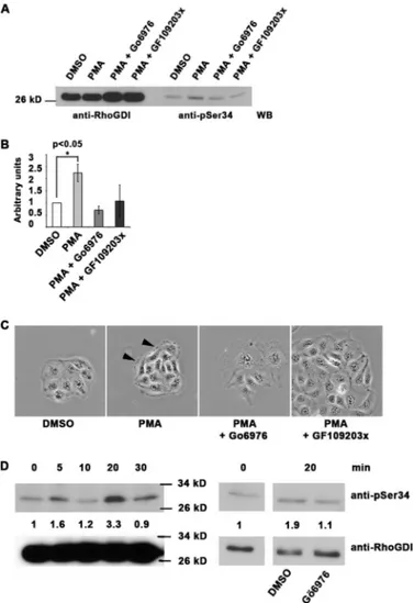
In a well characterized model of PKC␣ activation and trans-location (43), MDCK cells were treated with phorbol ester, which triggers membrane protrusion, and increased GTP-RhoA levels with activation of Rho kinases. This is accompa-nied by adherens junction loss and F-actin turnover, not microfilament assembly as seen in mesenchymal cells. West-ern blotting showed that levels of Ser-34-phosphorylated RhoGDI␣ rapidly increased with phorbol ester treatment, by 2– 4-fold, but this was suppressed in parallel cultures by treat-ment with Go¨6976 or GF109203X PKC inhibitors (Fig. 3, A and
B). The PKC␣ and - isoforms are present in MDCK cells (44). Both PKC inhibitors also suppressed the morphological changes associated with phorbol ester treatment (Fig. 3C). In further experiments, MDCK cells were transfected with empty vector, wild-type RhoGDI␣, or mutants with alanine (S34A) that cannot be phosphorylated or aspartic acid (S34D) as a phosphomimetic in place of serine 34. Cultures were untreated or phorbol ester-treated and then stained for E-cadherin or F-actin (Fig. 4) to follow adhesion changes. The most notable effect was that in untreated S34D cells, extensive protrusions were accompanied by poor adherens junction formation, unlike empty vector controls. On PKC activation by phorbol ester, there was an extensive spreading response with loss of F-actin containing microfilament bundles in control (empty vector) or wild-type RhoGDI-expressing cells (Fig. 4C). In phorbol ester-treated S34A cells, however, microfilament bundles persisted, compared with neighboring untransfected cells, with commen-surate lack of protrusions. Morphology and adherens junction formation in untreated S34A cells appeared normal. As a whole, the data suggest that RhoGDI (pseudo)phosphorylated at resi-due 34 has a strong impact on cell adhesion events, resembling the effects of PKC activation by phorbol ester.
FIGURE 3. Serine 34 phosphorylation of RhoGDI␣ is a regulator of
PKC-mediated adhesion modulation in MDCK cells. A–C, 10-min PMA
treat-ment (200 nM) of MDCK cells leads to phosphorylation of RhoGDI␣ on serine 34 that is inhibited by 1MGo¨6976 PKC␣ inhibitor or a broader spectrum PKC inhibitor (GF109203X). Total RhoGDI␣ is shown in the four left lanes. Quan-titation of triplicates is shown in B,⫾ S.D. Phase contrast images (C) show the characteristic ruffling induced by 200 nMPMA (arrowheads), but this was not seen where PKC inhibitors (1M) were present. Scale bar, 50m. D, MDCK cells were treated with 50 ng/ml HGF for 0 –30 min, and serine 34 phosphor-ylation of RhoGDI␣ was quantitated. Molecular mass marker positions are shown. On the right, sensitivity of this phosphorylation to PKC␣ inhibition at the 20-min time point is shown. Quantitation data for the experiment shown are presented but represent one of three repeated experiments. WB, Western blot.
Hepatocyte growth factor (HGF) is known to induce morphological and behavioral changes in MDCK cells, with the acquisition of a motile phenotype and cell scattering (45). Roles for GTP-RhoA with adducin as a downstream target have been reported (45, 46). This model was used in further experiments, where PMA is replaced with a physiologi-cal ligand. Treatment of cultures with 50 ng/ml HGF led to a rapid but transient increase in RhoGDI phosphorylation on Ser-34, with levels returning to base line in 30 min (Fig. 3D). This phosphorylation was sensitive to Go¨6976 PKC␣/ inhibitor (Fig. 3D). In total, the data show the widespread nature of RhoGDI␣ phosphorylation on Ser-34 in a variety of cell types, and its responsiveness to adhesion changes where there are roles for RhoA.
Acquisition of Negative Charge on Serine 34 Decreases RhoGDI␣ Binding to RhoA and Facilitates RhoA Activation—To characterize the molecular mechanisms regu-lated by RhoGDI␣ phosphorylation on Ser-34, RhoGDI␣ in complexes with Rac1 or RhoA were tested as potential PKC␣ substrates. For purification of RhoGDI␣-protein complexes, human RhoGDI and Rac1 cDNAs were co-expressed recombinantly in Drosophila S2 cells, which can prenylate GTPases, thus producing a functional complex. However, although S2 cells were able to express both RhoGDI␣ and Rac1 proteins, RhoA expression was mini-mal, and complexes with RhoGDI␣ were not obtainable. For this reason, recombinant RhoA-RhoGDI␣ com-plex was purified from yeast cells (24). Kinase assays with purified RhoGDI␣-RhoA complexes revealed a similar regulation of phosphoryla-tion to monomeric RhoGDI␣. Phos-phorylation did not occur when PtdSer/DL/Ca2⫹were used as activa-tors but was dependent on the pres-ence of PtdIns(4,5)P2(Fig. 5, A and B). Potentially, by partially opening the protein complex (30), PtdIns(4,5)P2, exposes the phosphoacceptor site thus making it available for phosphor-ylation. RhoGDI is therefore a
sub-strate for PKC␣ when complexed to either RhoA or Rac1, whereas the GTPases themselves were not phosphorylated (Fig. 5, A and B). The site of RhoGDI␣ phosphorylation when complexed to RhoA was confirmed to be Ser-34, as illustrated by Western blotting with the phosphospecific antibody (Fig. 5C). Therefore, bound GTPase does not affect the availability of the Ser-34 residue of RhoGDI␣ to be phosphorylated by PKC␣.
Confirmation of phosphoinositide dependence for RhoGDI␣ phosphorylation was obtained through the use of alternative inositides, PtdIns and PtdIns(3,4,5)P3. As shown in Fig. 5D, PtdIns(3,4,5)P3addition resulted in significant phosphoryla-tion of RhoGDI␣, whereas PtdIns only minimally induced RhoGDI phosphorylation. On the other hand, PtdIns and PtdIns(4,5)P2promoted phosphorylation of histone III-S (Fig. 5D), whereas PtdIns(3,4,5)P3was much less efficient, as re-ported previously (47). This again demonstrated that the nature of the substrate can influence the activation of the kinase and its subsequent phosphorylation. PtdIns(3,4,5) P3could act in an additive manner with PtdIns(4,5)P2for the phosphorylation of monomeric RhoGDI␣, but it could not promote the phosphor-ylation of RhoGDI␣ complexed with RhoA (Fig. 5E). This
sug-gests PtdIns(4,5)P2 as the physio-logically relevant regulator of the association between RhoA and RhoGDI and its ability to promote RhoGDI phosphorylation by PKC␣. To assess whether Ser-34 phos-phorylation regulates complex for-mation with GTPases, phospho-mimetic or -abolishing mutants of RhoGDI␣ were used in pulldown assay s from fibroblast lysates (Fig. 6A). When Ser-34 was mutated to Asp, there was significantly reduced RhoGDI␣ binding to RhoA (by around 75%) but not Rac1 or Cdc42. Mutation to Ala had minimal effect on the binding to any of the three GTPases. In a second assay, muta-tions of Ser-34 and Ser-62 to either Asp or Ala were compared. Single mutations of Ser-62 to either Asp or Ala had no effect on the binding of either RhoA or Rac1 (Fig. 6B).
The effect of RhoGDI␣ phosphor-ylation on its complex with RhoA was studied using ADP-ribosylation of RhoA by the C3 transferase (Fig. 6C). This approach has been employed before as an indication of partial release of RhoA by RhoGDI following incubation of the RhoA-RhoGDI complex with phosphoinositides (30). The C3 exotransferase is a bacterial toxin that ADP-ribosylates RhoA on Asn-41 (48) and results in RhoA inactivation through enhanced interaction with RhoGDI (49). When purified RhoA-RhoGDI complexes were incubated with TAT-C3, little ADP-ribosylation occurred either in the presence or absence of PtdIns(4,5)P2. In contrast, ADP-ribosylation was evident in samples that had been phosphorylated by PKC␣ (Fig. 6C). This result indicates that following phosphorylation of RhoGDI␣ by PKC␣, the RhoA switch I region is exposed sufficiently to allow rapid ADP-ribosylation by the C3 exotransferase, suggestive of activation of RhoA. This was further tested by measuring the incorporation of [35S]GTP␥S into RhoA complexed to RhoGDI␣ following magnesium depletion, an in vitro condi-tion that mimics the in vivo effects of guanine exchange factors (26). As shown in Fig. 6D, free RhoA rapidly exchanged its bound nucleotide (GDP) with GTP, and the reaction was satu-rated after 15 min, with an efficiency of⬃15%. RhoA com-plexed to RhoGDI␣ did not undergo nucleotide exchange, as
FIGURE 4. RhoGDI␣ status regulates MDCK cytoskeletal organization. MDCK epithelial cells were transfected with empty vector or RhoGDI cDNAs encod-ing His-tagged wild-type (WT RhoGDI), S34A, or S34D mutations. Transfected cells were detected by His antibodies or green fluorescent protein co-transfected with the empty vector. Cultures were stained for E-cadherin as a marker of adherens junctions (A and control in C) or phalloidin for F-actin (B and C). Micrographs in C show cultures treated for 10 min with 200 nMphorbol ester, which triggers loss of adherens junctions and reorganization of the actin cytoskeleton, in a Rho-dependent manner. In each case single fluorescence and merged images are shown. A, E-cadherin is abundant in cell-cell contact sites in all cases except S34D, where the cells exhibit protrusions (arrows) and do not have extensive E-cadherin on their surface. Staining for the actin cytoskeleton (B) shows that all transfected and untransfected cells have some F-actin containing microfilament bundles. Protrusions in S34D cells are labeled with arrows. In phorbol ester treated cultures (C), microfilament bundles are lost or are condensed into small aggregates (arrows) in all cases except S34A cells, where bundles persist (double arrows). Increased cell spreading is also characteristic of phorbol ester-treated cells, again not seen in the S34A transfected cells. Scale bar, 10m.
FIGURE 5. RhoGDI␣ can be phosphorylated on serine 34 when complexed with RhoA or Rac1. A and B, RhoGDI␣ was prepared in complex with either RhoA or Rac1 (0.5 g/assay) and subjected to phosphorylation by 3 ng of PKC␣ for 10 min. In both figures, a silver-stained SDS-polyacrylamide gel is shown on the left and an autoradiograph on the right. The positions of GDI and GTPase are marked. Lanes 1, no PKC activators; lanes 2, PtdSer/diolein/calcium (250/32/750M); and lanes 3, PtdIns(4,5)P2(50M). Neither GTPase is phosphorylated detectably by PKC␣. C, RhoGDI␣ complexed with RhoA (0.5 g of protein) was mixed with (50 M) PtdIns(4,5)P2⫾ 3 ng of PKC␣ for 10 min and in a Western blot (WB) by a phosphospecific antibody recognizing Ser(P)-34 RhoGDI. The result shows that the phosphospecific antibody can recognize the RhoGDI even when complexed with RhoA. D and E, three inositides (50 or 25Mof each where two were mixed) were compared as PKC␣␥ activators for RhoGDI␣, histone III-S. or RhoA/RhoGDI␣ complex phosphorylation. For complexed RhoGDI␣, PtdIns(4,5)P2was superior, with no additive effect of PtdIns(3,4,5)P3, although histone III-S phosphorylation was increased by all the inositides. Quantitation is shown below the autoradiograms.
expected, with only 3% incorporat-ing GTP␥S. Following phosphoryla-tion of RhoGDI␣ by PKC␣␥, a mean of 13% of RhoA exchanged to GTP (Fig. 6E), representing a 4-fold increase compared with the untreated control. A significant increase in GTP load could not be observed following incubation with PtdIns(4,5)P2 alone. These com-bined data indicate that RhoA can be sufficiently released from its inhibitory complex with RhoGDI following phosphorylation of the latter by PKC␣␥, to facilitate its activation.
Under these conditions of RhoGDI␣ phosphorylation by PKC␣␥, ⬃0.3 mol of phosphate were incorpo-rated per mol of RhoGDI molecule (Fig. 6F). As there appears to be only one major phosphorylation site, this would represent 30% of RhoGDI␣. Theoretically, 30% of the bound RhoA would therefore be able to exchange its bound nucleotide for GTP␥S. Under the experimental conditions employed, the data are consistent with approximately half of this potential pool of RhoA exchanging nucleotide for GTP␥S.
RhoGDI Phosphorylation in Fibroblasts Regulates GTP-RhoA Levels—Further experiments with fibroblasts tested the hypothesis that Ser-34 phosphorylation of RhoGDI␣ leads to elevated GTP-RhoA levels. Primary fibroblasts were transfected with the pRaichu 1502 cDNA, the protein acting as a sensitive indicator of GTP-RhoA levels when analyzed by FRET microscopy (supplemental Fig. 4A) (33). In this system, FRET efficiency is inversely proportional to RhoA-GTP levels. In a base-line character-ization of the fibroblast system, stress fiber and focal adhesion for-mation were manipulated through transfection of syndecan-4 cDNA constructs. In combination with the pRaichu 1502 cDNA, cells were transfected with wild-type (S4W) or cytoplasmically truncated (S4I) syn-decan-4 cDNA. Wild-type synde-can-4 promotes stress fiber and focal adhesion formation, although the S4I acts as a dominant negative, FIGURE 6. Phosphomimetic RhoGDI␣ at residue 34 leads to decreased retention of RhoA but no change
in binding of Rac1 or Cdc42. A, wild type (WT) and RhoGDI␣ mutated to alanine (S34A) or aspartate (S34D)
were compared in GTPase pulldown assays with lysate from rat embryo fibroblasts. In each Western blot, the right lane shows the GTPase detected in the lysate. Quantitation shows that phosphomimetic RhoGDI␣ has decreased ability to bind RhoA, although binding to Rac1 and Cdc42 is unaffected. Nonspecifically reacting polypeptides are present in the upper portion of each Western blot. B, serine 34 and 62 mutants were compared in their abilities to bind RhoA or Rac1 from rat embryo fibroblast lysates. Although mutation of serine 62 to alanine (S62A) or aspartate (S62D) had no impact on GTPase binding, mutation of serine 34 to aspartate (S34D) reduced RhoA binding specifically. The corresponding alanine mutant at serine 34 (S34A) bound both GTPases similarly to the wild-type (wt) RhoGDI␣. Quantitation of the Western blots is shown. C, C3 transferase ADP ribosylates RhoA once the RhoGDI␣-RhoA complex has been phosphorylated by PKC␣␥. Varying amounts of TAT-C3 transferase were added to GST-RhoA (upper panel). In the lower panel, 500 ng of RhoA in complex with RhoGDI␣ were untreated (⫺) or incubated with 50 mol of PtdIns(4,5)P2⫾ PKC␣␥ (lower panel). In both cases, 1Ci of [32P]NAD was included in a 5-min reaction at room temperature, and autoradiographs are shown from one of four independent experiments. D, exchange of GDP for GTP in RhoA is high in free GTPase but low when complexed to RhoGDI␣. Ten pmol of RhoA either alone (black bars) or complexed to RhoGDI␣ (white bars) was incubated with 30 pmol (0.5Ci) of GTP␥S for the indicated times, and radiolabel incorporation into the GTPase was measured by liquid scintillation spectroscopy. E, phosphorylation of RhoGDI␣ in complex with RhoA-GDP by PKC␣␥, in the presence of 50mol PtdIns(4,5)P2, leads to increased nucleotide exchange for GTP. PKC phosphorylation of 10 pmol of RhoGDI␣-RhoA complex for 1 h at room temperature was followed by a GTP exchange reaction in the presence of radiolabeled (0.5Ci) GTP␥S. The results are shown as mean ⫾ S.D. from three independent experiments. ** Significantly more nucleotide exchange occurs with RhoGDI␣ phos-phorylation (p⬍ 0.05) than without. F, stoichiometry of RhoGDI␣ phosphorylation. Left panel, 10 pmol of wild-type RhoGDI complexed with RhoA (left), wild-type RhoGDI (center), or S34A RhoGDI alone (right) were incubated with 10 ng of PKC␣␥ and 50 mol of PtdIns(4,5)P2for 1 h at room temperature, and radiolabel incorporation was measured. Values are mean⫾ S.D. from three experiments. Right panel, data from the left panel were recalculated to show stoichiometry of Ser-34 phosphorylation, subtracting background (S34A RhoGDI values)⫾ S.D. There was no significant difference in phosphorylation whether the RhoGDI was com-plexed with RhoA or not.
because it cannot bind PKC␣ (11, 50 –52). Stress fibers and focal adhesions are depleted in S4I-transfected cells. Stress fiber formation in these fibroblasts is sensitive to inhibition of GTP-RhoA by the C3 transferase or inhibition of Rho kinases by Y27632 (28). As expected, FRET efficiency de-creased significantly in S4W cells compared with control transfected or S4I cells, indicative of higher GTP-RhoA lev-els (supplemental Fig. 4). In S4I cells, FRET efficiency was increased significantly above control levels.
To manipulate RhoGDI␣ status in rat fibroblasts, endog-enous levels were first reduced by siRNA. Reduction of RhoGDI␣ levels in REF cells by siRNA techniques led to a drop in FRET efficiency, commensurate with a rise in GTP-RhoA levels as seen by FRET microscopy with the pRaichu
1502 probe (Fig. 7A). This was expected, because the GDI protein serves as a sink for GDP-bound Rho family members. In this background, wild-type RhoGDI cDNA or Ser-34 mutants (to alanine or aspartate) were introduced into the cells for further FRET analysis. Compared with an empty vector, expression of wild-type RhoGDI␣ was accompanied by decreased GTP-RhoA levels. This effect was enhanced by overexpression of the S34A mutant of RhoGDI␣ (Fig. 7B). Importantly, overexpression of the S34D phosphomimetic mutant had an opposite effect with elevated GTP-RhoA lev-els (Fig. 7B). These data confirm that Ser-34 can be a key RhoGDI␣ regulatory site in vivo. Upon phosphorylation, the GDI cannot retain RhoA efficiently, facilitating its activation by nucleotide exchange.
FIGURE 7. Serine 34 of RhoGDI␣ in fibroblasts regulates RhoA-GTP levels. A, rat embryo fibroblasts were cotransfected with pRaichu 1502 and either control siRNA or siRNA oligonucleotides for RhoGDI␣. After 48 h, cells were lysed and immunoblotted with anti-RhoGDI antibody. Vinculin was used as a loading control. FRET efficiency was measured after acceptor photobleaching and was quantitated (n⫽ 20). Efficiency was reduced after reduction in RhoGDI protein levels, indicative of higher GTP-RhoA levels. B, rat fibroblasts with endogenous levels of RhoGDI␣ reduced by siRNA were co-transfected with pRaichu1502 and one of His vector (Vec), His-tagged RhoGDI (WT), or His-tagged RhoGDI mutants (S34D or S34A). FRET efficiency was measured after acceptor photobleaching, and quantitation is shown (n⫽ 20). Expression of phosphomimetic RhoGDI␣ leads to decreased FRET efficiencies and therefore elevated RhoGTP levels. The converse was measured after expression of the nonphosphorylatable S34A mutant. p⬍ 0.01 indicates a FRET signal significantly different to the vector only control (t test).
DISCUSSION
RhoGDI proteins regulate the cycling and distribution of RhoGTPases (18, 19). Co-crystallization data of RhoGDI␣ bound to Cdc42 or Rac1 (38, 53) reveal that residues 34 –57 form an ordered helix-loop-helix motif characterized by hydro-phobic interactions. Acquisition of negative charge at Ser-34 may disrupt the stability of the helix-loop-helix motif, interfer-ing with the GTPase interaction and causinterfer-ing release of the GTPase from RhoGDI. In addition, there are extensive interac-tions between GDI N termini and switch I and II regions of the GTPases. GTPase binding establishes a bridge that brings regions of the N- and C-terminal domains of RhoGDI into proximity. A striking example is Arg-66 (Cdc42), the guani-dinium group of which exhibits hydrogen bonding with Asp-185 as well as Pro-30 and Ala-31 of the GDI. Additionally, hydrophobic interactions between the aliphatic portion of the Arg-66 with Gln-32 and Ile-122 of the GDI were observed (38). Because many of the key residues of the RhoGDI N-terminal region lie immediately adjacent to Ser-34, these interactions may be disturbed by phosphorylation. However, Arg-66 of Cdc42 is highly conserved in Rac1 and RhoA, and indeed there is high sequence conservation through the switch II region, consistent with its role in GTP hydrolysis regulation, so the RhoA-specific effect of Ser-34 phosphorylation deserves fur-ther analysis.
In vitro studies identifying the site and consequences of RhoGDI␣ phosphorylation are supported by in vivo studies with a Ser-34-phosphospecific antibody. Four widely differing cell types were investigated, and in each case endogenous phos-phorylation of the Ser-34 residue could be detected. In MDCK cells, phorbol ester stimulates cytoskeletal reorganization and PKC␣ translocation to the membrane (43), although integrin-mediated adhesion is promoted by phorbol ester in K562 cells, also associated with actin cytoskeletal reorganization (39). In both cell types, RhoGDI␣ phosphorylation on Ser-34 rapidly increased in concert with these adhesion events. Additionally, and consistent with in vitro data, RhoGDI␣ phosphorylation was blocked by the Go¨6976 inhibitor, which has highest activity against PKC␣ and PKC1 (41) The increase in Ser-34 phosphor-ylation of RhoGDI␣ in MDCK cells responding to phorbol ester, or HGF, is particularly interesting. Both stimulants acti-vate RhoA rather than Rac (45, 54, 55), although the molecular mechanism is unknown. The result is actin cytoskeletal reorga-nization and loss of cell-cell junctions. Moreover, two studies with mouse keratinocytes and KB cells showed that motility in response to these same agents was blocked both by C3 transfer-ase and overexpression of wild-type RhoGDI (55, 56). These studies therefore implicated RhoGDI, with which the current data are entirely consistent. A key result is that introduction of the S34D mutant of RhoGDI␣ into MDCK cells activated the motility responses of MDCK cells without a need for PKC acti-vation. In contrast, such responses were not seen in wild-type or S34A RhoGDI-expressing cells. However, we cannot rule out additional roles for guanine exchange factors and GTPase-ac-tivating proteins. Gentile et al. (57) showed, for example, that HGF-promoted invasion is associated with down-regulated expression of the RacGAP, Arhgap12.
Further experiments utilized the Raichu construct as a reporter for GTP-Rho levels (33). This sensitive indicator com-bined with FRET microscopy enabled the impact of RhoGDI␣ phosphorylation to be assessed. Expression of the S34D form of RhoGDI␣ in fibroblasts yielded a FRET decline (i.e. GTP-RhoA levels increased). This provided in vivo evidence that RhoGDI␣ phosphorylation regulates RhoGTP levels. In contrast, no ele-vation in GTP-RhoA levels was seen by transfection of wild-type RhoGDI␣ or an S34A form. In these cases, FRET was increased, suggesting that a form of RhoGDI␣ that cannot be phosphorylated sequesters GDP-RhoA. Although wild-type RhoGDI␣ can theoretically be phosphorylated when overex-pressed, increased protein levels alter the equilibrium in favor of decreased overall phosphorylation, consistent with Takaishi et al.(55).
Efficient Ser-34 phosphorylation of RhoGDI␣ by conven-tional PKC occurs in the presence of PtdIns(4,5)P2, not with the often used combination of phosphatidylserine, diacylglycerol, and calcium. This inositol phospholipid can mediate PKC␣ activation, and a lysine-rich-binding site in the C2 domain was identified (10). We, and others, showed that syndecan-4-medi-ated activation of PKC␣ in the presence of PtdIns(4,5)P2was calcium-independent (13, 35), and this mechanism of kinase activation corresponds well to the current data showing that the PKC␣/PtdIns(4,5)P2 combination efficiently phosphory-lated RhoGDI␣. The C2 domain of PKC␣ has a higher affinity for PtdIns(4,5)P2than phosphatidylserine, so the inositide may be a kinase-targeting site (15, 58). Recent work shows that PKC␣ C2 domain may adopt a different membrane orientation when bound to PtdIns(4,5)P2 compared with phosphatidyl-serine (59). In turn, this may modulate kinase activity, and our study provides some evidence that substrate specificity might be engendered this way. Therefore, we predict PKC␣ activation resulting from phospholipase C activity, i.e. the generation of diacylglycerol, would not trigger RhoGDI phosphorylation and RhoA activation. By the same token, because inositol 1,4,5-trisphosphate and Ca2⫹release from the endoplasmic reticu-lum are consequences of phospholipase C activity, the phos-phorylation of RhoGDI␣ is presumed to be independent of calcium fluxes. Consistent with this, we find that the PtdIns(4,5)P2-driven PKC␣ activation is indeed Ca2⫹ -inde-pendent. Moreover, RhoGDI␣ phosphorylation persisted in the presence of a characterized phospholipase C inhibitor, U73122 (60).
Our data differ from two reports suggesting that in endothe-lial cells Ser-96 of RhoGDI␣ was subject to phosphorylation by PKC␣, with a downstream increase in GTP-RhoA levels (61) or release of RhoG (62), a close relative of Rac1 (63). Although the methods and cells are different, we could not demonstrate phosphorylation of Ser-96 in vitro. A truncated form of RhoGDI␣(67–204), missing the N-terminal region but contain-ing Ser-96, was not phosphorylated by PKC␣. Knezevic et al. (61) utilized N-terminally green fluorescent protein-tagged RhoGDI␣ that may have affected the ability of PKC␣ to phorylate Ser-34. In contrast to that study, we prepared a phos-phospecific antibody that revealed both the widespread occur-rence of Ser-34 phosphorylation and regulation in response to PKC activation. Serine 96 lies in a consensus PKC
phosphory-lation site that is present in the immunoglobulin domain of mammalian RhoGDI␣ but not conserved across other verte-brates or isoforms. Based on structural data, the Ser-96 residue does not apparently form part of a contact site with GTPase or the hydrophobic pocket that captures the prenyl moiety. In contrast, Ser-34 is conserved across all vertebrate RhoGDI␣ isoforms and is present in all vertebrate and ␥ isoforms for which there are data. Although Ser-34 is a major PKC phosphor-ylation site, in vitro experiments suggested some residual phosphorylation after GDI mutation of Ser-34 to alanine or aspartate. The amounts varied but could represent another phosphorylation site, presumably also in the N-terminal por-tion of RhoGDI␣.
Because RhoGDI␣ is an abundant cytoplasmic protein, its role is potentially significant. Our work complements that of DerMardirossian et al. (64) where p21-activated kinase phos-phorylation of RhoGDI␣ on Ser-101 and Ser-174 causes spe-cific release of Rac1. In this case, the key sites are C-terminal and are close to each other and to the prenyl-binding cleft of the GDI (38). It is proposed that the negative charge may destabilize this hydrophobic interaction and cause release of the Rac1. This must be a very specific effect because Rho and Cdc42 are also prenylated yet are unaffected. A second model of RhoGDI reg-ulation involves Tyr-156 phosphorylation by Src, but only when not complexed to a GTPase (21). This not only reduced affinity of the GDI for RhoA, Rac1, and Cdc42, but it also triggered a membrane or cortical redistribution of the protein. The current data show that Ser-34 phosphorylation can occur in free or GTPase-bound RhoGDI␣. Phosphorylation on Tyr-156 was only noted in cells overexpressing a constitutively active form of Src (21), perhaps an indicator of its transient nature. Here, Ser-34 phosphorylation was observed in several different untransfected cell types, with levels changing in response to adhesion events.
Focal adhesion and microfilament bundle assembly is pro-moted by GTP-RhoA (65, 66), with activation of the Rho kinases, with ROCK I playing a major role in primary fibroblasts (28). Rho kinases phosphorylate myosin light chain and the myosin-binding subunit of myosin phosphatase, leading to myosin II-driven contraction. The transmembrane heparan sulfate proteoglycan, syndecan-4, supports focal adhesion assembly through the binding and persistent activation of PKC␣, in the presence of PtdIns(4,5)P2, an event upstream of GTP-RhoA (8, 11, 25). In MDCK cells, Rho kinase activity is required both for focal adhesion/stress fiber formation but also lamellipodial ruffle stabilization, possibly through phosphory-lation of adducin (29, 67). However, the upstream events that lead to the accumulation and targeting of GTP-RhoA were not known. Here, evidence suggests that a major downstream tar-get of PKC␣/PtdIns(4,5)P2is RhoGDI␣ with phosphorylation on Ser-34. The result of phosphorylation is release or decreased capture of the GTPase, leading to nucleotide exchange and interaction with downstream effectors.
REFERENCES
1. Ivaska, J., Kermorgant, S., Whelan, R., Parsons, M., Ng, T., and Parker, P. J. (2003) Biochem. Soc. Trans. 31, 90 –93
2. Larsson, C. (2006) Cell. Signal. 18, 276 –284
3. Suzuki, A., and Ohno, S. (2006) J. Cell Sci. 119, 979 –987
4. Reyland, M. E. (2009) Front. Biosci. 14, 2386 –2399
5. Mochly-Rosen, D., and Gordon, A. S. (1998) FASEB J. 12, 35– 42 6. Steinberg, S. F. (2008) Physiol. Rev. 88, 1341–1378
7. Jaken, S., Leach, K., and Klauck, T. (1989) J. Cell Biol. 109, 697–704 8. Dovas, A., Yoneda, A., and Couchman, J. R. (2006) J. Cell Sci. 119,
2837–2846
9. Bass-Zubek, A. E., Hobbs, R. P., Amargo, E. V., Garcia, N. J., Hsieh, S. N., Chen, X., Wahl, J. K., 3rd., Denning, M. F., and Green, K. J. (2008) J. Cell Biol. 181,605– 613
10. Corbala´n-García, S., García-García, J., Rodríguez-Alfaro, J. A., and Go´-mez-Ferna´ndez, J. C. (2003) J. Biol. Chem. 278, 4972– 4980
11. Keum, E., Kim, Y., Kim, J., Kwon, S., Lim, Y., Han, I., and Oh, E. S. (2004) Biochem. J. 378,1007–1014
12. Multhaupt, H. A., Yoneda, A., Whiteford, J. R., Oh, E. S., Lee, W., and Couchman, J. R. (2009) J. Physiol. Pharmacol. 60, Suppl. 4, 31–38 13. Oh, E. S., Woods, A., Lim, S. T., Theibert, A. W., and Couchman, J. R.
(1998) J. Biol. Chem. 273, 10624 –10629
14. Manna, D., Bhardwaj, N., Vora, M. S., Stahelin, R. V., Lu, H., and Cho, W. (2008) J. Biol. Chem. 283, 26047–26058
15. Marín-Vicente, C., Nicola´s, F. E., Go´mez-Ferna´ndez, J. C., and Corbala´n-García, S. (2008) J. Mol. Biol. 377, 1038 –1052
16. Guerrero-Valero, M., Marín-Vicente, C., Go´mez-Ferna´ndez, J. C., and Corbala´n-García, S. (2007) J. Mol. Biol. 371, 608 – 621
17. Bass, M. D., Morgan, M. R., Roach, K. A., Settleman, J., Goryachev, A. B., and Humphries, M. J. (2008) J. Cell Biol. 181, 1013–1026
18. DerMardirossian, C., and Bokoch, G. M. (2005) Trends Cell Biol. 15, 356 –363
19. Dovas, A., and Couchman, J. R. (2005) Biochem. J. 390, 1–9
20. Takahashi, K., Sasaki, T., Mammoto, A., Takaishi, K., Kameyama, T., Tsu-kita, S., and Takai, Y. (1997) J. Biol. Chem. 272, 23371–23375
21. DerMardirossian, C., Rocklin, G., Seo, J. Y., and Bokoch, G. M. (2006) Mol. Biol. Cell 17,4760 – 4768
22. Golovanov, A. P., Hawkins, D., Barsukov, I., Badii, R., Bokoch, G. M., Lian, L. Y., and Roberts, G. C. (2001) Eur. J. Biochem. 268, 2253–2260 23. Sheffield, P., Garrard, S., and Derewenda, Z. (1999) Protein Expr. Purif. 15,
34 –39
24. Read, P. W., and Nakamoto, R. K. (2000) Methods Enzymol. 325, 15–25 25. Lim, S. T., Longley, R. L., Couchman, J. R., and Woods, A. (2003) J. Biol.
Chem. 278,13795–13802
26. Self, A. J., and Hall, A. (1995) Methods Enzymol. 256, 3–10
27. Sebbagh, M., Renvoize´, C., Hamelin, J., Riche´, N., Bertoglio, J., and Bre´ard, J. (2001) Nat. Cell Biol. 3, 346 –352
28. Yoneda, A., Multhaupt, H. A., and Couchman, J. R. (2005) J. Cell Biol. 170, 443– 453
29. Fukata, Y., Kimura, K., Oshiro, N., Saya, H., Matsuura, Y., and Kaibuchi, K. (1998) J. Cell Biol. 141, 409 – 418
30. Faure´, J., Vignais, P. V., and Dagher, M. C. (1999) Eur. J. Biochem. 262, 879 – 889
31. Aktories, K., and Just, I. (1995) Methods Enzymol. 256, 184 –195 32. Bastiaens, P. I., Majoul, I. V., Verveer, P. J., So¨ling, H. D., and Jovin, T. M.
(1996) EMBO J. 15, 4246 – 4253
33. Yoshizaki, H., Ohba, Y., Kurokawa, K., Itoh, R. E., Nakamura, T., Mochi-zuki, N., Nagashima, K., and Matsuda, M. (2003) J. Cell Biol. 162, 223–232 34. Rodriguez, P., Mitton, B., and Kranias, E. G. (2005) Biotechnol. Lett. 27,
1869 –1873
35. Horowitz, A., and Simons, M. (1998) J. Biol. Chem. 273, 25548 –25551 36. Weekes, J., Barry, S. T., and Critchley, D. R. (1996) Biochem. J. 314,
827– 832
37. Ziegler, W. H., Tigges, U., Zieseniss, A., and Jockusch, B. M. (2002) J. Biol. Chem. 277,7396 –7404
38. Hoffman, G. R., Nassar, N., and Cerione, R. A. (2000) Cell 100, 345–356 39. Lub, M., van Vliet, S. J., Oomen, S. P., Pieters, R. A., Robinson, M., Figdor,
C. G., and van Kooyk, Y. (1997) Mol. Biol. Cell 8, 719 –728
40. Hocevar, B. A., Morrow, D. M., Tykocinski, M. L., and Fields, A. P. (1992) J. Cell Sci. 101,671– 679
41. Martiny-Baron, G., Kazanietz, M. G., Mischak, H., Blumberg, P. M., Kochs, G., Hug, H., Marme´, D., and Scha¨chtele, C. (1993) J. Biol. Chem.
42. Fujii, M., Ohtsubo, M., Ogawa, T., Kamata, H., Hirata, H., and Yagisawa, H. (1999) Biochem. Biophys. Res. Commun. 254, 284 –291
43. Sciorra, V. A., and Daniel, L. W. (1996) J. Biol. Chem. 271, 14226 –14232 44. Balboa, M. E., and Insel, P. A. (1998) Mol. Pharmacol. 53, 221–227 45. Kodama, A., Matozaki, T., Fukuhara, A., Kikyo, M., Ichihashi, M., and
Takai, Y. (2000) Mol. Biol. Cell 11, 2565–2575
46. Fukata, Y., Oshiro, N., Kinoshita, N., Kawano, Y., Matsuoka, Y., Bennett, V., Matsuura, Y., and Kaibuchi, K. (1999) J. Cell Biol. 145, 347–361 47. Kochs, G., Hummel, R., Fiebich, B., Sarre, T. F., Marme´, D., and Hug, H.
(1993) Biochem. J. 291, 627– 633
48. Lerm, M., Schmidt, G., and Aktories, K. (2000) FEMS Microbiol. Lett. 188, 1– 6
49. Genth, H., Gerhard, R., Maeda, A., Amano, M., Kaibuchi, K., Aktories, K., and Just, I. (2003) J. Biol. Chem. 278, 28523–28527
50. Longley, R. L., Woods, A., Fleetwood, A., Cowling, G. J., Gallagher, J. T., and Couchman, J. R. (1999) J. Cell Sci. 112, 3421–3431
51. Couchman, J. R. (2003) Nat. Rev. Mol. Cell Biol. 4, 926 –937
52. Mostafavi-Pour, Z., Askari, J. A., Parkinson, S. J., Parker, P. J., Ng, T. T., and Humphries, M. J. (2003) J. Cell Biol. 161, 155–167
53. Grizot, S., Faure´, J., Fieschi, F., Vignais, P. V., Dagher, M. C., and Pebay-Peyroula, E. (2001) Biochemistry 40, 10007–10013
54. Kitajo, H., Shibata, T., Nagayasu, H., Kawano, T., Hamada, J., Yamashita, T., and Arisue, M. (2003) Oncol. Res. 10, 1351–1356
55. Takaishi, K., Sasaki, T., Kato, M., Yamochi, W., Kuroda, S., Nakamura, T.,
Takeichi, M., and Takai, Y. (1994) Oncogene 9, 273–279
56. Nishiyama, T., Sasaki, T., Takaishi, K., Kato, M., Yaku, H., Araki, K., Mat-suura, Y., and Takai, Y. (1994) Mol. Cell. Biol. 14, 2447–2456
57. Gentile, A., D’Alessandro, L., Lazzari, L., Martinoglio, B., Bertotti, A., Mira, A., Lanzetti, L., Comoglio, P. M., and Medico, E. (2008) Oncogene
27,5590 –5598
58. Son, H., Lim, Y., Kim, J., Park, H., Choi, S., Han, I., Kim, W. S., Park, S., Bae, Y., and Oh, E. S. (2006) Arch. Biochem. Biophys. 454, 1– 6
59. Landgraf, K. E., Malmberg, N. J., and Falke, J. J. (2008) Biochemistry 47, 8301– 8316
60. Horowitz, L. F., Hirdes, W., Suh, B. C., Hilgemann, D. W., Mackie, K., and Hille, B. (2005) J. Gen. Physiol. 126, 243–262
61. Knezevic, N., Roy, A., Timblin, B., Konstantoulaki, M., Sharma, T., Malik, A. B., and Mehta, D. (2007) Mol. Cell Biol. 27, 6323– 6333
62. Elfenbein, A., Rhodes, J. M., Meller, J., Schwartz, M. A., Matsuda, M., and Simons, M. (2009) J. Cell Biol. 186, 75– 83
63. van Buul, J. D., Allingham, M. J., Samson, T., Meller, J., Boulter, E., García-Mata, R., and Burridge, K. (2007) J. Cell Biol. 178, 1279 –1293
64. DerMardirossian, C., Schnelzer, A., and Bokoch, G. M. (2004) Mol. Cell
15,117–127
65. Ridley, A. J., and Hall, A. (1994) EMBO J. 13, 2600 –2610 66. Machesky, L. M., and Hall, A. (1997) J. Cell Biol. 138, 913–926
67. Royal, I., Lamarche-Vane, N., Lamorte, L., Kaibuchi, K., and Park, M. (2000) Mol. Biol. Cell 11, 1709 –1725 .