Vol. 46, No. 2, pp. 228 - 232, 2005
Chondrosarcoma of the head and neck region is a rare disease, representing approximately 0.1% of all head and neck neoplasms. The 5-year survival rate of chondrosarcoma is 70-80%, showing relatively good prognosis; however, it is known to progress slowly and eventually cause multiple metastases. In this study, we reviewed chondrosarcoma cases experienced at Yonsei University Medical College during the last 15 years to investigate its clinical characteristics and treatment outcome. The medical records of 8 patients who were diagnosed with chondrosarcoma of the head and neck region and underwent surgical treatment between December 1990 and December 2002 were retrospectively reviewed. The primary sites were sinus, mastoid, jugular foramen and thyroid cartilage. In all patients, the initial treatment modality was surgery, and postoperative radiation therapy was performed in 4 cases. The pathological findings showed chondrosarcoma in 7 cases and mesenchymal chondrosarcoma in 1 case. The treatment out-come resulted in 3 cases of recurrence, of which 2 cases were treated successfully and the other case expired of disease, indicating a survival rate of 87.5%. In the case that resulted in death, complete excision could not be achieved. Therefore, we concluded that wide excision is a beneficial initial treatment of this rare disease.
Key Words: Chondrosarcoma, head and neck neoplasms, treatment outcome
INTRODUCTION
Chondrosarcoma of the head and neck region is
a rare disease, representing approximately 0.1% of all head and neck neoplasms.1 This malignant
tumor, in which the tumor cells form chondroid matrix, represents 10-20% of primary bone tu-mors and is the second most common sarcoma of bone origin.2,3 Due to the low incidence of this
disease, there have been few studies with large numbers of subjects, and therefore, various results have been reported.4-6 In this study, we reviewed
cases of chondrosarcoma experienced at Yonsei University Medical College during the last 15 years to determine the clinical manifestations, treatment methods and treatment results of this uncommon disease.
MATERIALS AND METHODS
Using the cancer registration for reference, patients who were admitted to our institute between 1990 and 2003, and whose final diagnosis at discharge was chondrosarcoma, were reviewed. There were 100 cases of chondrosarcoma and of those, the head and neck region was the primary site in only 8 cases. The charts were reviewed retrospectively in terms of chief complaint, pri-mary site, treatment modality, pathology finding, recurrence, follow-up and treatment results. RESULTS
2010 patients were diagnosed with head and neck neoplasms at our institute from 1990 to 2003 and of these, there were only 8 cases of
chon-Chondrosarcoma of the Head and Neck
Sei Young Lee1, Young Chang Lim2, Mee Hyun Song3, Jae Yeon Seok4, Won Sang Lee3, and
Eun Chang Choi3
1Department of Otorhinolaryngology-Head and Neck Surgery, Chung-Ang University College of Medicine, Seoul, Korea; 2Department of Otolaryngology-Head and Neck Surgery, Konkuk University College of Medicine, Seoul, Korea; 3Department of Otorhinolaryngology, Yonsei University College of Medicine, Seoul, Korea;
4Department of Pathology, Yonsei University College of Medicine, Seoul, Korea.
Received February 16, 2005 Accepted March 23, 2005
Reprint address: requests to Dr. Eun Chang Choi, Department of Otorhinolaryngology, Yonsei University College of Medicine, 134 Shinchon-dong, Seodaemun-gu, Seoul 120-752, Korea. Tel: 82-2-2228-3608, Fax: 82-2-393-0580, E-mail: eunchangmd@yumc. yonsei.ac.kr
Table 2. Chondrosarcoma of the Head and Neck by Site Site Number Paranasal sinuses 4 Temporal bone 3 Larynx 1 Total 8
drosarcoma, which represents 0.4% of all head and neck cancers. Of the 8 cases, 4 were male and 4 were female, with a mean age of 40 years ranging from 24 to 60. The follow-up period ranged from 25 months to 171 months with a mean of 76.5 months (Table 1). The primary sites were paranasal sinuses, temporal bone and larynx (Table 2). The chief complaints at the time of first visit were related to the primary sites, such that an ethmoid sinus origin tumor presented with ocular symptoms, maxillary and jugular foramen origin tumors with facial swelling, a mastoid origin tumor with facial paralysis, and a thyroid
cartilage origin tumor with neck mass. Patholo-gically, of the 8 cases of head and neck chondro-sarcoma, 7 were conventional chondrosarcoma and 1 was mesenchymal chondrosarcoma. Of the 7 cases of conventional chondrosarcoma, 2 were grade I, 4 were grade II and 1 was grade III (Fig. 1 and 2). Surgery was performed as the initial treatment modality in all cases. Radiotherapy was applied in 4 cases, which included 2 cases of recurrence and 2 cases with positive resection margin. The 2 cases with positive resection mar-gin were successfully treated after radiotherapy. Recurrence occurred in 3 cases. The pathological grade of all 3 cases was grade II and salvage treatment was performed in every case. Of the recurred cases, the case of mastoid origin tumor was successfully treated surgically with a transmastoid approach, whereas in the case of maxillary sinus origin, successful salvage was achieved 7 years from the point of diagnosis after receiving chemoradiation therapy and bilateral maxillectomy. However, the case of thyroid cartilage origin recurred in the tonsil 13 months Table 1. Clinical Characteristics of Head and Neck Chondrosarcoma
Case Age Sex CC Site Treatment Pathology Grade Margin Recur F/U(month) Outcome 1 34 M Epiphora Ethmoid
sinus Craniofacial resection& postop. RTx Chondrosarcoma II + No 111 NED
2 56 F Visual
loss Ethmoidsinus Medial maxillectomy& ethmoidectomy & postop. RTx
Chondrosarcoma III + No 30 NED
3 29 F Facial
swelling Maxillary sinus maxillectomySubtotal chondrosarcomaMesenchymal - No 25 NED
4 26 M Facial
swelling Maxillary sinus Partial maxillectomy Chondrosarcoma II - Yes 171 NED
5 24 F Facial
palsy Mastoidbone Mass excision viatransmastoid approach
Chondrosarcoma II - Yes 40 NED
6 49 F Facial
palsy Mastoidbone ITFA type C Chondrosarcoma I - No 51 NED
7 42 M Facial
swelling foramenJugular ITFA type A Chondrosarcoma I - No 130 NED
8 60 M Neck
mass cartilageThyroid Mass excision Chondrosarcoma II - Yes 54 DOD
after the initial treatment. Despite treatment with radiotherapy and two salvage operations, death occurred 54 months after diagnosis because of failure to completely excise the tumor at the last operation due to anatomical reasons. Seven of eight cases have no current evidence of disease, and the survival rate is 87.5% (Table 1).
DISCUSSION
Chondrosarcoma is a slow growing but malig-nant tumor with a relatively high local recurrence rate. Chondrosarcoma represents 10 to 20% of all malignant bone tumors and of these, 1 to 12% originate in the head and neck region.6,7 This tumor can originate from cartilage and soft tissue
as well as bone with common primary sites being the pelvic bone and long bones, such as the femur.8 The common primary sites in the head
and neck region include the mandible, nasal cavity, sinus, and maxilla; however, it is generally accepted that the tumor mostly arises from bony tissue of the head and neck.1,5,6,9In this study, the
8 cases were all of bone origin except for one case that originated from thyroid cartilage. The precipitating factors of this tumor are multiple hereditary exostosis, Ollier's disease, Maffucci's syndrome, previous intravenous thoratrast con-trast use, Paget's disease of bone, chondromyxoid fibroma, and previous irradiation, but none of these factors were found in this study.5
Chon-drosarcoma has a slight male predilection10 and
occurs most commonly between the 4th and 7th
decades.11 Chondrosarcomas of cartilaginous or soft tissue origin are more common in males and in patients over 50 years of age, whereas those of bone origin have higher incidence in females and in patients less than 50 years of age.1 According to one report, the rates of lymph node and distant metastasis were relatively low: 5.6% and 6.7%, respectively. In addition, metastasis was usually observed in patients who had undergone multiple operations over a long period of time.1 In this study, no case of metastasis was seen among the 8 cases, including the 3 patients who received multiple salvage operations.
Chondrosarcomas show various histological patterns ranging from benign chondroid tumor to undifferentiated neoplasm, which make them difficult to diagnose pathologically.10 Evans et al.
classified chondrosarcomas into 3 grades, from grade I to grade III, according to cellular density, nuclear differentiation, and the size of nucleus. This classification is still currently used.12
Never-theless, there have been efforts to classify chon-drosarcomas simply into high grade and low grade for better correlation with prognosis.1 In
this study, no recurrence occurred in grade I tumors, showing a relatively good correlation between tumor grade and prognosis. Chondrosar-coma has several histological types, of which the conventional type is the most common. Other types include clear cell, myxoid, mesenchymal and dedifferentiated variants.13 The clear cell variant is otherwise called malignant
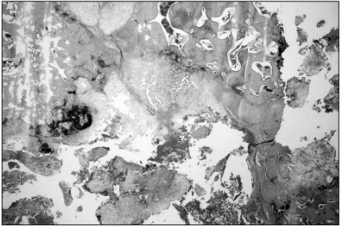
chondro-Fig. 1.Chondrosarcoma infiltrating the marrow space and surrounding preexisting bony trabeculae. Hematoxylin & Eosin, ×14.
Fig. 2. Conventional chondrosarcoma grade II.
blastoma, but there has been controversy over whether to classify it as a variant of chondro-sarcoma or to separately categorize it as malignant chondroblastoma.14 Although there differences
exist among reports, the myxoid variant is known to originate from soft tissues rather than bone tissues.15 The mesenchymal variant is also called
the aggressive variant, since 2/3 of cases present before the age of 30, and in advanced stages and grades.16 The only patient with mesenchymal
chondrosarcoma in this study was a young female aged in the 3rd decade. Dedifferentiated chondro-sarcoma characteristically contains an anaplastic component histologically. In addition, chondro-sarcomas of the larynx have several characteristics that distinguish them from those of different origin: they are low grade tumors, presentation occurs after 50 years of age, and they have a better prognosis compared to bone origin chondro-sarcomas of the head and neck region.17
Surgical treatment is known as the most effective treatment modality for chondrosarcoma. The most important point in performing surgery is to ensure an adequate safety margin histolo-gically since residual disease is known to be an important cause of recurrence. In general, the principle technique for extralaryngeal chondro-sarcoma is a wide bloc resection, whereas that for laryngeal chondrosarcoma is conservative resec-tion. The reason for this is that laryngeal chon-drosarcoma is usually of low grade, showing a relatively favorable prognosis so that mass excision without total laryngectomy is sufficient to achieve an acceptable survival through salvage surgery.1,18 Neck dissection is not routinely
per-formed because of a low incidence of lymph node metastasis. Concerning the radiosensitivity of chondrosarcoma, diverse opinions have been re-ported, ranging from radioresistant to radiosen-sitive and even curative.19However, it is generally
accepted that radiotherapy should be used for palliative purpose in unresectable cases, or as an adjuvant therapy in cases of residual disease rather than as an initial treatment.20,21
Chemo-therapy has a limited role in chondrosarcoma, but can be applied as an adjuvant therapy in high grade mesenchymal chondrosarcomas, in cases of rapid local recurrence with aggressive behavior, or in cases with potential for metastasis.6 In this
study, all 8 cases were initially treated with surgery and of these, 4 cases were treated suc-cessfully with surgery alone. The 2 patients who had residual disease after initial treatment re-ceived radiotherapy and are disease free at the present day, suggesting that radiotherapy does have some role in controlling the disease.
The survival rate of chondrosarcoma is reported as 44-87%,4,11,22 and there are two possible
expla-nations for this wide range. One is that it is hard to perform large-scale studies due to the rarity of this disease. The other is that the survival rate has improved a great deal recently thanks to the advancement in radiographic imaging and sur-gical technique. The survival rate in this study was 87.5%, which is relatively high compared with other reports. The prognostic factors of chondrosarcoma are resectability, stage, grade and primary site, while myxoid and mesenchymal variants are known for their poor prognosis.1,5The
most important prognostic factor is resectability, which makes complete excision of the tumor the single most significant factor in determining the prognosis. This is also supported by the fact that the most common cause of death in chondrosar-coma is local recurrence, not metastasis. The cause of death of the case in this study was residual disease that remained because of an inability to ensure an adequate safety margin at the last operation, despite multiple previous operations. In cases of recurrence, chondrosarcoma grows progressively for more than 2 years from recur-rence to death. Aggressive surgical treatment should be considered, even for recurrent tumors, since the usual pattern of recurrence is local failure and not metastasis. In this study, all 3 cases of recurrence received aggressive treatment including surgery, and 2 of the 3 cases were suc-cessfully salvaged. One of the recurred cases was salvaged 7 years after receiving multiple treat-ments due to multiple recurrences. There was no evidence of metastasis during the 7 years, proved the slow-growing nature of the disease with low incidence of metastasis. Moreover, this example demonstrates the importance of local control during treatment.
Chondrosarcoma of the head and neck region is such an uncommon disease that we only experi-enced 8 cases during a 15-year period. This
dis-ease has a slow-growing nature with low in-cidence of metastasis. Therefore, we conclude that complete excision of the primary site is of utmost importance for successful treatment. Although the number of cases was small, and despite consider-able controversy, we speculate that postoperative radiotherapy may have some role in the treatment of chondrosarcoma, considering the 2 cases re-ported here with positive margin that were suc-cessfully treated with postoperative radiotherapy.
REFERENCES
1. Koch BB, Karnell LH, Hoffman HT, Apostolaskis LW, Robinson RA, Zhen W, et al. National cancer database report on chondrosarcoma of the head and neck. Head Neck 2000;22:408-25.
2. Fechner RE, Mills SE. Atlas of tumor pathology. 3rd series. Fascicle 8: Armed Forces Institute of Pathology; 1993.
3. Barnes R, Catto M. Chondrosarcoma of bone. J Bone Joint Surg 1966;48:729-64.
4. Finn DG, Goepfert H, Batsakis JG. Chondrosarcoma of the head and neck. Laryngoscope 1984;94:1539-44. 5. Burkey BB, Hoffman HT, Baker SR, Thornton AF,
McClatchey KD. Chondrosarcoma of the head and neck. Laryngoscope 1990;100:1301-5.
6. Ruark DS, Schlehaider UK, Shah JP. Chondrosarcomas of the head and neck. World J Surg 1992;16:1010-6. 7. Garrington GE, Collett WK. Chondrosarcoma I.
litera-ture review. J Oral Pathol 1988;17:1-11.
8. McCoy JM, McConnel F. Chondrosarcoma of the nasal septum. Arch Otolaryngol 1981;107:125-7.
9. Weiss WW, Bennett JA. Chondrosarcoma: a rare tumor of the jaws. J Oral Maxilllofac Surg 1986;44:73-9. 10. Batsakis JG, Solomon AR, Rice DH. The pathology of
head and neck tumors: neoplasm of cartilage, bone, and the notochord, part 7. Head Neck Surg 1980;3:43-57.
11. Arlen M, Toleefsen HF, Huvos AG, Marcove RC. Chondrosarcoma of head and neck. Am J Surg 1970; 120:456-60.
12. Evans HL, Ayala AG, Romsdahl MM. Prognostic factors in chondrosarcoma of bone. Cancer 1977;40:818-31.
13. Huvos AG, Marcove RC. Chondrosarcoma in the young: A clinicopathologic analysis of 79 patients younger than 21 years of age. Am J Surg Pathol 1987; 11:930-42.
14. Bjornsson J, Unni KK, Dahlin DC, Beabout JW, Sim FH. Clear cell chondrosarcoma of bone. Observation in 47 cases. Am J Surg Pathol 1984;8:223-30.
15. Abramavoci LC, Steiner GC, Bonar F. Myxoid chon-drosarcoma of soft tissue and bone: A retrospective study of 11 cases. Hum Pathol 1995;26:1215-20. 16. Lichtenstein L, Bernstein D. Unusual benign and
malignant chondroid tumors of bone: A survey of some mesenchymal cartilage tumors and malignant chon-droblastic tumors, including a few multicentric ones as well as many atypical benign chondroblastomas and chondromyxoid fibromas. Cancer 1983;52:533-41. 17. Kozelsky TF, Bonner JA, Foote RL, Olsen KD,
Kasperbauer JL, Mc Caffrey TV, et al. Laryngeal chon-drosarcomas: the Mayo clinic experience. J Surg Oncol 1997;65:269-73.
18. Lewis JE, Olsen KD, Inwards CY. Cartilaginous tumors of the larynx: clinicopathologic review of 47 cases. Ann Otol Rhinol Laryngol 1997;106:94-100.
19. Harwood AR, Cummings BJ, Fitzpatrick PJ. Radio-therapy for unusual tumors of the head and neck. J Otolaryngol 1984;13:391-4.
20. Harwood AR, Krajbich JI, Fornasier VL. Radiotherapy of chondrosarcoma of bone. Cancer 1980;45:2769-77. 21. McNaney D, Lindberg RD, Ayala AG, Barkley HT Jr,
Hussey DH. Fifteen year radiotherapy experience with chondrosarcoma of bone. Int J Radiat Oncol Biol Physiol 1982;8:187-90.
22. Fu YS, Perzin KH. Non-epithelial tumors of the nasal cavity, paranasal sinuses, and nasopharynx: A clinico-pathologic study. III. Cartilaginous tumors. Cancer 1974;34:453-63.