Structural Basis for Proficient Incorporation of dTTP Opposite
O
6
-Methylguanine by Human DNA Polymerase
*
□SReceived for publication, September 9, 2010Published, JBC Papers in Press, October 20, 2010, DOI 10.1074/jbc.M110.183665 Matthew G. Pence‡, Jeong-Yun Choi§, Martin Egli‡, and F. Peter Guengerich‡§1
From the‡Department of Biochemistry and Center in Molecular Toxicology, Vanderbilt University School of Medicine, Nashville, Tennessee 37232-0146 and§Department of Pharmacology, School of Medicine, Ewha Womens University, 911-1, Mok-5-dong, Yangcheon-gu, Seoul 158-710, Republic of Korea
O6-Methylguanine (O6-methylG) is highly mutagenic and is
commonly found in DNA exposed to methylating agents, even physiological ones (e.g. S-adenosylmethionine). The efficiency of a truncated, catalytic DNA polymerasecore enzyme was determined for nucleoside triphosphate incorporation opposite O6-methylG, using steady-state kinetic analyses. The
results presented here corroborate previous work from this laboratory using full-length pol, which showed that dTTP incorporation occurs with high efficiency opposite O6-methylG.
Misincorporation of dTTP opposite O6-methylG occurred
with⬃6-fold higher efficiency than incorporation of dCTP. Crystal structures of the truncated form of polwith
O6-methylG as the template base and incoming dCTP or dTTP were solved and showed that O6-methylG is rotated into the syn conformation in the polactive site and that dTTP misin-corporation by polis the result of Hoogsteen base pairing with the adduct. Both dCTP and dTTP base paired with the Hoogsteen edge of O6-methylG. A single, short hydrogen bond
formed between the N3 atom of dTTP and the N7 atom of O6 -methylG. Protonation of the N3 atom of dCTP and bifurcation of the N3 hydrogen between the N7 and O6atoms of O6-methylG allow base pairing of the lesion with dCTP. We conclude that dif-ferences in the Hoogsteen hydrogen bonding between nucleo-tides is the main factor in the preferential selectivity of dTTP op-posite O6-methylG by human pol, in contrast to the mispairing
modes observed previously for O6-methylG in the structures of
the model DNA polymerases Sulfolobus solfataricus Dpo4 and
Bacillus stearothermophilus DNA polymerase I.
Alkylating agents damage DNA by reacting with the nitro-gen and oxynitro-gen atoms in DNA bases. Human exposure to
alkylating agents arises from endogenous (e.g. food-derived nitrosamines) and exogenous (e.g. tobacco-specific nitro-soamines and chemotherapeutic agents including temozolo-mide and streptozotocin) sources (1). The endogenously pro-duced compound S-adenosylmethionine also methylates DNA (2). Many alkylating agents that react with DNA form the mutagenic and cytotoxic DNA lesion O6-alkylguanine (3),
along with other methylated bases. The mutagenicity of O6 -alkylated DNA is a factor in human diseases such as cancer, teratogenic defects, and premature aging (1). Work focused on the adduct O6-methylG2has shown that it causes
G:C3 A:T transition mutations (4).
The mutagenic potential of O6-methylG arises from the
ability of DNA polymerases to incorporate dTTP opposite the lesion efficiently (5– 8). This incorporation occurs, although H1NMR and Tmstudies have demonstrated that the O
6
-methylG:T base pair is less stable than the O6-methylG:C base
pair in duplex DNA (9 –12). Two possible base-pairing mech-anisms, isosteric Watson-Crick pairing and wobble pairing, have been proposed for the O6-methylG:C base pair from
re-sults obtained in NMR experiments with oligonucleotides and crystal structures of DNA polymerase ternary complexes (12– 15). A crystal structure of the Bacillus stearothermophilus DNA polymerase I showed cytosine paired opposite O6 -meth-ylG in an isosteric Watson-Crick geometry (14). The crystal structure of the Y-family DNA polymerase Sulfolobus
solfa-taricusDpo4 revealed wobble base pairing of cytosine
oppo-site O6-methylG (13). The NMR results and crystal structures
demonstrate that the O6-methylG:T base pair forms a pseudo
Watson-Crick pair, with a single hydrogen bond formed between the O2 atom of T and the N2 atom of O6-methylG (11, 13, 14,
16). The shape of the Watson-Crick-like O6-methylG:T base pair fits in the active site of the DNA polymerase without distorting the DNA, thereby contributing to the incorporation of dTTP opposite the lesion (17). Also, the sequence context of O6
-meth-ylG has been shown to influence the extent of dTTP incorpora-tion by DNA polymerases opposite the lesion (18).
Replicative DNA polymerases catalyze the misincorpora-tion of dTTP opposite O6-alkylG with similar or even higher
efficiency than incorporation of the correct dCTP (4, 7, 14, 18). Similarly, the Y-family DNA polymerases, , and all show poor nucleotide discrimination when bypassing O6
-alkylG (5, 8). Pols and have similar efficiencies for dTTP
*This work was supported, in whole or in part, by National Institutes of Health Grants R01 ES010375 (to F. P. G.), P01 ES005355 (to M. E.), P30 ES000267 (to F. P. G. and M. E.), and T32 ES007028 (to F. P. G. and M. G. P.). Vanderbilt University is a member institution of LS-CAT at the Advanced Photon Source (APS), Argonne, Illinois. Use of the APS was supported by the U.S. Department of Energy, Office of Science, Office of Basic Energy Sciences under Contract DE-AC02-06CH11357.
□S
The on-line version of this article (available at http://www.jbc.org) con-tainssupplemental Fig. S1.
The atomic coordinates and structure factors (codes 3NGD and 3OSN) have been deposited in the Protein Data Bank, Research Collaboratory for Structural Bioinformatics, Rutgers University, New Brunswick, NJ (http://www.rcsb.org/).
1To whom correspondence should be addressed: Dept. of Biochemistry and
Center in Molecular Toxicology, Vanderbilt University School of Medicine, 638 Robinson Research Bldg., 2200 Pierce Ave., Nashville, TN 37232-0146. Tel.: 615-322-2261; Fax: 615-322-3141; E-mail: f.guengerich@vanderbilt.edu.
2The abbreviations used are: O6-methylG, O6-methylguanine; MBP,
malt-ose-binding protein; pol, DNA polymerase .
at Ewha Medical Library on October 24, 2016
http://www.jbc.org/
Downloaded from
at Ewha Medical Library on October 24, 2016
http://www.jbc.org/
Downloaded from
at Ewha Medical Library on October 24, 2016
http://www.jbc.org/
Downloaded from
at Ewha Medical Library on October 24, 2016
http://www.jbc.org/
Downloaded from
at Ewha Medical Library on October 24, 2016
http://www.jbc.org/
and dCTP incorporation opposite O6-methylG; however, pol
incorporates dTTP with⬃10-fold higher efficiency than dCTP opposite the lesion (5). Differences in the catalytic mechanisms of the Y-family DNA polymerases, inferred from kinetic and structural analyses, offer insights and possible ex-planations for the different abilities of the Y-family DNA polymerases to bypass O6-alkylG DNA lesions, in particular O6-methylG. Pols and mainly use Watson-Crick base
pairing during nucleotide incorporation (19 –23). Pol, how-ever, has been shown to use a Hoogsteen base-pairing mecha-nism for efficient nucleotide incorporation opposite unad-ducted and some adunad-ducted template purines (24 –27).
The high potential for mutation from misincorporation of nucleotides opposite O6-methylG and the potential roles of
Y-family DNA polymerases in bypassing the adduct in vivo motivated our structural analyses of pol and the O6-methylG
lesion. The results provide an understanding of the molecular mechanism facilitating the high efficiency of misincorpora-tion by DNA pol opposite O6-methylG. The high efficiency
of dTTP incorporation arising from aberrant Watson-Crick-like base pairing with O6-methylG is well established (4, 5, 7, 8, 14, 18). The increasing evidence that pol utilizes rotation of the purine template into a syn conformation led to a hy-pothesis that the increased efficiency of dTTP over dCTP in-corporation opposite O6-methylG by pol arises from a more
stable Hoogsteen base pairing of T opposite O6-methylG than
C in the pol active site (24–27). Here, we report that the crystal structure of pol, in complex with a template O6
-methylG, demonstrates that the lesion adopts a syn conforma-tion in the pol active site. Important differences in the hy-drogen bonding and base positioning of dCTP or dTTP opposite O6-methylG contribute to the increased efficiency of
dTTP misincorporation opposite O6-methylG by pol. EXPERIMENTAL PROCEDURES
Oligonucleotides—The self-complementary 18-mer DNA
oligonucleotides 5⬘-TCTXGGGTCCTAGGACC(ddC)-3⬘ (X:
O6-methylG and ddC: dideoxy CMP), 5
⬘-TCTXGGGTC-CTAGGACCC-3⬘, and 5⬘-TCTGGGGTCCTAGGACCC-3⬘
were purchased from Midland Certified Reagent Co. (Mid-land, TX) and purified using reversed-phase HPLC by the manufacturer.
Expression and Purification of Human Pol—The
recombi-nant catalytic fragment of pol (amino acids 1–420) was ex-pressed as an GST:pol or MBP:pol (gift from F. W. Perrino, Wake Forest University, Winston-Salem, NC) fusion protein (GST or MBP positioned at the N terminus of pol) with a PreScission Protease (GE Healthcare) cut site (LEVLFQG) four (GST) or seven (MBP) residues from the pol N-terminal methionine. The PreScission Protease recognition sequence and pol coding sequence were verified by nucleotide se-quence analysis. The plasmid constructs were transfected into
Escherichia coliBL21 Gold (DE3) cells (Stratagene, La Jolla,
CA) for overexpression. Cells were grown at 37 °C to O.D.600
0.5. After induction with 1 mMisopropyl-D
-thiogalactopyr-anoside, the cells were allowed to grow for 15 h at 17 °C. Cell extracts were prepared, and the GST-pol fusion protein was bound to a 1-ml GSTrap HP column (GE Healthcare) in 50
mMTris-HCl buffer (pH 7.5) containing 1 mMEDTA, 150 mM
NaCl, and 1 mMdithiothreitol. The fusion protein was cleaved directly on the column by the addition of PreScission Protease (2 units/ml) and incubation at 4 °C for 12– 48 h. An eluted fraction containing human pol was collected and concen-trated using a Centriprep威 centrifugal concentrator (Amicon, Beverly, MA;supplemental Fig. S1). A280measurements
(⑀280⫽ 16,390M⫺1cm⫺1) (28) indicated a yield of⬃10–30
mg of pol per 4-liter preparation.
Primer Extension and Steady-state Kinetic Assays—For primer extension and kinetic assays, the G- and O6
-methylG-containing DNA oligonucleotides were 5⬘ [␥-32P]ATP
end-labeled (PerkinElmer Life Sciences) and annealed before being added to reactions containing 20 mMTris-HCl buffer (pH 7.5)
and 2 mMdithiothreitol, 0.1 mg/ml BSA, 200MdNTPs, and
the amount of pol indicated (in the figure legends). Incuba-tions were done for 10 min at 37 °C, and reacIncuba-tions were quenched with 30l of C2H5OH. Samples were dried and
resuspended in 6l of a 95% formamide/0.02% (w/v) brom-phenol blue/0.02% (w/v) xylene cyanol dye solution. Exten-sion products were separated on 8Murea/23% (w/v) poly-acrylamide gels, visualized using a phosphorimaging system, and quantified using Quantity OneTMsoftware (Bio-Rad). For
steady-state kinetic assays, the site-specific insertion proce-dure of Boosalis et al. (29) was used. The reaction conditions were the same as for the primer extension assays except that the concentration of pol was 5 nM, and only single
nucleo-side triphosphates were used, either dCTP or dTTP. The amount of pol in the reactions yielded ⱕ20% extended prod-uct. Incubations were for 10 min at 37 °C, and reactions were processed as described above. All extended product bands were used to determine kinetic parameters (Kmand kcat) by nonlinear regression using GraphPad Prism 5.0 software (GraphPad, San Diego, CA). Relative insertion frequencies were calculated as (kcat/Km)incorrect/(kcat/Km)correct.
Crystallization of Pol:O6-MethylG Ternary Complexes— The purified pol catalytic core was concentrated to ⬃11 mg of protein/ml using a Centriprep威 centrifugal concentrator. The pol was mixed at a 1:1.2 molar ratio with the 3⬘ dideoxyC-terminated O6-methylG-containing
oligonucleo-tide. To study ternary complexes, MgCl2and dCTP (or dTTP)
were added to final concentrations of 10 and 20 mM, respec-tively. Crystals grew from solutions prepared as described by Nair et al. (25), i.e. containing 0.2– 0.4M(NH4)2SO4, 12.5–
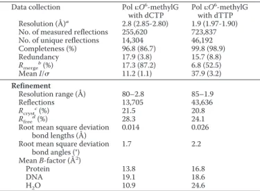
15% PEG 5000 monomethyl ether (w/v), and 0.1MMES (pH 6.5). Crystal trays were incubated at 4 °C, and diffraction qual-ity crystals appeared in 1–3 days. The crystals belonged to space group P6522 and had cell dimensions of a⫽ b ⫽ 97.9 Å, c⫽ 202.5 Å for dCTP-containing crystals and a ⫽ b ⫽ 97.9 Å, c⫽ 202.7 Å for dTTP-containing crystals, with␣ ⫽  ⫽ 90°, ␥ ⫽ 120°. For data collection, crystals were step-soaked for 5 min in mother liquor solutions containing 0 –25% glycerol (w/v) and flash frozen in liquid nitrogen.
Structure Determination and Refinement—X-ray diffraction data were collected at a wavelength of 1.54 Å using a Bruker Microstar microfocus rotating-anode x-ray generator with a Bruker Proteum PT135 CCD detector (Bruker AXS, Madison, WI) in the Vanderbilt Center for Structural Biology or on the
at Ewha Medical Library on October 24, 2016
http://www.jbc.org/
21-ID-D beam line at the Advanced Photon Source (APS) at Argonne National Laboratory (Argonne, IL). The data were indexed, integrated, and scaled using HKL2000 (30). Phases were calculated using molecular replacement with Phaser software, which generated a unique solution when pol (Pro-tein Data Bank code 2ALZ) minus DNA was used as a search model (31). Electron density maps (calculated to 2.8 Å (dCTP) and 1.9 Å (dTTP)) showed clear density around the O6 -methylG lesion and both incoming dCTP and dTTP. Models were built using COOT software and refined using REFMAC5 software using TLS refinement (32–34). The refined model converged to an Rcryst⫽ 21.5% and Rfree⫽ 28.3% for the
dCTP-containing complex and an Rcryst⫽ 20.8% and Rfree⫽
24.1% for the dTTP-containing complex. Ramachandran plots for the refined models show good stereochemistry, with 90.0 (dCTP-containing) and 92.5% (dTTP-containing) of residues in the favored regions and 0.0 (dCTP-containing) and 0.0% (dTTP-containing) in the disallowed regions. Figures were prepared using PyMOL (35).
RESULTS
Extension of G- and O6-MethylG-containing
Oligonucleo-tide Primers by Human Pol—Human pol incorporated (the
correct) dCTP opposite template G but preferentially misin-corporated dTTP opposite template O6-methylG. The ability
of the pol catalytic core to extend a primer annealed to a
G-or O6-methylG-containing DNA template was determined in
a primer extension experiment with a self-complementary 18-mer oligonucleotide used as both the primer and template in the reaction (self-annealing of the oligonucleotide gener-ated 4-base overhangs at both ends of the duplex DNA) (Fig. 1A). The results of the primer extension reaction demon-strated that pol incorporated nucleotides opposite template G and extended the primer by one nucleotide using this sub-strate (Fig. 1B). The lack of further extension is consistent
with the previously demonstrated low processivity of pol (5, 24, 36 –38). Pol incorporated a single nucleotide opposite
O6-methylG but was unable to extend from the nascent O6
-methylG base pair (Fig. 1B). The 19-mer product bands ran faster in the gel when G was the template base compared with the 19-mer product bands formed using O6-methylG. This
result suggests that a different incoming nucleotide was incor-porated opposite G than O6-methylG. Based on primer
exten-sion experiments performed in the presence of single nucleo-tides, it was determined that the 19-mer product bands had dCTP incorporated opposite G and dTTP misincorporated opposite O6-methylG (data not shown). Quantification of the
primer extension data demonstrated that O6-methylG
inhib-ited extension catalyzed by pol compared with that using template G (Fig. 1C). The results of the primer extension ex-periments using the pol catalytic fragment (amino acids 1– 420) are consistent with previous results of primer exten-sion opposite template G and O6-methylG using full-length
pol (amino acids 1–715) (5).
Steady-state Kinetics of Nucleotide Incorporation Opposite
G and O6-MethylG by Pol—To quantify more precisely the
results of the primer extension experiment, steady-state ki-netic analyses were performed using the pol catalytic frag-ment (Table 1). Pol was more efficient at misincorporation of dTTP opposite O6-methylG than correct incorporation of dCTP. Steady-state kinetic analyses using full-length enzyme show that pol has a ⬃10-fold higher efficiency for dTTP in-corporation compared with dCTP inin-corporation opposite
O6-methylG (5). The catalytic core of pol was similar to the
full-length enzyme and maintained a high efficiency of dTTP misincorporation opposite O6-methylG; dTTP
misincorpora-tion efficiency was⬃6-fold higher than dCTP incorporation opposite O6-methylG (Table 1). The increased efficiency of dTTP misincorporation opposite O6-methylG is mainly due
to a Kmeffect; no changes were observed in the kcatvalues for
dCTP or dTTP incorporation (Table 1). Thus, the different efficiencies of dCTP or dTTP incorporation opposite O6
-methylG might result from binding and positioning of the nucleotide differently in the pol active site.
Crystal Structures of Human Pol:O6-MethylG Ternary
Complexes—Several crystal structures of pol ternary
com-plexes with normal and adducted DNA templates have dem-onstrated the ability of pol to rotate template purines to the
synconformation (24 –27). Thus, the high efficiency of pol for dTTP misincorporation opposite O6-methylG may be due
to dCTP and dTTP base pairing in different modes with the
FIGURE 1. Primer extension of pol opposite G- and O6
-methylG-con-taining DNA templates. A, the 18-mer oligonucleotide was
self-comple-mentary and generated two extendable 3⬘ termini at both ends of the du-plex. X was either G or O6-methylG. B, a primer extension experiment was
performed using G- and O6-methylG-containing DNA templates (50 n
M) and increasing pol concentrations in the presence of MgCl2(2 mM). C,
quanti-fied primer extension data is shown.
TABLE 1
Steady-state kinetics of nucleotide incorporation opposite G and O6-methylG by pol
Kmand kcatvalues were determined by quantifying gel band intensities using QuantityOne software (Bio-Rad) and nonlinear regression analysis of product versus关dNTP兴 curves using GraphPad Prism 5.0.
Template dNTP Km kcat kcat/Km finc a M s⫺1 s⫺1mM⫺1 O6-MethylG C 1200⫾ 700 0.03⫾ 0.007 2.5⫻ 10⫺2 1 T 140⫾ 60 0.02⫾ 0.002 1.4⫻ 10⫺1 5.6 G C 90⫾ 10 0.04⫾ 0.002 4.4⫻ 10⫺1 1 T 860⫾ 360 0.01⫾ 0.003 1.2⫻ 10⫺2 0.27 a
fincwas calculated as (kcat/Km)incorrect/(kcat/Km)correct.
at Ewha Medical Library on October 24, 2016
http://www.jbc.org/
Hoogsteen edge of O6-methylG. To test this hypothesis, the
crystal structures of pol:O6-methylG ternary complexes were solved with incoming dTTP or dCTP (Fig. 2, A and B, and Table 2). The structural data indicate that pol bound O6
-methylG in a ternary complex with incoming dCTP or dTTP without major changes in the protein fold or the position of specific amino acid side chains around the active site. The structures of the pol:O6-methylG complex were similar,
re-gardless of whether dCTP or dTTP was the incoming nucleo-tide and superimpose with a root mean square deviation value of 0.34 Å (Fig. 2C). Also, the structure of the pol:O6-methylG
complex is similar to the pol:G ternary complex (2ALZ), and the structures superimpose with a root mean square deviation value of 0.42 Å (Fig. 2D). These data indicate that the prefer-ence of pol for dTTP incorporation opposite O6-methylG
does not result from major conformational changes in the protein, therefore suggesting that preferential dTTP incorpo-ration occurs as a result of differences in the base pairing of
dTTP and dCTP with O6-methylG.
The electron density around the O6-methylG lesion
indi-cates that the base is rotated from the anti to the syn confor-mation in the pol active site. The ability of pol to rotate template purine bases into a syn conformation was shown previously in crystal structures of pol complexed with a nor-mal G template (2ALZ), a 1,N6-ethenodeoxyadenosine lesion
(2DPJ), and an N2-ethylG lesion (3EPG) (24 –26). These crys-tal structures have demonstrated a Hoogsteen base pairing mechanism for pol (24–26). The Hoogsteen edge of the ad-ducted template base forms hydrogen bonds with the incom-ing dCTP and dTTP, and clear electron density is seen around
O6-methylG and each incoming dCTP or dTTP (Fig. 3, A and B). Interestingly, for minor groove DNA adducts, e.g. at the N2 position of G, the ability of pol to form Hoogsteen base pairs results in high efficiency of correct nucleotide incorpo-ration (24, 39). However, this may not be the case with O6
-methylG, where an increased efficiency of misincorporation is observed (Table 1) (5).
Hydrogen Bonding Between dCTP and (syn) O6-MethylG—
Preferential incorporation of dTTP opposite O6-methylG is
the result of a more energetically favored base pair formed with the Hoogsteen edge of O6-methylG compared with
dCTP. Different hydrogen bonds were formed whether dCTP or dTTP pair with O6-methylG in the pol active site (Fig. 4).
The N3 atom of cytosine must be protonated to function as a hydrogen donor to the N7 and O6 atoms of O6-methylG. A
positively charged cytosine has been hypothesized to bind in the active site of pol, as a hydrogen atom at the N3 position
FIGURE 2. Crystal structures of pol in complex with O6-methylG and
incoming dCTP and dTTP. A, pol with template O6-methylG and
incom-ing dCTP. B, pol with template O6-methylG and incoming dTTP. C,
compar-ison of the pol complexes containing template O6-methylG and incoming
dCTP (pink) and dTTP (blue). D, comparison of the pol with template G (2ALZ; yellow) and pol with template O6-methylG and incoming dCTP (red).
TABLE 2
Data collection and refinement statistics Data collection Pol:O6-methylG
with dCTP
Pol:O6-methylG with dTTP Resolution (Å)a 2.8 (2.85-2.80) 1.9 (1.97-1.90)
No. of measured reflections 255,620 723,837 No. of unique reflections 14,304 46,192 Completeness (%) 96.8 (86.7) 99.8 (98.9) Redundancy 17.9 (3.8) 15.7 (8.8) Rmerge b(%) 17.3 (87.2) 6.8 (52.5) Mean I/ 11.2 (1.1) 37.9 (3.2) Refinement Resolution range (Å) 80–2.8 85–1.9 Reflections 13,705 43,636 Rcryst c (%) 21.5 20.8 Rfree d (%) 28.3 24.1
Root mean square deviation bond lengths (Å)
0.014 0.026 Root mean square deviation
bond angles (°) 1.7 2.2 Mean B-factor (Å2 ) Protein 13.8 16.8 DNA 19.1 18.6 H2O 10.9 24.6 a
Values for outermost shells are given in parentheses.
b
Rmerge⫽ ⌺兩I ⫺ 具I典兩⌺I, where I is the integrated intensity of a given reflection.
c
Rcryst⫽ ⌺储Fobserved⫺ Fcalculated储/⌺兩Fobserved兩.
d
Rfreewas calculated using 5% random data omitted from the refinement.
FIGURE 3. Pol active site. A, electron density map showing the template
O6-methylG in the syn conformation paired with incoming dCTP. B, electron
density map showing the template O6-methylG in the syn conformation
paired with incoming dTTP.
at Ewha Medical Library on October 24, 2016
http://www.jbc.org/
is needed to form a base pair with the syn G- and N2
-ethylG-template bases (24, 25). The geometry of cytosine, bound in the active site of pol, suggests that the N3 atom opposite
O6-methylG is protonated and donates its hydrogen to the
N7 and O6 acceptor atoms of O6-methylG in a bifurcated
hy-drogen bond (Fig. 4A). The distances between the N3 atom of dCTP and the two hydrogen acceptors of O6-methylG are 2.9
Å and 3.0 Å, respectively. A second hydrogen bond is formed
between the N4 atom of dCTP and the O6 atom of O6
-meth-ylG, with a distance of 3.2 Å (Fig. 4A). The hydrogen bond network between dCTP and O6-methylG is different from the
hydrogen bonds formed between dCTP and the unadducted G. The structure of pol with unmodified G (2ALZ) showed
two hydrogen bonds formed between the N7䡠N3 and O6䡠N4
atoms of G and dCTP, respectively (Fig. 4B).
Hydrogen Bonding Between dTTP and (syn) O6-MethylG—
A single hydrogen bond, with a distance of 2.7 Å, forms
be-tween the N7 and N3 atoms of O6-methylG and dTTP,
re-spectively (Fig. 4C). The length of the bond provides some indication of its strength, and the short distance of the
hydro-gen bond formed between dTTP and O6-methylG suggests a
strong hydrogen bonding interaction that is potentially fa-vored over the relatively weak bifurcated hydrogen bond formed with dCTP. The possibility that dTTP may also form a bifurcated hydrogen bond with O6-methylG is unlikely given
the position of the base and the asymmetrical distances of the bond lengths (the bond between the O6atom and the N3
at-oms is 3.1 Å; Fig. 4D).
Positioning of dCTP and dTTP Opposite O6-MethylG in the
Pol Active Site—Different hydrogen bond networks form
depending on whether the dCTP or dTTP base is paired with
O6-methylG in the pol active site. These differences affect
how the incoming dCTP or dTTP binds in the active site of pol when paired with O6-methylG. Superposition of the O6 -methylG bases demonstrates how the incoming dCTP and dTTP are accommodated and positioned differently opposite the lesion. Comparison of the positions of the incoming nu-cleotides relative to the position of O6-methylG revealed that
the dCTP:O6-methylG base pair is sheared relative to the
dTTP:O6-methylG base pair by⬃1.0 Å (Fig. 5A). The N3
atom of dTTP is positioned to be directly in line with the N7 atom of O6-methylG, whereas the N3 atom of dCTP is posi-tioned equidistant between the N7 and O6 atoms of O6
-methylG (Fig. 5A). The comparison showed that the dCTP base has a different rise relative to the dTTP base when paired opposite O6-methylG where the dTTP base lies in a plane
⬃1.2 Å below the base of the incoming dCTP (Fig. 5B).
DISCUSSION
Utilization of the Hoogsteen edge of adducted and unad-ducted template purines, when base pairing with incoming nucleotides, provides the basis for the higher efficiency of
dTTP misincorporation opposite O6-methylG observed with
pol compared with other Y-family DNA polymerases (5). The Hoogsteen edge of O6-methylG presents a unique
hydro-gen bond platform for the C and T nucleoside triphosphates. Differences in the hydrogen bonds that form (observed in the crystal structures) between dTTP or dCTP paired opposite
O6-methylG appear to contribute to the⬃10-fold higher
steady-state efficiency of dTTP misincorporation by human pol (Table 1) (5). To form a hydrogen bond between dCTP and (syn) O6-methylG, the N3 atom of dCTP must be
proto-nated (40). The pKaof C in solution is 4.5, thus the N3 atom
of C is not protonated at physiological pH (41). However, the pKaof C has been reported to increase to 7.5 when paired in a
hemiprotonated C:C(H⫹) base pair (42). Also, the possibility that C has an elevated pKawhen bound in the active site of pol has been suggested (25). The crystal structure of pol and O6-methylG indicates that the protonated N3 atom of
FIGURE 4. Hoogsteen edge hydrogen bonding in the pol active site. A, template O6-methylG forms a bifurcated hydrogen bond with the protonated
dCTP. B, template G forms two hydrogen bonds with the protonated dCTP (2ALZ). C, single hydrogen bond forms between template O6-methylG and
in-coming dTTP. D, bifurcated hydrogen bond does not form between O6-methylG and dTTP.
FIGURE 5. Positions of dCTP and dTTP bases paired opposite O6-methylG. A, template O6-methylG aligned to show how dCTP and dTTP position
differ-ently opposite the adduct in the pol active site. B, rotated view showing differ-ences in the rise between the O6-methylG:dCTP and dTTP base pairs.
at Ewha Medical Library on October 24, 2016
http://www.jbc.org/
dCTP acts as a hydrogen donor to both the N7 and O6 accep-tor atoms of O6-methylG to form a bifurcated hydrogen bond. Splitting the proton between two acceptor atoms lowers the energy of the bifurcated hydrogen bond compared with the simple hydrogen bond formed between a single donor and acceptor pair (43). Thus, the protonation of the N3 atom of dCTP and formation of a bifurcated hydrogen bond between
O6-methylG and dCTP may factor together in lowering the
efficiency of DNA pol-catalyzed dCTP incorporation oppo-site O6-methylG. Moreover, it is likely that this bifurcated hydrogen bond is only formed in a fraction of the encounters with dCTP. In many cases cytosine is not protonated, and the resulting repulsion will then prevent formation of a produc-tive complex. The crystal structure of pol and O6-methylG:
dTTP indicates that a single hydrogen bond forms between the N3 and N7 atoms of dTTP and O6-methylG, respectively.
The short length of the hydrogen bond (2.7 Å) suggests that the strength of the O6-methylG:dTTP bond may be greater than typical N–H–N hydrogen bonds (2.9 –3.0 Å) that normally form (with a bond energy of⬃5 kcal/mol). The in-creased strength of the O6-methylG:dTTP hydrogen bond
may stabilize the bound nucleoside triphosphate in the pol active site and contribute to the high efficiency of dTTP mis-incorporation opposite the lesion.
The thermodynamic and energetic properties of the hydro-gen bonds formed with the Hoogsteen edge of O6-methylG appear to be more important to the aberrant incorporation of dTTP opposite O6-methylG than the geometry of the base
pair fit into the active site of pol. Comparison of the pol :O6-methylG structures with structures of pol and template
G paired with dCTP (2ALZ) (25) or template A paired with dTTP (2FLL) (44) reveal a relatively undistorted geometry for all of the base pairs (Fig. 6). An exception is observed in the glycosidic bond angle (73°) of dCTP when paired opposite
O6-methylG, which may contribute to the lower efficiency of
dCTP incorporation opposite O6-methylG (Fig. 6B). Yet, the
similarity of the geometry and fit of the unadducted and ad-ducted Hoogsteen base pairs in the pol active site suggests an important role for the hydrogen bond in the base
selectiv-ity for this Y-family DNA polymerase. Similarly in the case of
S. solfataricusDpo4, hydrogen bonding is more important
than base pair geometry for nucleotide selectivity opposite
O6-methylG. The crystal structures of Dpo4 with O6-methylG
and O6-benzylG have shown that a wobble base pair forms
when dCTP pairs opposite the lesion (13, 16). However, un-like pol, Dpo4 utilizes wobble base pairing to preferentially incorporate dCTP opposite O6-methylG, thus maintaining
high fidelity during lesion bypass (13). When incorporating nucleotides opposite O6-methylG, the Y-family DNA poly-merases differ from replicative DNA polypoly-merases, which rely on Watson-Crick geometry for nucleotide selectivity and which have been proposed to tolerate the fit of the less ther-mally stable Watson-Crick-like O6-methylG:dTTP base pair
better than the more thermally stable O6-methylG:dCTP
wobble base pair with distorted Watson-Crick geometry (10). The structure of the B. stearothermophilus DNA polymerase I (BF) revealed that a O6-methylG:dTTP base pair with pseudo-Watson-Crick geometry fits in the active site of the DNA po-lymerase (14). Interestingly, the structure showed that an isosteric O6-methylG:dCTP base pair was also accommodated
in the active site. The authors suggested that, upon binding the adducted base pair, the DNA polymerase introduced changes to the shape of the DNA so that Watson-Crick geom-etry was maintained despite energetic barriers (14).
The results of the structural analysis with human pol pro-vide epro-vidence that the differences in the hydrogen bonds formed between the incoming nucleotides dCTP or dTTP paired opposite O6-methylG are important to facilitating the
increased misincorporation of dTTP opposite the lesion. For-mation of a Hoogsteen base pair during nucleotide incorpora-tion is a unique property of pol, allowing it to bypass minor groove DNA adducts efficiently, e.g. the N2 position of G. The
N2-ethylG lesion, for example, is rotated out of the active site of pol and does not interfere with hydrogen bond formation between the template base and incoming nucleotide (24). In contrast, the major groove O6-methylG adduct, when rotated
to the syn conformation, remains on the hydrogen bonding face of the template base and influences the hydrogen
bond-FIGURE 6. Geometric properties of Hoogsteen base pairs G:dCTP, O6-methylG:dCTP, A:dTTP, and O6-methylG:dTTP in the active site of pol. The
C1⬘–C1⬘ distances (Å) and glycosidic bond angles (°) are shown for the G:dCTP (2ALZ) (A), O6-methylG:dCTP (B), A:dTTP (2FLL) (C), and O6-methylG:dTTP (D)
base pairs.
at Ewha Medical Library on October 24, 2016
http://www.jbc.org/
ing properties of the adduct. The modified hydrogen bond
pattern formed between O6-methylG and dCTP lowers the
potential for binding of dCTP in the pol active site and re-sults in decreased efficiency of correct nucleotide incorpora-tion. The ability of DNA polymerases to misincorporate dTTP opposite O6-methylG results in a high number of
G:C3 A:T transition mutations caused by O6-alkylG DNA
adducts. The reason for such low discrimination by DNA polymerases when replicating O6-methylG is not well
under-stood despite substantial biochemical and structural work done to address this problem. The structure of pol (in com-plex with O6-methylG) shows that the adduct does not
change the shape of the active site, suggesting that hydrogen bonding has an important role in the selection of incorpo-rated nucleotides during pol-catalyzed bypass of O6-methylG.
Differences in the hydrogen bonds formed between O6
-meth-ylG and dTTP or dCTP demonstrate how small changes to the geometry and strength of the hydrogen bond can affect base pairing and thereby influence the efficiency and fidelity of nucleotide incorporation by DNA polymerases.
Acknowledgments—We thank F. W. Perrino for the purified MBP-pol and S. Anderson for assistance with x-ray data collection.
REFERENCES
1. Drabløs, F., Feyzi, E., Aas, P. A., Vaagbø, C. B., Kavli, B., Bratlie, M. S., Pen˜a-Diaz, J., Otterlei, M., Slupphaug, G., and Krokan, H. E. (2004) DNA
Repair 3,1389 –1407
2. Shank, R. C. (1987) Arch. Toxicol. Suppl. 10, 204 –216 3. Loveless, A. (1969) Nature 223, 206 –207
4. Loechler, E. L., Green, C. L., and Essigmann, J. M. (1984) Proc. Natl.
Acad. Sci. U.S.A. 81,6271– 6275
5. Choi, J. Y., Chowdhury, G., Zang, H., Angel, K. C., Vu, C. C., Peterson, L. A., and Guengerich, F. P. (2006) J. Biol. Chem. 281, 38244 –38256 6. Snow, E. T., Foote, R. S., and Mitra, S. (1984) J. Biol. Chem. 259,
8095– 8100
7. Woodside, A. M., and Guengerich, F. P. (2002) Biochemistry 41, 1027–1038
8. Haracska, L., Prakash, S., and Prakash, L. (2000) Mol. Cell. Biol. 20, 8001– 8007
9. Gaffney, B. L., Marky, L. A., and Jones, R. A. (1984) Biochemistry 23, 5686 –5691
10. Gaffney, B. L., and Jones, R. A. (1989) Biochemistry 28, 5881–5889 11. Patel, D. J., Shapiro, L., Kozlowski, S. A., Gaffney, B. L., and Jones, R. A.
(1986) Biochemistry 25, 1036 –1042
12. Patel, D. J., Shapiro, L., Kozlowski, S. A., Gaffney, B. L., and Jones, R. A. (1986) Biochemistry 25, 1027–1036
13. Eoff, R. L., Irimia, A., Egli, M., and Guengerich, F. P. (2007) J. Biol. Chem.
282,1456 –1467
14. Warren, J. J., Forsberg, L. J., and Beese, L. S. (2006) Proc. Natl. Acad. Sci.
U.S.A. 103,19701–19706
15. Ginell, S. L., Kuzmich, S., Jones, R. A., and Berman, H. M. (1990)
Bio-chemistry 29,10461–10465
16. Eoff, R. L., Angel, K. C., Egli, M., and Guengerich, F. P. (2007) J. Biol.
Chem. 282,13573–13584
17. Tan, H. B., Swann, P. F., and Chance, E. M. (1994) Biochemistry 33, 5335–5346
18. Singer, B., Chavez, F., Goodman, M. F., Essigmann, J. M., and Dosanjh, M. K. (1989) Proc. Natl. Acad. Sci. U.S.A. 86, 8271– 8274
19. Vasquez-Del Carpio, R., Silverstein, T. D., Lone, S., Swan, M. K., Choudhury, J. R., Johnson, R. E., Prakash, S., Prakash, L., and Aggarwal, A. K. (2009) PLoS One 4, e5766
20. Lone, S., Townson, S. A., Uljon, S. N., Johnson, R. E., Brahma, A., Nair, D. T., Prakash, S., Prakash, L., and Aggarwal, A. K. (2007) Mol. Cell 25, 601– 614
21. Irimia, A., Eoff, R. L., Guengerich, F. P., and Egli, M. (2009) J. Biol. Chem.
284,22467–22480
22. Hwang, H., and Taylor, J. S. (2005) Biochemistry 44, 4850 – 4860 23. Wolfle, W. T., Washington, M. T., Kool, E. T., Spratt, T. E., Helquist,
S. A., Prakash, L., and Prakash, S. (2005) Mol. Cell. Biol. 25, 7137–7143 24. Pence, M. G., Blans, P., Zink, C. N., Hollis, T., Fishbein, J. C., and
Per-rino, F. W. (2009) J. Biol. Chem. 284, 1732–1740
25. Nair, D. T., Johnson, R. E., Prakash, L., Prakash, S., and Aggarwal, A. K. (2005) Structure 13, 1569 –1577
26. Nair, D. T., Johnson, R. E., Prakash, L., Prakash, S., and Aggarwal, A. K. (2006) Nat. Struct. Mol. Biol. 13, 619 – 625
27. Nair, D. T., Johnson, R. E., Prakash, S., Prakash, L., and Aggarwal, A. K. (2004) Nature 430, 377–380
28. Gasteiger, E., Gattiker, A., Hoogland, C., Ivanyi, I., Appel, R. D., and Bai-roch, A. (2003) Nucleic Acids Res. 31, 3784 –3788
29. Boosalis, M. S., Petruska, J., and Goodman, M. F. (1987) J. Biol. Chem.
262,14689 –14696
30. Otwinowski, Z., and Minor, W. (1997) Methods Enzymol. 276, 307–326 31. McCoy, A. J., Grosse-Kunstleve, R. W., Storoni, L. C., and Read, R. J.
(2005) Acta Crystallogr. D Biol. Crystallogr. 61, 458 – 464
32. Emsley, P., and Cowtan, K. (2004) Acta Crystallogr. D Biol. Crystallogr.
60,2126 –2132
33. Murshudov, G. N., Vagin, A. A., and Dodson, E. J. (1997) Acta
Crystal-logr. D Biol. CrystalCrystal-logr. 53,240 –255
34. Winn, M. D., Murshudov, G. N., and Papiz, M. Z. (2003) Methods
Enzy-mol. 374,300 –321
35. DeLano, W. L. (2002) The PyMOL Molecular Graphics System, DeLano Scientific LLC, San Carlos, CA
36. Tissier, A., McDonald, J. P., Frank, E. G., and Woodgate, R. (2000) Genes
Dev. 14,1642–1650
37. Zhang, Y., Yuan, F., Wu, X., and Wang, Z. (2000) Mol. Cell. Biol. 20, 7099 –7108
38. Johnson, R. E., Washington, M. T., Haracska, L., Prakash, S., and Prakash, L. (2000) Nature 406, 1015–1019
39. Washington, M. T., Minko, I. G., Johnson, R. E., Wolfle, W. T., Harris, T. M., Lloyd, R. S., Prakash, S., and Prakash, L. (2004) Mol. Cell. Biol. 24, 5687–5693
40. Williams, L. D., and Shaw, B. R. (1987) Proc. Natl. Acad. Sci. U.S.A. 84, 1779 –1783
41. Dawson, R. M. C., Elliott, D. C., Elliott, W. H., and Jones, K. M. (eds) (1986) Data for Biochemical Research, 3rd Ed., pp. 80 – 85, Clarendon Press, Oxford
42. Gray, D. M., Cui, T., and Ratliff, R. L. (1984) Nucleic Acids Res. 12, 7565–7580
43. Afonin, A. V., and Vashchenko, A. V. (2010) Magn. Reson. Chem. 48, 309 –317
44. Nair, D. T., Johnson, R. E., Prakash, L., Prakash, S., and Aggarwal, A. K. (2006) Structure 14, 749 –755
at Ewha Medical Library on October 24, 2016
http://www.jbc.org/
S-1
Structural Basis for Proficient Incorporation of dTTP Opposite O
6-Methylguanine by Human DNA
Polymerase ι
Matthew G. Pence, Jeong-Yun Choi, Martin Egli, and F. Peter Guengerich
J. Biol. Chem. 285, XXX-XXX (2010)
CONTENTS
S-2
profile showing peaks from PreScission Protease incubation (a) and after washing with 20 mM
glutathione (b). B, SDS-PAGE of purified human pol ι. Lane 1: Markers (M), lane 2: Pre-induction
sample (Pre), lane 3: Pol ι (a, corresponds to peak a from Fig. S1A), lane 4: empty, and lane 5:
glutathione wash (b, corresponds to peak b from Fig. S1A).
Matthew G. Pence, Jeong-Yun Choi, Martin Egli and F. Peter Guengerich
ι
by Human DNA Polymerase
doi: 10.1074/jbc.M110.183665 originally published online October 20, 2010 2010, 285:40666-40672.
J. Biol. Chem.
10.1074/jbc.M110.183665 Access the most updated version of this article at doi:
Alerts:
When a correction for this article is posted •
When this article is cited •
to choose from all of JBC's e-mail alerts Click here
http://www.jbc.org/content/285/52/40666.full.html#ref-list-1
This article cites 42 references, 18 of which can be accessed free at
at Ewha Medical Library on October 24, 2016
http://www.jbc.org/