Sonographic Features of Axillary
Lymphadenopathy Caused by
Kikuchi Disease
Ji Hyun Youk, MD, Eun-Kyung Kim, MD, Kyung Hee Ko, MD, Min Jung Kim, MD
Received January 31, 2008, from the Department of Radiology, Bundang CHA Hospital, Pochon CHA University, Sungnam, Korea (J.H.Y., K.H.K.); and Department of Radiology, Research Institute of Radiological Science, Yonsei University College of Medicine, Seoul, Korea (E.-K.K., M.J.K.). Revision requested February 28, 2008. Revised manuscript accepted for publication March 6, 2008.
Address correspondence to Eun-Kyung Kim, MD, Department of Radiology, Yonsei University College of Medicine, Seodaemun-ku, Shinchon-dong 134, Seoul 120-752, Korea.
E-mail: ekkim@yuhs.ac Abbreviations
S/L, shortest axis/longest axis
Objective. The purpose of this study was to document the sonographic findings of axillary lym-phadenopathy in Kikuchi disease. Methods. The medical records and sonographic findings of 7 patients with a pathologic diagnosis of Kikuchi disease in the axillary lymph node by sonographically guided core needle biopsy (n = 6) or excisional biopsy (n = 1) were reviewed. On sonograms, lymph nodes were assessed for their distribution, size, shape, border, echogenicity, and internal architecture, and those sonographic features of each node were evaluated to determine whether the findings favored malignant or benign lymphadenopathy. Results. Of the 7 patients (1 man and 6 women; mean age ± SD, 34.3 ± 7.7 years), 29 affected lymph nodes (5–38 mm; mean, 14.8 ± 7.2 mm) were identified on sonograms. The sonographic characteristics were as follows: the shortest axis/longest axis ratio of the node (mean, 0.595) was 0.5 or greater in 22 nodes (76%); the border was sharp in 16 (55%); the cortex was hypoechoic in 20 (69%) and isoechoic in 9 (31%); the hilum was narrow in 1 (4%) and absent in 16 (55%); and cortical thickening was found in 13 (45%, concentric in 6 and eccentric in 7). Nineteen lymph nodes (66%) were classified as having malignant-favoring features, and 10 (34%) were classified as having benign-favoring features. Conclusions. Many axillary lymph nodes in Kikuchi disease look suspicious sonographically. When lymph nodes in the axilla show suspi-cious findings on sonograms of relatively young patients, Kikuchi disease can be considered a possible differential diagnosis, and image-guided percutaneous biopsy should be done. Key words: axilla; Kikuchi disease; lymph node; sonography.
ikuchi disease, also known as Kikuchi-Fujimoto disease or histiocytic necrotizing lymphadenitis, is an uncommon cause of benign lymphadenopa-thy that was first described independently by Kikuchi1and Fujimoto et al2in 1972. They described the
distinctive histologic appearance of the lymph nodes as “lymphadenitis showing reticulum cell hyperplasia with nuclear debris and phagocytosis” and “cervical subacute necrotizing lymphadenitis,” respectively. It is a self-limit-ing disease that revolves spontaneously, generally within 1 to 4 months, although in rare cases lymphadenopathy may persist for up to 1 year after diagnosis.3–5
Initially regarded as a unique type of lymphadenitis occurring predominantly in cervical lymph nodes of young Southeast Asian women, it is now recognized that this disease can affect patients regardless of age, sex, or ethnic background and can involve any anatomic site, including nodal and extranodal locations such as the skin, bone marrow, liver, spleen, and gastrointestinal
K
tract.3Involvement of nodal areas other than
cer-vical has been described, including axillary, tho-racic, abdominal, and pelvic.6
There have been a few reports about radiologic findings of Kikuchi disease, which described cervi-cal lymph node involvement and were primarily focused on computed tomographic findings.7–11
To our knowledge, however, there has been no published study of the sonographic findings of axillary lymph node involvement in Kikuchi dis-ease, although the sonographic findings of cervi-cal lymph node involvement have been reported in a few studies.13We found only scarce mention
of axillary lymph node involvement in a few case reports.14–18In this study, we retrospectively
ana-lyzed the sonographic findings of axillary lym-phadenopathy in Kikuchi disease.
Materials and Methods
We conducted a database search of pathologic records from February 2000 through August 2007 and found 7 patients with a histologically proven diagnosis of Kikuchi disease in axillary lymph nodes. We retrospectively reviewed the medical records and the radiologic examinations of these patients. The Institutional Review Board approved this retrospective study, and informed consent was not required.
All patients underwent gray scale sonographic examinations of the axilla, which were performed with high-resolution sonography units with 7.5- or 12-MHz linear array transducers (HDI 3500 and iU22 systems, Philips Medical Systems, Bothell, WA). The diagnosis of Kikuchi disease was reached by means of sonographically guided core needle biopsy in 6 patients and excisional biopsy in 1 patient. Sonographically guided biopsy was performed on the largest node by a freehand tech-nique using a 14- or 16-gauge spring-loaded Tru-Cut needle with a 22-mm throw (Pro-Mag 2.2, Manan Medical Products, Inc, Northbrook, IL; Magnum, C. R. Bard, Inc, Tempe, AZ; and SACN biopsy needle, Medical Device Technologies, Gainesville, FL). We assumed that other nodes in the axilla with gray scale appearances similar to those of the sampled node would be the same pathologically, and these nodes were also includ-ed in the study. All sonographic examinations and biopsy procedures were performed by the 1 of 4
radiologists with extensive clinical experience in sonography and biopsy.
On sonography, lymph nodes were assessed for their distribution, size, shape, border demarca-tion, echogenicity, and internal architecture. The distribution of lymphadenopathy in the axilla was classified into 3 levels relative to the pec-toralis minor muscle19: level I consisted of nodes
below the lateral border of the muscle; level II consisted of nodes behind the muscle; and level III consisted of nodes above the medial border of the muscle. The size of the lymph node was defined as its length along the longest axis as measured on the images. The shape of the lymph node was assessed by its shortest axis/longest axis (S/L) ratio. An S/L ratio of less than 0.5 indi-cated a long or an elliptical node, whereas an S/L ratio of 0.5 or greater indicated a round node.20,21
The nodal border was classified as sharp or unsharp compared with surrounding fatty tis-sue. The echogenicity of the lymph node was compared with that of adjacent muscles and was categorized as hypoechoic, isoechoic, or hyperechoic. The internal architecture of the node was evaluated for the echogenic fatty hilum (wide, narrow, or absent) and cortical thickening (concentric or eccentric).21 When the hilum
appeared elliptical in the longitudinal plane, generally conforming to the shape of the whole node, it was classified as wide. A flat, slitlike hilum was considered narrowed. Those nodes that showed no central zone of hyperechogenic-ity were classified as having no hilum. For evalu-ation of cortical thickening, the cortex was considered thickened when the thickness at its broadest point was half the transverse diameter of the hilum or greater in the longitudinal plane. Those nodes that showed focal cortical widening of at least 100% (ie, the thickness of the cortex at one site was at least double that at its narrowest point) were classified as having eccentric cortical thickening. If the nodal hilum (the reference structure) was absent, cortical thickening could not be assessed.21In addition, the sonographic
findings of each axillary lymph node were analyzed to determine whether the findings favored malignant or benign lymphadenopa-thy. Abnormal sonographic findings favoring malignant axillary lymph nodes were a round shape, a sharp border, an abnormal sonographic
appearance of the cortex, which included diffuse or focal thickening of the cortex or hypoe-chogenicity, and a narrow or absent fatty hilum. If at least 3 of those findings were shown, the lymph node was classified as having malignant-favoring features.22–24All images were analyzed
by 2 experienced radiologists with consensus. Results
Patient Characteristics
The patients’ clinical characteristics are summa-rized in Table 1. The patients’ ages ranged from 22 to 42 years (mean ± SD, 34.3 ± 7.7 years), and 6 of them were women. All patients had swelling in the axilla (4 right, 2 left, and 1 bilateral). The duration of symptoms ranged from 5 days to 3 months (mean, 24.6 ± 31.1 days). Physical exam-inations revealed palpable axillary masses in all 7 patients and a low-grade fever in 1. On labora-tory evaluation, the patient with the fever had leukopenia (white blood cell count, 2600/mm3),
but the others had nonspecific results. For all patients, there was no clinical or radiologic evi-dence of Kikuchi disease involvement in other organs, and their clinical symptoms resolved without any specific treatment by follow-up.
Sonographic Characteristics of Lymph Nodes
In the 7 patients, 29 affected lymph nodes (size, 5–38 mm; mean, 14.8 ± 7.2 mm) identified on sonograms were included in this study. For the distribution of lymphadenopathy in the axilla, in 4 patients, the lymphadenopathy was confined to level I, and in the other 3, it was detected from level I through level II or III (Table 1).
Table 2 shows the results of sonographic assess-ment in each node. The S/L ratio ranged from 0.15 to 1 (mean, 0.60 ± 0.22). In 7 (24%) of 29
lymph nodes, the S/L ratio (mean, 0.595) was less than 0.5 (Figures 1 and 2), and in 22 (76%), the S/L ratio was 0.5 or greater (Figures 3 and 4). The nodal border was sharp in 16 nodes (55%) and unsharp in 13 (45%; Figure 2). The nodal cortex was hypoechoic in 20 nodes (69%) and isoechoic in 9 (31%). The hilum was wide in 12 nodes (41%; Figures 1 and 2), narrow in 1 (4%; Figure 3), and absent in 16 (55%; Figure 4). Cortical thickening was found in 13 nodes (45%), and the thickening was concentric in 6 (46%; Figures 1 and 3) and eccentric in 7 (54%; Figure 2). In the combined evaluation of abnormal sonographic findings, 19 lymph nodes (66%) were classified as having malignant-favoring features, and 10 (34%) were classified as having benign-favoring features. Discussion
Kikuchi disease classically presents as painful cervical lymphadenopathy in young adults, usu-ally female. Cervical lymph nodes are the prima-ry sites of this disease in 70% to 98% of cases.14
However, virtually any nodal chain can be involved, and axillary, supraclavicular, mediasti-nal, celiac, peripancreatic, and inguinal loca-tions have been reported.9 Most reports about
radiologic findings of Kikuchi disease described cervical lymph node involvement. Fulcher8
reported the sonographic findings of Kikuchi disease in the neck, consisting of multiple enlarged lymph nodes that were homogeneous and surrounded by a hyperechoic rim. These sonographic findings were similar to those described by Ogawa et al,13except for the
hetero-geneous texture found in the latter study. Ying et al12reported the sonographic findings of 6
cervi-cal lymph nodes in Kikuchi disease. They were hypoechoic (6 [100%]), were predominantly
Table 1. Characteristics of 7 Patients With Kikuchi Disease in the Axillary Lymph Nodes
Patient 1 2 3 4 5 6 7
Sex M F F F F F F
Age, y 38 28 30 38 42 22 42
Site* R, I B, I R, I–III L, I, II R, I L, I–III R, I
Palpation Yes Yes Yes Yes Yes Yes Yes
Duration of symptoms, d 5 5 7 5 30 30 90
Involved lymph nodes, n 4 5 5 4 3 6 2
B indicates bilateral; F, female; L, left; M, male; and R, right.
elliptical or oval with an S/L ratio of less than 0.5 (5 [83.3%]), tended to have an echogenic hilum (4 [66.7%]) and an unsharp border (4 [66.7%]), and were found to be similar to reactive nodes, for which there was no significant difference in sonographic findings compared with Kikuchi disease. Of axillary lymph node involvement in Kikuchi disease, only a few case reports or series have been published, and the information about the radiologic findings has been limited. The sonographic findings of a conglomerate of a few enlarged hypoechoic lymph nodes16and an oval
predominantly hypoechoic lymph node with cortical thickening14 were described in 2 case
reports.
According to Esen23and Lernevall,25there are
several sonographic characteristics of malignant lymph nodes in the axilla. Pathologic nodes tend to become more rounded; therefore, a high S/L ratio (>0.5) increases suspicion of malignancy,
whereas a low S/L ratio (<0.5) indicates that a node is benign. Malignant lymph nodes usually have sharp borders, and their cortex tends to become markedly hypoechoic compared with adjacent muscles. Eccentric enlargement with focal thickening of the cortex, at least doubling, is a strong indicator of malignant transformation. Compression of the hilum and especially absence of the hilum are highly suggestive of malignancy. In this study, the sonographic findings of axillary lymph nodes in Kikuchi disease were a predomi-nantly round shape with an S/L ratio of 0.5 or greater (22 [76%] of 29), a tendency to show a hypoechoic cortex (20 [69%] of 29), a sharp border (16 [55%] of 29), a narrow or an absent echogenic hilum (17 [59%] of 29), and eccentric cortical thickening (7 [54%] of 13). All lymph nodes showed at least 1 malignant-favoring feature, and approximately two-thirds of them (66%) had 3 or more suspicious malignant findings. These results,
Table 2. Sonographic Characteristics of Axillary Lymph Nodes in Kikuchi Disease
Cortical Favoring
Node Size, mm S/L Ratio Border Echogenicity Hilum Widening Features
1 38 0.36 Sharp Hypoechoic Narrow Concentric Malignant
2 25 0.73 Sharp Hypoechoic Narrow Concentric Malignant
3 18 0.73 Sharp Hypoechoic Wide Eccentric Malignant
4 20 0.57 Sharp Hypoechoic Wide Eccentric Malignant
5 21 0.43 Sharp Hypoechoic Wide Eccentric Benign
6 18 0.38 Sharp Hypoechoic Wide Concentric Benign
7 18 0.59 Sharp Hypoechoic Wide Concentric Malignant
8 17 0.68 Sharp Hypoechoic Wide Concentric Malignant
9 12 0.57 Unsharp Isoechoic Wide Concentric Benign
10 21 0.5 Sharp Hypoechoic Absent NA Malignant
11 8 0.81 Unsharp Hypoechoic Absent NA Malignant
12 9 0.9 Unsharp Hypoechoic Absent NA Malignant
13 7 0.71 Sharp Isoechoic Absent NA Malignant
14 5 0.52 Sharp Isoechoic Absent NA Malignant
15 17 0.15 Sharp Hypoechoic Absent NA Benign
16 12 0.78 Unsharp Hypoechoic Absent NA Malignant
17 10 0.88 Sharp Hypoechoic Absent NA Malignant
18 7 0.52 Unsharp Hypoechoic Absent NA Malignant
19 15 0.56 Sharp Isoechoic Absent NA Malignant
20 7 0.9 Unsharp Isoechoic Absent NA Malignant
21 6 0.8 Unsharp Isoechoic Absent NA Malignant
22 17 0.24 Unsharp Hypoechoic Wide Eccentric Benign
23 12 0.57 Unsharp Hypoechoic Wide Eccentric Benign
24 12 0.5 Unsharp Isoechoic Absent NA Benign
25 11 1 Unsharp Hypoechoic Absent NA Malignant
26 10 0.7 Sharp Hypoechoic Absent NA Malignant
27 12 0.59 Unsharp Isoechoic Absent NA Benign
28 25 0.18 Unsharp Hypoechoic Wide Eccentric Benign
29 20 0.43 Sharp Isoechoic Wide Eccentric Benign
considered to favor the appearance of malignant lymphadenopathy, are contrary to those of Ying et al12mentioned above. Such a discrepancy may
be attributable to the small sample size and dif-ferences in the anatomic location, study popula-tion, methods, and radiologic details from which to draw conclusions. Also, the malignant-favor-ing sonographic features of Kikuchi disease found in this study could not have been distinct because more than a few lymph nodes showed benign-favoring features, and there was an over-lap between reactive and malignant nodes in the sonographic criteria described above.25 The
results did not show any specific sonographic features of Kikuchi disease in the axilla; therefore, further investigation with a larger patient data -base is necessary to validate these results.
In the axilla, lymph node enlargement is undoubtedly the most common pathologic find-ing.26Most cases in which lymphadenopathy is
associated with visible breast cancer represent metastatic breast disease. In those with no visi-ble breast disease, such as this study population, various conditions such as connective tissue disease (commonly rheumatoid arthritis), infec-tions, benign reactive nodes, hematologic malig-nancies, occult carcinomas, and metastatic tumors other than breast cancer can be consid-ered as differential diagnoses.27,28However, even
these conditions usually present as sonographi-cally nonspecific lymphadenopathy. Therefore, it would be a challenge to differentiate the cause of axillary lymphadenopathy by imaging studies alone, prompting histopathologic examinations. Sonographically guided fine-needle aspiration biopsy of lymph nodes is common and has been an accurate method for diagnosing malignancy. For the diagnosis of Kikuchi disease, this proce-dure may show the characteristic pathologic features, but it is frequently nonspecific or non-diagnostic, and excisional biopsy is needed for a definitive diagnosis.5,29In this study, 1 patient
who had undergone excisional biopsy initially underwent sonographically guided fine-needle aspiration biopsy, which yielded a nonspecific diagnosis negative for malignancy. For sono-graphically guided core needle biopsy, Kim et al30
reported that Kikuchi disease in cervical lymph nodes was definitely diagnosed by that procedure in all 25 of their patients, which prevented most
of the patients from undergoing unnecessary excision biopsy. Also, in this study, 6 patients who underwent sonographically guided core biopsy had definitive diagnoses. Therefore, sonograph-ically guided core needle biopsy may be effi-cient for diagnosing Kikuchi disease in the axilla and could be used as an alternative to excisional biopsy.
Figure 1. Gray scale sonogram of an axillary lymph node classified as benign in a 28-year-old woman showing a sharp margin, a hypoechoic cortex, a wide hilum, concentric cortical thickening, and an S/L ratio of 0.38.
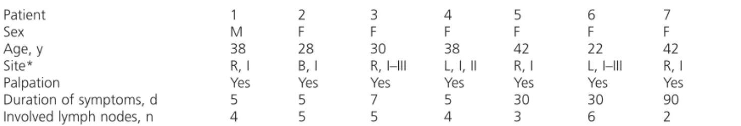
Figure 2. Gray scale sonogram of an axillary lymph node classified as benign in a 42-year-old woman showing an unsharp margin, a hypoechoic cortex, a wide hilum, eccentric cortical thickening, and an S/L ratio of 0.18.
Our study had some limitations. First, this ret-rospective analysis was limited by the small sample size. A larger study would be helpful to characterize the sonographic findings of this disease. Second, there could have been a selec-tion bias because only cases that had undergone sonographic evaluation and biopsy were includ-ed; patients who did not undergo biopsy were not included. Most cases with biopsy tend to have imaging findings suspicious for malignancy;
therefore, more malignant-favoring cases could have been included in this study. Third, not all lymph nodes underwent biopsy, and even among those that did, all but 1 had a diagnosis by core biopsy and not excisional biopsy.
Kikuchi disease in the axilla can be challenging to diagnose, and the differential diagnoses are quite broad. Moreover, the sonographic findings of Kikuchi disease in the axilla were nonspecific and could not be differentiated sonographically from those of malignant lymphadenopathy. When lymph nodes in the axilla show suspicious findings sonographically, Kikuchi disease can be considered one of several possible differential diagnoses, and pathologic examination should be done. To avoid an incorrect diagnosis and prevent resulting unnecessary interventions and subse-quent patient morbidity and mortality, it is impor-tant to be aware of the characteristic clinical and morphologic findings.
References
1. Kikuchi M. Lymphadenitis showing focal reticulum cell hyperplasia with nuclear debris and phagocytosis. Nippon Ketsueki Gakkai Zasshi 1972; 35:379–380.
2. Fujimoto Y, Kozima Y, Yamaguchi K. Cervical subacute necrotizing lymphadenitis: a new clinicopathologic entity. Naika 1972; 20:920–927.
3. Onciu M, Medeiros LJ. Kikuchi-Fujimoto lymphadenitis. Adv Anat Pathol 2003; 10:204–211.
4. Lin HC, Su CY, Huang CC, Hwang CF, Chien CY. Kikuchi’s disease: a review and analysis of 61 cases. Otolaryngol Head Neck Surg 2003; 128:650–653.
5. Poulose V, Chiam P, Poh WT. Kikuchi’s disease: a Singapore case series. Singapore Med J 2005; 46:229–232. 6. Bennie MJ, Bowles KM, Rankin SC. Necrotizing cervical
lymphadenopathy caused by Kikuchi-Fujimoto disease. Br J Radiol 2003; 76:656–658.
7. Kwon SY, Kim TK, Kim YS, Lee KY, Lee NJ, Seol HY. CT find-ings in Kikuchi disease: analysis of 96 cases. AJNR Am J Neuroradiol 2004; 25:1099–1102.
8. Fulcher AS. Cervical lymphadenopathy due to Kikuchi dis-ease: US and CT appearance. J Comput Assist Tomogr 1993; 17:131–133.
9. Miller WT Jr, Perez-Jaffe LA. Cross-sectional imaging of Kikuchi disease. J Comput Assist Tomogr 1999; 23:548–551. 10. Kim TA, Lupetin AR, Graham C. CT appearance of
Kikuchi-Fujimoto disease. Clin Imaging 1995; 19:1–3.
11. Na DG, Chung TS, Byun HS, Kim HD, Ko YH, Yoon JH. Kikuchi disease: CT and MR findings. AJNR Am J Neuroradiol 1997; 18:1729–1732.
Figure 3. Gray scale sonogram of an axillary lymph node classified as malignant in a 38-year-old man showing a sharp margin, a hypoechoic cortex, a narrow hilum, concentric cortical thickening, and an S/L ratio of 0.73.
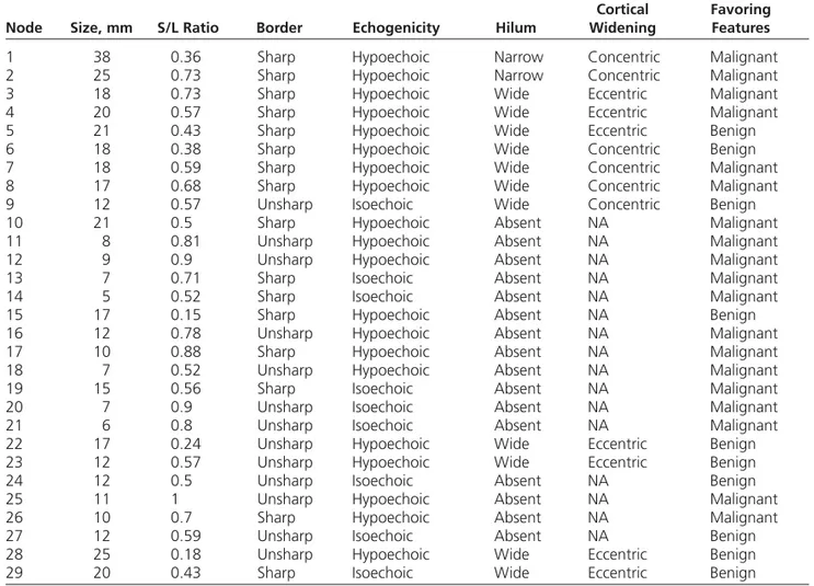
Figure 4. Gray scale sonogram of an axillary lymph node assessed as malignant in a 30-year-old woman showing a sharp margin, a hypoechoic cortex, absence of an echogenic fatty hilum, and an S/L ratio of 0.5.
12. Ying M, Ahuja AT, Yuen HY. Grey-scale and power Doppler sonography of unusual cervical lymphadenopathy. Ultrasound Med Biol 2004; 30:449–454.
13. Ogawa M, Ueda S, Ohto M, Fujita M, Kubosawa T. Ultrasonography of cervical lymphadenopathy in a patient with Kikuchi’s disease. Acta Radiol 1991; 32:260–261. 14. Kovacs S, Friedman PD, Kuehn A. Unilateral axillary
adenopathy caused by Kikuchi-Fujimoto disease. Breast J 2006; 12:77–79.
15. Ziegler T, Robiller F, Schau A, Winkler C. Unidirectional axil-lary lymph node swelling in a 31-year-old woman [in German]. Med Klin (Munich) 2002; 97:624–627. 16. Madle-Samardzija N, Turkulov V, Vukadinov J, Stajnic S,
Canak G. Histiocytic necrotizing lymphadenitis (Kikuchi-Fujimoto disease) [in Croatian]. Med Pregl 2000; 53:513– 516.
17. Heldenberg D, Amar M, Ben-Arie Y, Iuchtman M. Axillary involvement in pediatric Kikuchi’s disease. Eur J Pediatr Surg 1996; 6:32–34.
18. Aqel NM, Peters EE. Kikuchi’s disease in axillary lymph nodes draining breast carcinoma. Histopathology 2000; 36:280–281.
19. Berg JW. The significance of axillary node levels in the study of breast carcinoma. Cancer 1955; 8:776–778.
20. Tohnosu N, Onoda S, Isono K. Ultrasonographic evaluation of cervical lymph node metastases in esophageal cancer with special reference to the relationship between the short to long axis ratio (S/L) and the cancer content. J Clin Ultrasound 1989; 17:101–106.
21. Vassallo P, Wernecke K, Roos N, Peters PE. Differentiation of benign from malignant superficial lymphadenopathy: the role of high-resolution US. Radiology 1992; 183:215– 220.
22. Shetty MK, Carpenter WS. Sonographic evaluation of iso-lated abnormal axillary lymph nodes identified on mam-mograms. J Ultrasound Med 2004; 23:63–71.
23. Esen G. Ultrasound of superficial lymph nodes. Eur J Radiol 2006; 58:345–359.
24. Tateishi T, Machi J, Feleppa EJ, et al. In vitro B-mode ultra-sonographic criteria for diagnosing axillary lymph node metastasis of breast cancer. J Ultrasound Med 1999; 18: 349–356.
25. Lernevall A. Imaging of axillary lymph nodes. Acta Oncol 2000; 39:277–281.
26. Abdsaleh S, Azavedo E, Lindgren PG. Ultrasound-guided large needle core biopsy of the axilla. Acta Radiol 2004; 45:193–196.
27. Patel T, Given-Wilson RM, Thomas V. The clinical impor-tance of axillary lymphadenopathy detected on screening mammography: revisited. Clin Radiol 2005; 60:64–71. 28. Ikeda DB. Clinical breast problems and unusual breast
con-ditions. In: Breast Imaging: The Requisites. 1st ed. Philadelphia, PA: Elsevier Mosby; 2004; 303–305.
29. Dorfman RF, Berry GJ. Kikuchi’s histiocytic necrotizing lym-phadenitis: an analysis of 108 cases with emphasis on dif-ferential diagnosis. Semin Diagn Pathol 1988; 5:329–345. 30. Kim BM, Kim EK, Kim MJ, Yang WI, Park CS, Park SI. Sonographically guided core needle biopsy of cervical lym-phadenopathy in patients without known malignancy. J Ultrasound Med 2007; 26:585–591.