Ⅰ. INTRODUCTION
우식이나 외상 등으로 치아 조직이 손상되었을 때 보존적 인 방법으로 치아를 수복할 수 없다면 근관 치료를 시행하게 된다. 근관 치료란 치아 내의 치수나 감염된 조직, 이물질, 세균 등을 제거하고 인공 재료가 잘 들어갈 수 있도록 근관을 확장 및 소독 후, 인공 재료로 근관을 채워 넣어 감염원이 다시 침투하지 못하도록 하는 치료를 말하며, 이후 상부 보철 물로 치아의 실질적인 기능을 회복하는 과정을 거친다 . 현재 는 일반적으로 다양한 크기의 거타-퍼챠 콘을 근관 내에 채워 넣은 후 상방의 수복물을 통해 완벽한 밀폐를 얻고자 한다. 이 때 거타-퍼챠의 끝부분과 옆면에 근관용 실러를 묻혀 근관 내에 적용하는데, 근관용 실러는 보통 시간에 따른 경화성 액 체 재료이며 치아벽과 거타-퍼챠와 같은 충전용 인공 재료 사이의 공간을 메꾸는 동시에 치아 내벽의 미세한 부근관 조직 등 틈 부위를 막는 역할을 한다 (Branstetter와 von Fraunhofer, 1982; MarMartins 등, 2013). 이처럼 실러는 그 용어 그대로 대한치과재료학회지 43(1) : 91-100, 2016ISSN:2384-4434 (Print); 2384-3268 (Online) Available online at http://www.kadm.org http://dx.doi.org/10.14815/kjdm.2016.43.1.91
혼합 직후 또는 경화된 근관용 실러의 한천중층시험
김미주
1, 강효진
1, 홍주희
1, 김광만
1*연세대학교 치과대학 치과생체재료공학교실 및 연구소1
<Abstract>
Agar Overlay Test of Root Canal Sealers Before and After Setting Procedures
Mijoo Kim
1, Hyojin Kang
1, Ju-Hee Hong
1, Kwang-Mahn Kim
1* Department and Research Institute of Dental Biomaterials and Bioengineering,College of Dentistry, Yonsei University, Seoul 03722, Republic of Korea1
This study was performed to evaluate the cytotoxicity of root canal sealers before and after setting procedures by an agar overlay test. Six kinds of root canal sealers were used in this experiment: AH Plus, AH 26, ADSEAL, DIA-PROSEAL, Tubli-Seal, and Sealapex. Test materials were prepared as either freshly mixed type of provided components or hardened specimens after setting procedures in each Teflon ring-shaped holder. Agar overlay test was done with L929 mouse fibroblast cells and positive, negative and test materials were set on the solidified agar medium. After 24 hrs, decolorization index and lysis index were evaluated and its cell response was interpretated. Decolorization zones and lysis values by all root canal sealers used in this study showed index 5 when freshly mixed root canal sealers were placed. Decolorization zones by AH Plus and AH 26 were larger than those by other two kinds of resin sealers after setting procedures. Zinc oxide eugenol-based sealer, Tubli-Seal, showed decolorization index 3 and lysis index 5, otherwise, calcium hydroxide based-sealer, Sealapex, did decolorization index 2 and lysis index 4 after hardening procedures. Among test materials, DIA-PROSEAL had the least cytotoxicity after hardening, followed by ADSEAL and Tubli-Seal. In conclusion, all root canal sealers had the severe cytotoxic effects when freshly mixed materials were tested. However, cytotoxicity was reduced after sealer hardening, especially in case of DIA-PROSEAL.
Key words: Agar Overlay Test, Cytotoxicity, Root Canal Sealer.
* Correspondence: 김광만 (ORCID ID: 0000-0002-5235-0294) 서울 서대문구 연세로 50-1 연세대학교 치과대학 치과생체재료 공학교실
Tel: +82-10-4498-3082, Fax: +82-364-9961 E-mail: kmkim@yuhs.ac
밀폐성 (sealability)을 높이는 역할을 하며 전통적으로는 산화 아연유지놀 계열 실러, 수산화칼슘 계열 실러, 그리고 레진 계열 실러의 3가지로 크게 나뉜다. 근관용 실러는 치아에 충 분한 밀폐력을 제공해야 하고, 외력이나 온도 변화 등에 의해 재료 자체나 계면 부위가 깨지는 것이 없어야 하며, 충분한 작업시간 후에 견고히 굳어 추후 방사선 촬영 시 불투과성을 나타내야 한다 (Kim 등, 2008). 그리고 골 재생을 유도할 수 있는 성분을 함유하여 접촉하는 세포 또는 조직의 경조직 재생 활성을 증가시키기도 한다 (Bernabe 등, 2005; Gomes-Filho 등, 2009; Gomes-Filho 등, 2013). 또한 바로 혼합하여 적용할 때와 경화한 후 조직과 직접/간접적으로 접촉할 때 최소한의 염증 또는 괴사 반응을 일으키는, 다시 말해, 우수한 생체적합 성을 지녀야 한다 (Grossman, 1958). 이런 실러의 생체적합성을 평가할 때 실험실 내 방법 또는 동물이식시험 등의 방법을 사용하는데, 전자의 경우 경화된 실러의 용출물을 추출하여 MTT, XTT, WST 등의 세포생존율 시험검사를 시행하기도 하고, 한천중층시험법과 같은 간접적 인 세포독성평가법이 쓰이기도 한다. 한천중층시험법은 고체 배지를 사용하기 때문에 재료의 원형 그대로 경화/미경화된 형태를 적용할 수 있으며, 독성 성분에 의해 일정한 영역까지 영향 받은 세포의 범위를 육안으로 쉽게 확인할 수 있다는 장점이 있다. 이 시험법은 ISO 7405에 제시되어 있으며, 세포 배양용기에 부착한 세포 위의 액체 배지를 제거하고 고체 한 천 배지를 넣어 굳힌 후 상부에 시험 물질을 올려놓아 일정 시간동안 배양시켜 세포가 영향 받는 범위와 재료에 의한 세 포의 형태학적 변화를 평가한다. 즉, 시험물질에 의해 영향 받은 변색 부위를 측정하고 세포사멸 지수를 평가하여 적용된 재료의 독성 정도를 평가하는 방법이다. 현재의 근관용 실러 는 대부분 2가지의 성분을 섞어 근관 내 시적하면 수 분~수 시간 후 경화되는 형태로 제공된다. 따라서 ISO 7405에서 제 시된 사용 재료의 특성에 따라, 혼합 직후 근관 내에 넣었을 때, 그리고 경화된 후 치근단 밖의 환경에 영향을 미칠 수 있는 상황에서의 세포독성 둘 다 평가해야 한다. 그러나 대부분의 기존 연구에서는 혼합한 실러를 경화시켜 시편을 만들고 이를 직접 세포에 적용하거나, 경화 시편에서 용출액을 추출 후 희석하여 세포에 적용한 독성 평가를 주로 하였다 (Huang 등, 2002; Schwarze 등, 2002; Bouillaguet 등,
2004; Susini 등, 2006; Eldeniz 등, 2007; Bryan 등, 2010; Scelza 등, 2012; Silva 등, 2013). 이는 근관용 실러의 실제 적용법과 상이한 실험 설계이며 임상 적용 시 세포 독성 결과 와 차이를 보일 수 있다. 따라서 현 연구에서는 한천중층시험 법으로 6종 근관용 실러의 세포 독성을 혼합 직후 그리고 경 화과정 이후로 나누어 평가하고자 하였다.
Ⅱ. MATERIALS AND METHODS
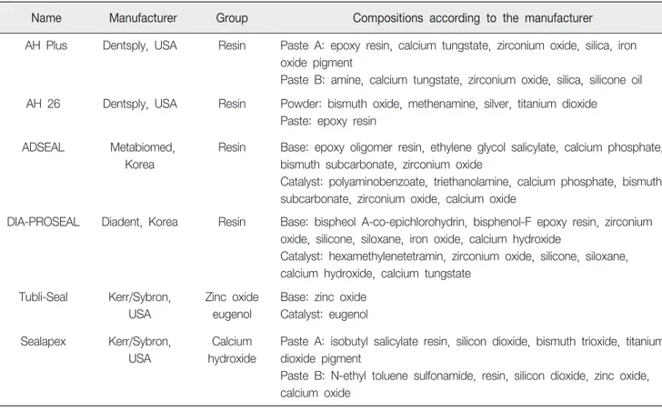
1. 연구재료 본 연구에서는 2가지의 연고형 또는 분말형 제재를 섞은 후 시간이 지남에 따라 경화되는 근관용 실러를 대상으로 시 험하였으며, 시험 물질에 대한 제조자 제공 정보는 표1에 기 재하였다. 한천중층시험에 적용한 세포는 L929 mouse fibroblast (한 국세포주은행, KCLB No.10001)였으며 RPMI 1640 배지 (Gibco, Grand island, USA)에 10% fetal bovine serum (FBS, Gibco), 1% 페니실린 (Gibco)을 혼합하여 배양액으로 사용하 였다. 2. 연구방법 1) 시편준비 시험 재료는 내경 5 mm, 높이 2 mm의 테플론 링 안에 넣어 한천중층시험용 시편을 준비하였다. 표 1에 기재된 6가 지 근관용 실러를 제조자의 지시에 따라 혼합한 직후 또는 37 ℃, 100% 상대습도 용기 내에서 24시간 경화시킨 것으로 나누어 시험하였다. 혼합 직후의 근관용 실러를 적용할 때는 한천 배지 위에 테플론 링을 먼저 놓고 내부를 재료로 채워 넣었다. 경화된 시편을 적용할 때는 페트리디시에 근관용 실 러로 채운 테플론 링을 놓고 주어진 온도와 습도 조건에서 경화시킨 후, 본 시험 시 한천 배지 위에 시편을 올려 놓았다. ISO 10993-12의 제시에 따라 양성 및 음성 대조군은 라텍스 고무와 폴리에틸렌 필름으로 설정하였으며, 각각 직경 5 mm 로 잘라 고압멸균소독 후 시험에 사용하였다.Table 1. Root canal sealers used in this study
Name Manufacturer Group Compositions according to the manufacturer
AH Plus Dentsply, USA Resin Paste A: epoxy resin, calcium tungstate, zirconium oxide, silica, iron oxide pigment
Paste B: amine, calcium tungstate, zirconium oxide, silica, silicone oil AH 26 Dentsply, USA Resin Powder: bismuth oxide, methenamine, silver, titanium dioxide
Paste: epoxy resin ADSEAL Metabiomed,
Korea
Resin Base: epoxy oligomer resin, ethylene glycol salicylate, calcium phosphate, bismuth subcarbonate, zirconium oxide
Catalyst: polyaminobenzoate, triethanolamine, calcium phosphate, bismuth subcarbonate, zirconium oxide, calcium oxide
DIA-PROSEAL Diadent, Korea Resin Base: bispheol A-co-epichlorohydrin, bisphenol-F epoxy resin, zirconium oxide, silicone, siloxane, iron oxide, calcium hydroxide
Catalyst: hexamethylenetetramin, zirconium oxide, silicone, siloxane, calcium hydroxide, calcium tungstate
Tubli-Seal Kerr/Sybron, USA
Zinc oxide eugenol
Base: zinc oxide Catalyst: eugenol Sealapex Kerr/Sybron,
USA
Calcium hydroxide
Paste A: isobutyl salicylate resin, silicon dioxide, bismuth trioxide, titanium dioxide pigment
Paste B: N-ethyl toluene sulfonamide, resin, silicon dioxide, zinc oxide, calcium oxide
Table 2. Definition of zone/lysis index values
Index Description
Zone index 0 No detectable zone around or under sample 1 Zone limited to area under sample
2 Zone not > 0.5 cm in extension from sample 3 Zone not > 1 cm in extension from sample 4 Zone > 1 cm in extension from sample 5 Zone involving entire plate
Lysis index 0 No observable lysis
1 Up to 20% of zone lysed 2 20-40% of zone lysed 3 40-60% of zone lysed 4 60-80% of zone lysed 5 Over 80% lysed within zone
2) 한천중층시험 시험 방법은 ISO 7405의 한천중층시험법을 따랐다. 2 X 10⁶개의 L929세포를 직경 100 mm 세포배양용기에서 24시간 단층배양한다. 액체배지를 걷어내고 100 ℃에서 멸균 후 48 ℃로 식힌 3% 한천액과 2배로 농축된 세포배양액을 1:1로 혼 합한 후 48 ℃로 가열한다. 이 혼합액 10 mL를 세포 위에 놓고 약 30분간 굳힌다. 10 mL의 뉴트럴레드 (neutral red, Sigma, St. Louis, USA) 염색시약을 15분 가량 놓고 세포를 염색시킨다. 굳은 한천 배지 위에 양성대조군, 음성대조군, 그리고 시험재료를 서로 20 mm 이상 간격을 두고 올려놓는 다. 24시간이 지난 후 세포배양용기를 백지 위에 놓고 재료 하방의 붉은 염색약이 변색된 범위를 자 (Mitutoyo, Kanagawa, Japan)로 측정하여 표 2에 따라 탈색지수 (Decoloration index) 를 정한다. 또한 역위상차현미경 (Nikon ECLIPSE TS100-F, Tokyo, Japan)으로 탈색된 범위 내에서 세포가 사멸된 비율을 측정하여 사멸지수 (Lysis index)를 구한다. 탈색지수와 세포사 멸지수 각각의 세포반응지수를 표 3에 따라 기재하고 세포독성 을 평가하였다. 동일한 시험을 독립적인 조건에서 4번씩 반복 실험하였다.
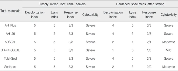
Ⅲ. RESULTS
한천중층시험 후 6가지 근관용 실러의 탈색지수, 세포사멸 지수 및 세포반응지수와 이의 해석결과를 표 4에 기재하였다. 혼합 직후에 적용한 근관용 실러는 모두 탈색지수 5, 세포사멸 지수 5를 나타내었다. 그러나 경화 후 시편으로 시험하였을 때는 AH Plus, AH 26, 그리고 Tubli-Seal은 모두 탈색지수 4,Table 3. Cell response
Scale Cell response Interpretation
0 0 Non cytotoxic
1 1 Mildly cytotoxic
2 2 to 3 Moderately cytotoxic
3 4 to 5 Severly cytotoxic
Table 4. Agar overlay test results of six kinds of root canal sealers before and after setting procedures
Test materials
Freshly mixed root canal sealers Hardened specimens after setting Decolorization index Lysis index Response index Cytotoxicity Decolorization index Lysis index Response index Cytotoxicity
AH Plus 5 5 3/3 Severe 4 5 3/3 Severe
AH 26 5 5 3/3 Severe 4 5 3/3 Severe
ADSEAL 5 5 3/3 Severe 2 1 2/1 Moderate
DIA-PROSEAL 5 5 3/3 Severe 1 0 1/0 Mild
Tubli-Seal 5 5 3/3 Severe 4 5 3/3 Severe
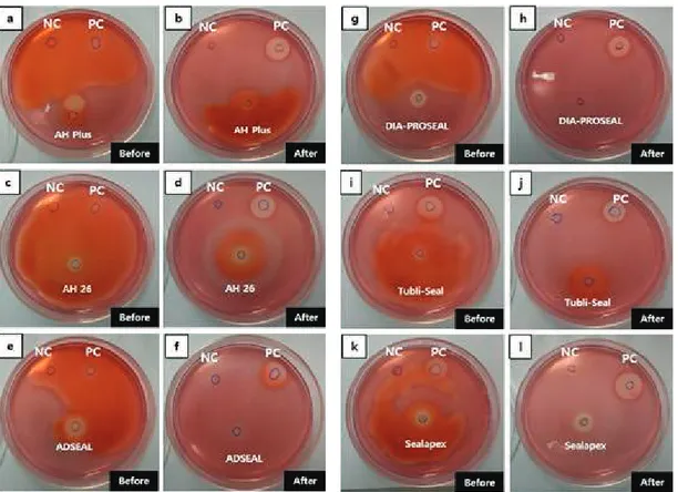
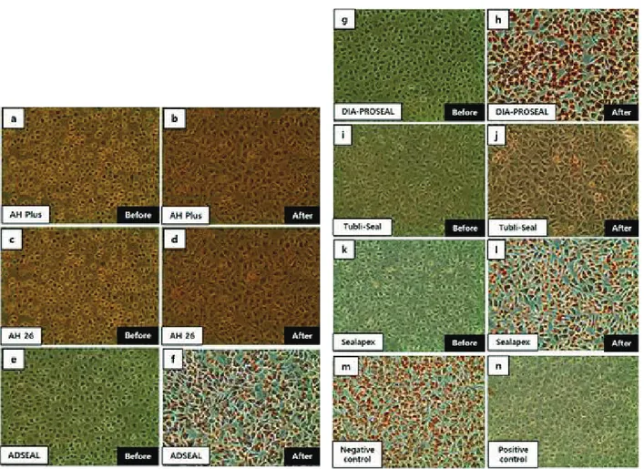
세포사멸지수 5였고, ADSEAL과 DIA-PROSEAL은 각각 2와 1, 그리고 1과 0의 수치를 보였다. Sealapex는 ADSEAL과 유사하 게 탈색지수가 2였으나, 세포사멸지수는 3을 나타냈다. 따라 서 혼합 직후의 근관용 실러는 모두 심한 (severe) 세포 독성으 로 판단되었고, 경화 후에는 DIA-PROSEAL이 경미 (mild)한 세포 독성, ADSEAL과 Sealapex가 중등도 (moderate) 세포 독 성을 나타냈다. 그 외의 3가지 근관용 실러는 경화 후에도 모 두 심한 세포 독성 상태로 판단되었다. 한천중층시험 후 탈색 된 양상은 그림 1에 나타내었다. 그림 2에 나타난 세포의 현미 경 사진에서는, 탈색지수 및 세포사멸지수가 큰 재료를 적용할 때 세포가 탈색되고 원형에 가까운 모양을 나타냈으며, 시편 경화 후 적용한 DIA-PROSEAL (그림 2, h)은 음성대조군과 유사한 세포 형태를 나타내었다.
Ⅳ. DISCUSSION
한천중층시험법은 일정한 두께의 고체 한천 배지를 투과하 는 시험물질의 독성 성분으로 하방의 세포가 영향을 받아 염 색된 핵 부위가 탈색되고 세포사멸현상을 일으키는 정도를 평가하여 재료의 세포독성을 가늠하는 시험법이다. 고체 시 편인 경우에는 동일한 크기와 형태를 가진 양성 그리고 음성 대조군과 함께 시험 시 세포 독성을 평가할 수 있고, 액체일 경우에는 일정한 면적을 가진 흡습지를 이용하여 재료가 충분 히 스며들게 한 후 다른 재료와 비교 가능하다. 그러나 시간이 지남에 따라 경화되는 재료는 굳기 전과 후의 독성 정도가 다르게 평가될 수 있으며, 근관용 실러의 경우에는 적용되는 임상적 상황을 고려한 시험 방법이 필요하기 때문에 한천중층 시험을 재료 혼합 직후 그리고 충분히 경화시킨 후로 나누어 시험하는 것이 적합하다. 근관용 실러는 제품에 따라 경화시 간이 수 분에서 수 시간으로 다양하고 이로 인해 미경화 및 경화된 실러에 의한 세포 독성이 나타날 수 있다 (Lodiene 등, 2013; Silva 등, 2013). 또한 술자가 근관용 실러를 적용하 는 술식과 실러 적용 시 근단부에 가하는 힘, 재료의 경화 전후 물리적 특성 등에 따라 미경화 또는 경화된 실러가 세포 및 조직에 영향을 끼치게 된다. 근관 충전 과정 중 미경화 실러가 근단공을 빠져나가면 직접적으로 치근단 세포나 인접 조직에 영향을 줄 수 있고, 경화된 실러는 조직액에 의해 지속 적인 용출 작용이 일어나 하방 조직에 영향을 줄 수 있다 (Tanaka 등, 1991; Ferracane, 1994; Willershausen 등, 2000; Huang 등, 2002). 따라서 본 연구에서는 혼합 직후 재료의 적용부터 근관 내에서 경화 후까지의 두 가지 조건의 경우를 모두 적용할 수 있는 한천중층시험을 세포 독성 평가방법으로 선택하였다. 시험 결과에서 혼합 직후의 근관용 실러는 모두 심한 세포 독성 양상을 보였다. 제조자 지시 정보에 의하면 현재 사용된 실러의 경우 최소 50분 (ADSEAL)에서 최대 8시간 (AH Plus) 까지의 경화 시간이 필요하고, 혼합 직후 한천중층시험을 시 행하면 표 1에 기재된 각 성분의 화합물 또는 혼합물이 액체 상태로 고체 한천 배지를 통과할 수 있다. 현재 사용된 6종의 근관용 실러 성분 중, 각종 단량체, 유지놀, 수산화칼슘, 그 외 무기 및 유기 화합물 등이 기존 연구에서 세포 독성을 유발 한다고 알려져 있다 (Gulati 등, 1991; Reyes-Carmona 등, 2011; Lodiene 등, 2013; Silva 등, 2013; Mohammadi 등, 2014; Dianat 등, 2015; Kaur 등, 2015). 그리고 혼합 직후 실러는 점성을 가진 액체 형태이기 때문에 하방 세포의 넓은 부위까 지 퍼져나갈 수 있어서 독성을 나타내는 범위가 더욱 크게 나타났다. 경화 후 시편을 적용할 때는 경도에서 심한 정도의 다양한 세포 독성 결과를 보였다. 완전히 경화된 레진 계열 실러는 중합의 정도나 잔류 단량체의 용출 정도에 따라 다양한 세포 독성을 보인 것으로 분석된다. 특히 DIA-PROSEAL은 경화 후 용출되는 성분의 세포 독성이 타 레진 제품에 비해 낮았으며, 이의 용해도 및 성분 분석 시험이 추가적으로 이루어져야 할 것이다. 산화아연유지놀 계열 실러는 경화 과정에서 산화아 연이 유지놀과 혼합되면서 Zinc eugenolate를 형성한다. 치아 의 수분은 이를 다시 분해시켜 유지놀 성분을 상아세관으로 방출시킨다. 또한 경화 후에도 유지놀은 약산인 페놀 성분을 함유하고 있으며 진정, 마취 작용 효과를 나타낸다고 알려져 있지만, 이의 세포 독성도 다수 보고되었다 (Gulati 등, 1991; Markowitz 등, 1992; Azar 등, 2000). 따라서 경화된 Tubli-Seal 은 이런 유지놀 성분이 경화 전후 모두 하방 세포에 영향을 끼쳐 심한 세포 독성을 보인 것으로 추정된다. Sealapex은 수산화칼슘 계열로 분류하지만, 레진 성분도 동시에 함유하고있는 것으로 알려져 있다. 따라서 경화 후에도 수산화칼슘을 포함한 용출 성분과 잔류 레진 단량체가 세포 독성을 일으킨 것으로 사료된다. 수산화칼슘은 pH를 11정도의 알칼리성 상 태로 만들어 세포 독성을 야기할 수 있지만, 경조직 재생을 증가시킨다고 알려진 재료이기 때문에 이런 치료적인 효과를 지닌 재료는 골 재생과 관련한 추가 실험으로 종합적인 재료 의 위해성 판단이 필요하다 (Ji 등, 2010; Paula-Silva 등, 2010). 경화된 근관용 실러의 경우 재료 종류에 상관 없이 심한 세포 독성을 나타냈으며, 경화 후에는 각 재료의 용출 성분에 의해 세포 독성 정도가 달라졌다. 따라서 시적할 수 있는 형태 로 혼합된 미경화 실러의 각 성분에 대한 독성 평가 및 성분의 개선이 필요하며, 경화 후에 용출되는 성분에 대한 장기적인 영향의 검토가 필요하다. 또한 실험에 사용된 모든 근관용 실러는 경화 후에 탈색지수 또는 세포사멸지수가 감소하였기 때문에 제품의 경화 시간을 짧게 만드는 것이 세포 및 조직에 대한 독성 물질의 영향을 감소시킬 수 있을 것이다. 그러나 본 연구에서 사용한 한천중층시험법은 치료과정 중에 근관용 실러가 하방 세포나 조직과 접하는 표면적을 고려하지 않고, 조직액의 흐름에 의한 용출물 희석 효과를 간과한다는 단점이 있다. 그리고 재료와 세포의 거리는 고체 한천 배지 10 mL의 두께만큼 일괄적으로 정해 놓기 때문에 치과 치료 중 상황을 반영하지 못한다는 한계도 있다. 따라서 앞으로 이런 임상 환경을 실험실 내에서 더욱 잘 재현할 수 있도록 추가적인 실험 모델의 제작이 필요하고, 치과 치료의 특수성을 고려한 재료의 독성 평가 방법에 대한 연구가 이루어져야 할 것이다.
Figure 1. Decolorization zones of six root canal sealers before and afterREM수면setting. (a) AH Plus before hardening, (b) AH Plus after hardening, (c) AH 26 before hardening, (d) AH 26 after hardening, (e) ADSEAL before hardening, (f) ADSEAL after hardening, (g) DIA-PROSEAL before hardening, (h) DIA-PROSEAL after hardening, (i) Tubli-Seal before hardening, (j) Tubli-Seal after hardening, (k) Sealapex before hardening, (l) Sealapex after hardening. NC: negative control, PC: positive control.
Ⅴ. CONCLUSION
본 연구에서는 한천중층시험법으로 혼합 직후 또는 경화된 형태의 산화아연유지놀 계열, 수산화칼슘 계열, 그리고 레진 계열 실러의 세포 독성을 평가하였다. 이 시험법을 통해 탈색 지수와 세포사멸지수 및 이에 따른 세포 반응을 평가하였고 다음과 같은 결과를 얻었다. 1. 모든 종류의 근관용 실러는 혼합 직후 적용하였을 때 심한 세포 독성을 나타냈지만, 경화 후 적용하였을 때는 탈색 또는 세포사멸지수가 감소하였다.2. 경화 후 세포 독성은 DIA-PROSEAL < ADSEAL, Sealapex < AH PLUS, AH 26, Tubli-Seal의 순서로 높아졌다. 본 연구에서 사용된 재료 중 DIA-PROSEAL은 경화 후 최종 산물에 의한 세포 독성이 가장 낮은 결과를 보였고, 현 6종 재료 중 가장 생체친화성이 높은 재료로 사용 가능하다. 근관 용 실러의 개발과 평가에 있어서 임상적 상황을 고려한 경화 전/후의 독성 평가는 모두 이루어져야 하며, 앞으로 치과 재료 의 실제 적용법을 고려한 독성 평가용 실험 모델의 개발이
Figure 2. Cellular changes after agar overlay test of six root canal sealers before and after setting. (a) AH Plus before hardening, (b) AH Plus after hardening, (c) AH 26 before hardening, (d) AH 26 after hardening, (e) ADSEAL before hardening, (f) ADSEAL after hardening, (g) DIA-PROSEAL before hardening, (h) DIA-PROSEAL after hardening, (i) Tubli-Seal before hardening, (j) Tubli-Seal after hardening, (k) Sealapex before hardening, (l) Sealapex after hardening, (m) negative control, (n) positive control. (X 100 microscopic examinations)
이루어져야 할 것이다. * 본 연구는 2016년 연세대학교 치과생체재료공학연구소 지원 하에 수행되었습니다. * ㈜신흥, ㈜메타바이오메드, ㈜베리콤이 실험 재료를 제공 하였으며 이에 감사드립니다.
Ⅵ. REFERENCES
Azar NG, Heidari M, Bahrami ZS, Shokri F (2000). In vitro cytotoxicity of a new epoxy resin root canal sealer. J Endod 26(8), 462-465.
Bernabe PF, Holland R, Morandi R, de Souza V, Nery MJ, Otoboni Filho JA, Dezan Junior E, Gomes-Filho JE (2005). Comparative study of MTA and other materials in retrofilling of pulpless dogs' teeth. Braz Dent J 16(2), 149-155.
Bouillaguet S, Wataha JC, Lockwood PE, Galgano C, Golay A, Krejci I (2004). Cytotoxicity and sealing properties of four classes of endodontic sealers evaluated by succinic dehydrogenase activity and confocal laser scanning microscopy. Eur J Oral Sci 112(2), 182-187. Branstetter J, von Fraunhofer JA (1982). The physical
properties and sealing action of endodontic sealer cements: a review of the literature. J Endod 8(7), 312-316. Bryan TE, Khechen K, Brackett MG, Messer RL, El-Awady
A, Primus CM, Gutmann JL, Tay FR (2010). In vitro osteogenic potential of an experimental calcium silicate-based root canal sealer. J Endod 36(7), 1163- 1169.
Dianat O, Azadnia S, Mozayeni MA (2015). Toxicity of calcium hydroxide nanoparticles on murine fibroblast cell line. Iran Endod J 10(1), 49-54.
Eldeniz AU, Mustafa K, Orstavik D, Dahl JE (2007). Cytotoxicity of new resin-, calcium hydroxide- and silicone-based root canal sealers on fibroblasts derived
from human gingiva and L929 cell lines. Int Endod J 40(5), 329-337.
Ferracane JL (1994). Elution of leachable components from composites. J Oral Rehabil 21(4), 441-452.
Gomes-Filho JE, Watanabe S, Bernabe PF, de Moraes Costa MT (2009). A mineral trioxide aggregate sealer stimulated mineralization. J Endod 35(2), 256-260. Gomes-Filho JE, Watanabe S, Cintra LT, Nery MJ,
Dezan-Junior E, Queiroz IO, Lodi CS, Basso MD (2013). Effect of MTA-based sealer on the healing of periapical lesions. J Appl Oral Sci 21(3), 235-242.
Grossman LI (1958). An improved root canal cement. J Am Dent Assoc 56(3), 381-385.
Gulati N, Chandra S, Aggarwal PK, Jaiswal JN, Singh M (1991). Cytotoxicity of eugenol in sealer containing zinc-oxide. Endod Dent Traumatol 7(4), 181-185. Huang FM, Tai KW, Chou MY, Chang YC (2002).
Cytotoxicity of resin-, zinc oxide-eugenol-, and calcium hydroxide-based root canal sealers on human periodontal ligament cells and permanent V79 cells. Int Endod J 35(2), 153-158.
Ji YM, Jeon SH, Park JY, Chung JH, Choung YH, Choung PH (2010). Dental stem cell therapy with calcium hydroxide in dental pulp capping. Tissue Eng Part A 16(6), 1823-1833.
Kaur A, Shah N, Logani A, Mishra N (2015). Biotoxicity of commonly used root canal sealers: A meta-analysis. J Conserv Dent 18(2), 83-88.
Kim EC, Lee BC, Chang HS, Lee W, Hong CU, Min KS (2008). Evaluation of the radiopacity and cytotoxicity of Portland cements containing bismuth oxide. Oral Surg Oral Med Oral Pathol Oral Radiol Endod 105(1), e54-57.
Lodiene G, Kopperud HM, Orstavik D, Bruzell EM (2013). Detection of leachables and cytotoxicity after exposure to methacrylate- and epoxy-based root canal sealers in vitro. Eur J Oral Sci 121(5), 488-496.
Lopez-Lopez J, Estrugo-Devesa A, Jane-Salas E, Segura-Egea JJ (2012). Inferior alveolar nerve injury resulting from overextension of an endodontic sealer: non-surgical management using the GABA analogue pregabalin. International Endodontic Journal 45(1), 98-104. Love RM, Firth N (2009). Histopathological profile of
surgically removed persistent periapical radiolucent lesions of endodontic origin. Int Endod J 42(3), 198- 202.
Markowitz K, Moynihan M, Liu M, Kim S (1992). Biologic properties of eugenol and zinc oxide-eugenol. A clinically oriented review. Oral Surg Oral Med Oral Pathol 73(6), 729-737.
Martins VJ, Lins RX, Berlinck TC, Fidel RA (2013). Cytotoxicity of root canal sealers on endothelial cell cultures. Braz Dent J 24(1), 15-20.
Mohammadi Z, Karim Soltani M, Shalavi S, Yazdizadeh M, Jafarzadeh M (2014). Calcium hydroxide-based root canal sealers: an updated literature review. Compend Contin Educ Dent 35(5), 334-339; quiz 340.
Paula-Silva FW, Ghosh A, Arzate H, Kapila S, da Silva LA, Kapila YL (2010). Calcium hydroxide promotes cementogenesis and induces cementoblastic differentiation of mesenchymal periodontal ligament cells in a CEMP1- and ERK-dependent manner. Calcif Tissue Int 87(2), 144-157.
Reyes-Carmona JF, Santos AR, Figueiredo CP, Felippe MS, Felippe WT, Cordeiro MM (2011). In vivo host interactions with mineral trioxide aggregate and
calcium hydroxide: inflammatory molecular signaling assessment. J Endod 37(9), 1225-1235.
Sari S, Duruturk L (2007). Radiographic evaluation of periapical healing of permanent teeth with periapical lesions after extrusion of AH Plus sealer. Oral Surg Oral Med Oral Pathol Oral Radiol Endod 104(3), e54-59. Scelza MZ, Coil J, Alves GG (2012). Effect of time of
extraction on the biocompatibility of endodontic sealers with primary human fibroblasts. Braz Oral Res 26(5), 424-430.
Schwarze T, Leyhausen G, Geurtsen W (2002). Long-term cytocompatibility of various endodontic sealers using a new root canal model. Journal of Endodontics 28(11), 749-753.
Silva EJ, Santos CC, Zaia AA (2013). Long-term cytotoxic effects of contemporary root canal sealers. J Appl Oral Sci 21(1), 43-47.
Susini G, About I, Tran-Hung L, Camps J (2006). Cytotoxicity of Epiphany and Resilon with a root model. Int Endod J 39(12), 940-944.
Tanaka K, Taira M, Shintani H, Wakasa K, Yamaki M (1991). Residual monomers (TEGDMA and Bis-GMA) of a set visible-light-cured dental composite resin when immersed in water. J Oral Rehabil 18(4), 353-362. Willershausen B, Marroquin BB, Schafer D, Schulze R
(2000). Cytotoxicity of root canal filling materials to three different human cell lines. J Endod 26(12), 703-707.