저작자표시-비영리-변경금지 2.0 대한민국 이용자는 아래의 조건을 따르는 경우에 한하여 자유롭게 l 이 저작물을 복제, 배포, 전송, 전시, 공연 및 방송할 수 있습니다. 다음과 같은 조건을 따라야 합니다: l 귀하는, 이 저작물의 재이용이나 배포의 경우, 이 저작물에 적용된 이용허락조건 을 명확하게 나타내어야 합니다. l 저작권자로부터 별도의 허가를 받으면 이러한 조건들은 적용되지 않습니다. 저작권법에 따른 이용자의 권리는 위의 내용에 의하여 영향을 받지 않습니다. 이것은 이용허락규약(Legal Code)을 이해하기 쉽게 요약한 것입니다. Disclaimer 저작자표시. 귀하는 원저작자를 표시하여야 합니다. 비영리. 귀하는 이 저작물을 영리 목적으로 이용할 수 없습니다. 변경금지. 귀하는 이 저작물을 개작, 변형 또는 가공할 수 없습니다.
A Dissertation for the Degree of Master of Science
Antioxidant hesperetin improves the
quality of porcine oocytes during
aging in vitro
Won-Jae Kim
Department of Biotechnology
GRADUATE SCHOOL
JEJU NATIONAL UNIVERSITY
항산화제 헤스페레틴이 돼지
난자의 노화기간 동안 난자의
미치는 영향 연구
지도교수 박세필
김 원 재
이 논문을 이학 석사학위 논문으로 제출함
제주대학교 대학원
2019 년 2 월
Antioxidant hesperetin improves the
quality of porcine oocytes during
aging in vitro
Won-Jae Kim
(Supervised by Professor Se-Pill Park)
A thesis submitted as a Qualified Dissertation
for the Degree of Master of Science
February, 2019
Department of Biotechnology
GRADUATE SCHOOL
JEJU NATIONAL UNIVERSITY
Approved as a Qualified Dissertation
for the Degree of Master of Science
Dissertation Committee:
Chairperson of the supervising committee
Professor Key-Zung Riu, Ph.D., College of Applied Life Sciences, Jeju National University
Professor Eun-Young Kim, Ph.D., College of Applied Life Sciences, Jeju National University
Professor Se-Pill Park, Ph.D., College of Applied Life Sciences, Jeju National University
Department of Biotechnology
GRADUATE SCHOOL
JEJU NATIONAL UNIVERSITY
i
CONTENTS
C O N T E N T S … … … . i
LIST OF FIGURES……… .ⅲ
LIST OF TABLES……… ...ⅳ
AB S T RACT … … … …… … …… … … … …… … …… … … … . . .1
1. INTRODUCTION……… .2
2
. M A T E R I A L S A N D M E T H O D S … … … . . 4
2.1. Chemicals and reagents……….………...4
2.2. IVM and aging of porcine oocytes ……….……….4
2.3. Hesperetin treatment ………..………..4
2.4. PA and embryo culture ………...5
2.5. Measurement of intracellular ROS and GSH levels .……….5
2.6. Terminal deoxynucleotidyl transferase dUTP nick end labeling (TUNEL) assay and
Hoechst staining ………..6
2.7. mRNA extraction and cDNA synthesis …………..………...6
2.8. Real-time RT-PCR ………..……….…...……….7
2.9. Western blot analysis ………...………8
2.9. Statistical analysis………..………...…………. .9
3 . R E S U L T S … … … . . . 1 0
3.1 Hesperetin enhances the in vitro development of aging porcine oocytes ………....10
3.2. Hesperetin reduces the level of ROS in aging porcine oocytes in vitro
………..……….12
3.3. Hesperetin prevents aberrant spindle organization and chromosome misalignment in
aging porcine oocytes in vitro ………...………….………..14
3.4. Hesperetin increases expression of cytoplasmic maturation markers in aging porcine
oocytes in vitro ……….………...16
3.5. Hesperetin increases expression of estrogen receptor genes in aging porcine oocytes
in vitro ………..…….18
ii
3.6.
Hesperetin improves the developmental capacity and quality of embryos derived from agingporcine oocytes in vitro………..20
4. DISCUSSION……… ...22
REFERENCES……… ..………25
ABSTRACT IN KOREAN………..……….30
iii
LIST OF FIGURES
Fig. 1. Effect of hesperetin on the level of oxidative stress in porcine oocytes during aging in vitro. Fig. 2. Effect of hesperetin on spindle morphology in porcine oocytes during aging in vitro.
Fig. 3. Effect of hesperetin on MAPK activity and maternal gene expression in porcine oocytes during
aging in vitro.
Fig. 4. Effect of hesperetin on expression of estrogen receptor genes in porcine oocytes during aging
in vitro.
Fig. 5. Effect of hesperetin treatment during aging of porcine oocytes in vitro on subsequent embryo
iv
LIST OF TABLES
Table 1. Primers used for real-time RT-PCR
Table 2. Effect of hesperetin treatment during aging of porcine oocytes in vitro on subsequent embryo
1
ABSTRACT
The citrus flavonoid hesperetin has a variety of pharmacological actions, including antioxidant, anti-inflammatory, and anticancer activities. This study investigated whether hesperetin prevents aging of oocytes in vitro in which it determined the maturation of nuclear and cytoplasm and the developmental capacity of embryo by modulating the ROS level. Porcine oocytes were matured in
vitro for 44 h (control) and for an additional 24 h in the presence of 0, 1, 10, 100, and 250 μM
hesperetin (aging, H-1, H-10, H-100, and H-250, respectively). Although there was no difference in the rate of maturation among all the groups, both the control and H-100 groups significantly increased in the rate of cleavage and blastocyst formation compared to the aging group (p < 0.01). The H-100 group significantly decreased reactive oxygen species (ROS) activity and increases the level of glutathione (GSH) and expression of the antioxidant genes (PRDX5, NFE2L, SOD1 and SOD2) compared to the aging group. The H-100 groups prevented aberrant spindle organization and chromosomal misalignment, blocked the decrease in the level of phosphorylated-p44/42 mitogen-activated protein kinase (MAPK) and increased the mRNA expression of cytoplasmic maturation factor genes (GDF9, CCNB1, BMP15 and MOS). Subsequently, both the control and H-100 groups significantly increased the total cell number and decreased the apoptosis cells at the blastocyst stage compared to aging group. The results indicate that hesperetin improves the quality of porcine oocytes by protecting them against oxidative stress during aging in vitro.
2
1. INTRODUCTION
In vitro maturation (IVM) is a technique used to mature oocytes. It can be offered to women with
infertility problems, combined with IVF, offering women pregnancy without ovarian stimulation. A healthy oocyte is important for successful fertilization, embryo development, and embryo implantation after transfer (Q. Wang & Sun, 2007). Mature mammalian oocytes are arrested in metaphase of the second meiosis (MII) following ovulation. Despite great advances in assisted reproductive technologies, these approaches often fail due to oocyte aging (Miao, Kikuchi, Sun, & Schatten, 2009). Aging oocytes frequently display spindle aberrations, disturbances in chromosome congression, mitochondrial alterations, and changes in gene and protein expression (Eichenlaub-Ritter, 2012). Consequently, many researchers have sought to develop methods that can protect oocytes against aging in vitro.
Oocyte aging is characterized by a deterioration in oocyte quality, and negatively affects fertilization and subsequent embryo development (Miao et al., 2009). Aging in vitro is associated with increased levels of ROS such as hydrogen peroxide, superoxide, and the hydroxyl radical (Chaube, Prasad, Thakur, & Shrivastav, 2005; Goud, Goud, Diamond, Gonik, & Abu-Soud, 2008). The level of ROS correlates with oocyte age (Lim & Luderer, 2011; Takeo, Kimura, Shirasuna, Kuwayama, & Iwata, 2017). Accumulation of ROS influences apoptosis (Orrenius, Gogvadze, & Zhivotovsky, 2007), spindle assembly (Rajani et al., 2012), and lipid, protein, and DNA damage (Bomfim et al., 2017; Patra, Rautray, & Swarup, 2011). Consequently, ROS may affect follicular oocyte growth. ROS damage oocytes and thereby decrease their developmental competence (Goto, Noda, Mori, & Nakano,
1993). The balance between ROS and antioxidants influences aging in vitro. Accumulation of ROS
promotes aging (Finkel & Holbrook, 2000). ROS may be a major cause of oocyte aging and poor oocyte quality in patients stimulated with ovulation drug therapies for long durations.
3
Hesperetin is a naturally occurring flavanone-glycoside and is the main flavonoid in lemons and sweet oranges. This compound inhibits oxidases more strongly than its glycosylate derivatives (de Souza et al., 2016). Hesperidin is structurally a flavanone β‐glycoside at C‐7 of hesperetin aglycone,
which is 3′, 5, 7‐trihydroxy‐4′‐methoxy flavanone.Hesperetin acts protects against oxidative stress via
estrogen receptor mediated actions (Hwang, Lin, Shih, Yeh, & Yen, 2012). This compound has a wide range of pharmacological actions, including antioxidant (J. Wang, Zhu, Yang, & Liu, 2013), anti‐ inflammatory (Hirata, Murakami, Shoji, Kadoma, & Fujisawa, 2005), and anticancer (Zarebczan, Pinchot, Kunnimalaiyaan, & Chen, 2011) activities. However, it is unknown whether hesperetin acts as an antioxidant in porcine oocytes.
This study investigated the effects of appropriate concentrations of hesperetin on antioxidate porcine oocytes during aging in vitro. We analyzed the level of ROS, expression of antioxidant, maternal, and estrogen receptor genes, and spindle morphology in aging porcine oocytes treated with or without hesperetin. In addition, we determined the developmental competence and quality of embryos derived from these oocytes. Our results demonstrate that hesperetin protects oocytes against oxidative stress during aging in vitro and thereby prevents deterioration in their quality. These findings may be applicable to in vitro fertilization and help prevent aging of oocytes in vitro.
4
2. MATERIALS & METHODS
2.1. Chemicals and reagents
All chemicals and reagents were purchased from Sigma (St. Louis, MO, USA) unless stated otherwise.
2.2. IVM and aging of porcine oocytes
Pre-pubertal porcine ovaries were collected from a local slaughterhouse and transported to the laboratory in saline supplemented with 75 μg/mL penicillin G and 50 μg/mL streptomycin sulfate within 2 h at 30–33°C. Cumulus-oocyte complexes (COCs) were aspirated from follicles with a diameter of 2–8 mm using an 18-gauge needle and a disposable 10 mL syringe. COCs were washed three times in tissue culture medium (TCM)-199-HEPES containing 0.1% (w/v) bovine serum albumin (BSA). Thereafter, COCs were matured in groups of 50 in 500 μL TCM-199 (Gibco, Grand Island, NY, USA) containing Earle’s salts, 0.57 mM cysteine, 10 ng/mL epidermal growth factor, 0.5 μg/mL follicle-stimulating hormone, 0.5 μg/mL luteinizing hormone, and 10% (v/v) porcine follicular fluid under mineral oil for 44 h at 38.8°C in 5% CO2 and 95% air. Oocyte aging was induced by
culturing COCs for an additional 24 h in TCM-199 (Sigma).
2.3. Hesperetin treatment
Mature oocytes were covered with mineral oil and cultured in wells of a four-well multi-dish containing 500 mL TCM-199 at 38.8°C in a humidified atmosphere of 5% CO2 and 95% air. After
5
maturation, MII stage oocytes were transferred into TCM-199 containing 0, 1, 10, 100, and 250 μM hesperetin (Sigma) and cultured for 24 (68 for in vitro maturation [IVM]) as described above. After treatment, oocytes were collected and aging was assessed.
2.4. PA and embryo culture
Following maturation, cumulus cells were removed by pipetting in the presence of 1 mg/mL hyaluronidase for 2–3 min. PA was induced by treating oocytes in porcine zygote medium (PZM)-5 containing 0.4% (w/v) BSA (IVC medium) with 5 μM Ca2+ ionomycin (Sigma) for 5 min. After 3 h of culture in IVC medium containing 7.5 μg/mL cytochalasin B (Sigma), embryos were washed three times in the same medium and cultured for 7 days at 38.8°C in a humidified atmosphere of 5% CO2 and 95% air. Oocytes and embryos were washed in Dulbecco’s phosphate-buffered saline (DPBS) and eitherfixed in 3.7% (w/v) paraformaldehyde for 20 min and stored at 4°C, or snap-frozen in liquid nitrogen and stored at 70°C, depending on the experiment.
2.5. Measurement of intracellular ROS and GSH levels
DCFHDA and CMF2HC were used to determine the intracellular levels of ROS and GSH,
respectively, as previously described (H. W. Yang et al., 1998; You, Kim, Lim, & Lee, 2010) with slight modifications. Briefly, cumulus cells were removed from COCs by pipetting in the presence of 0.1% (w/v) hyaluronidase. Denuded oocytes were incubated in DPBS containing 50 μM DCFHDA or 100 μM CMF2HC in the dark for 20 min at 38.8°C. Thereafter, oocytes were washed more than five
times with DPBS containing 0.1% (w/v) BSA to completely remove excess dye and immediately analyzed by epifluorescence microscopy (Olympus, Tokyo, Japan). The ROS level was measured using excitation and emission wavelengths of 450–490 nm and 515–565 nm, respectively. The excitation and emission wavelengths of CMF2HC are 371 and 464 nm, respectively. Grayscale images
6
were acquired with a digital camera (Nikon, Tokyo, Japan) attached to the microscope, and mean grayscale values were calculated using Image J software (NIH, Bethesda, MD, USA). Background fluorescence values were subtracted from the final values prior to statistical analysis. The experiment was independently repeated 6-7 times with 10–20 oocytes per experiment.
2.6. Terminal deoxynucleotidyl transferase dUTP nick end labeling (TUNEL) assay and Hoechst staining
At 7 days after PA, blastocysts were fixed, washed more than three times with PBS containing 0.1% BSA, and then incubated with 0.1% Triton X-100 at 38.8°C for 30 min. Blastocysts were incubated with fluoresceinconjugated dUTP and terminal deoxynucleotidyl transferase (In Situ Cell Death Detection Kit, Roche, Manheim, Germany) in the dark for 1 h at 38.8°C. Mitotic and apoptotic cells were scored. Nuclei were stained with Hoechst 33342 (1 μg/ml) for 30 min, and embryos were washed with PBS containing 0.1% BSA. Blastocysts were mounted onto glass slides and examined under an inverted Olympus IX-71 fluorescence microscope. The experiment was independently repeated four times.
2.7. mRNA extraction and cDNA synthesis
mRNA was isolated from more than three biological replicates, with 30–40 oocytes per replicate, using a Dynabeads mRNA Direct Kit (Invitrogen, Carlsbad, CA, USA) according to the manufacturer’s instructions. mRNA was collected in 10 µL elution buffer provided with the kit. Eluted RNA was reverse-transcribed into cDNA using an oligo (dT)20 primer and SuperScript II
7
2.8. Real-time RT-PCR
The protocol used was basically the same as that described previously (S. E. Lee, Sun, Choi, Uhm, & Kim, 2012). Real-time RT-PCR was performed using the primer sets listed in Table 2 and a StepOne Plus Real-time PCR System (Applied Biosystems, Warrington, UK) with a final reaction volume of 20 µL containing SYBR Green PCR Master Mix (Applied Biosystems). The PCR conditions were as follows: 10 min at 95°C, followed by 39 cycles of 15 sec at 95°C and 60 sec at 54°C or 60°C. Samples were then cooled to 12°C. Relative gene expression levels were analyzed by the 2-ΔΔCt method (Livak and Schmittgen, 2001) after normalization against the expression level of a housekeeping gene (glyceraldehyde-3-phosphate dehydrogenase or β-actin). The experiment was independently repeated three times.
Table 1. Primers used for real-time RT-PCR Gene GeneBank
accession no. Primer sequence
Annealing temp (°C) Product size (bp) GAPDH AF017079.1 F: CATGGTCTACATGTTCCAGTATG R: GCTGTTGTCATACTTCTCATGGT 60 304 BMP15 NM_001005155 F: CCCTCGGGTACTACACTATG R: GGCTGGGCAATCATATCCT 60 192 CCNB1 NM_001170768.1 F: CCAACTGGTTGGTGTCACTG R: GCTCTCCGAAGAAAATGCAG 60 195 GDF9 XQ68750.1 F: GTCTCCAACAAGAGAGAGATTC R: CTGCCAGAAGAGTCATGTTAC 54 109 MOS NM_001113219 F: TGGGAAGAAACTGGAGGACA R: TTCGGGTCAGCCCAGTTCA 60 121 SOD1 GU9444822.1 F: GCCACTGTGTACATCGAAGAT R: GTGATCCCAATTACACCACAG 54 173 SOD2 NM_214127.2 F: AGACCTGATTACCTGAAAGC 54 110
8 R: CTTGATGTACTCGGTGTGAG NFE2L2 XM_005671981.2 F: ACAACTCAGCACCTTGTACC R: CCTTACTCTCCAAGTGAGTACTC 54 81 PRDX5 AF110735.2 F: GGCATGTCTGAGTGTTAATGAC R: CAAAGAGAGACACCAAGGAATC 54 152 ESR1 NM_214220.1 F: TGGAGTGTACACGTTTCTGT R: GTGTCTGTGATCTTGTCCAG 54 87 ESR2 NM_001001533.1 F: AACTCTCCTGTCTCCTACAACT R: GGCAGCTTTCTACATAGGAG 54 91 F, forward; R, reverse.
2.9. Western blot analysis
The protocol was basically the same as that described previously (S. E. Lee et al., 2012). In brief, oocytes (40 per sample) were solubilized in 20 μL of 1× sodium dodecyl sulfate (SDS) sample buffer (62.5 mM Tris-HCl, pH 6.8, containing 2% (w/v) SDS, 10% (v/v) glycerol, 50 μM dithiothreitol, and 0.01% (w/v) bromophenol blue or phenol red) and heated for 5 min at 95°C. Proteins were resolved on 5–12% Tris SDS-PAGE gels for 1.5 h at 80–100 V. Samples were then transferred to Hybond-ECL nitrocellulose membranes (Amersham, Buckinghamshire, UK) at 300 mA for 2 h in transfer buffer (25 mM Tris, pH 8.5, containing 200 mM glycine and 20% (v/v) methanol). After blocking with 5% (w/v) nonfat milk prepared in PBS for 1 h, the membranes were incubated for at least 2 h with an anti-p44/42 MAPK or anti-phospho-anti-p44/42 MAPK antibody diluted 1:500 in blocking solution (1× Tris-buffered saline, pH 7.5, containing 0.1% (v/v) Tween-20 and 5% (w/v) nonfat milk). Thereafter, the membranes were washed three times in TBST (20 mM Tris-HCl, pH 7.5, containing 250 mM NaCl and 0.1% (v/v) Tween-20) and incubated for 1 h with anti-rabbit IgG-horseradish peroxidase diluted 1:2,000 in blocking solution. After three washes with TBST, immunoreactive protein bands were visualized with a chemiluminescent reagent (Invitrogen). The experiment was independently repeated three times.
9
2.10. Statistical analysis
The general linear model procedure within the Statistical Analysis System (SAS User’s Guide, 1985, Statistical Analysis System Inc., Cary, NC, USA) was used to analyze data from all experiments. The paired Student’s t-test was used to compare relative gene expression. P values <0.05 were considered significant.
10
3. Results
3.1. Hesperetin enhances the in vitro development of aging porcine oocytes
To determine the optimal concentration of hesperetin, porcine oocytes were matured for 44 h (control) or for an additional 24 h (total of 68 h) in the presence of 0, 1, 10, 100, and 250 μM hesperetin (aging, H-1, H-10, H-100, and H-250, respectively; Table 1). The percentage of oocytes that reached MII did not differ between the groups. Following parthenogenetic activation (PA), the percentage of oocytes that became cleavage and reached the 2–4-cell stage did not differ between the H-1, H-100, and control groups. However, it was significantly lower (p<0.01) in the H-10, H-250, and aging groups than in the control group (control, 58.2±1.8%; aging, 40.2±0.8%; H-1, 55.2±1.2%; H-10, 43.9±0.7%; H-100, 54.7±1.2%; and H-250, 43.5±1.0%). The percentage of cleaved oocytes that reached the blastocyst stage on day 7 was significantly higher (p<0.01) in the H-100 group than in the other hesperetin-treated groups and was similar in the H-100 and control groups (control, 38.1±0.8%; aging, 23.2±0.8 %; H-1, 19.7±1.3%; H-10, 26.7±0.6%; H-100, 37.9±1.1%; and H-250, 18.4±1.6%; Table 1). Therefore, the control, aging, and H-100 groups were compared in subsequent experiments.
11
Table. 2. Effect of hesperetin treatment during aging of porcine oocytes in vitro on subsequent
embryo development Treatment group Hesperetin concentration (μM ) No. of germinal vesicle oocytes No. (%) of Surviving oocytes Cleaved oocytes on day 2 Blastocysts on day 7 Control 0 300 275 (91.7±0.7) 160 (58.2±1.8)b 61 (38.1±0.8)b Aging 0 300 246 (82.0±2.8) 99 (40.2±0.8)a 23 (23.2±0.8)a H-1 1 300 248 (82.7±1.0) 137 (55.2±1.2)b 27 (19.7±1.3)a H-10 10 300 264 (88.0±1.3) 116 (43.9±0.7)a 31 (26.7±0.6)a H-100 100 300 265 (88.3±1.1) 145 (54.7±1.2)b 55 (37.9±1.1)b H-250 250 300 262 (87.3±1.0) 114 (43.5±1.0)a 21 (18.4±1.6)a H, hesperetin. a-bp<0.05
12
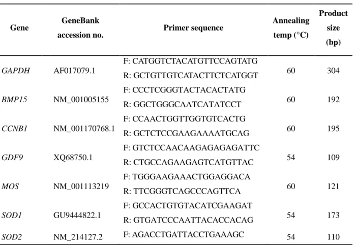
3.2. Hesperetin reduces the level of ROS in aging porcine oocytes in vitro
The effects of hesperetin on the levels of ROS and glutathione (GSH) were analyzed by staining oocytes with dichlorohydrofluorescein diacetate (DCFHDA) and CellTracker™ Blue 4-chloromethyl-6,8-difluoro-7-hydroxycoumarin (CMF2HC), respectively. The staining intensity of ROS was significantly lower (p<0.001) in the control and H-100 groups than in the aging group (control, 53.3±1.3 pixels/oocyte; aging, 73.5±1.6 pixels/oocyte; and H-100, 67.2±1.9 pixels/oocyte; Fig. 1B). The staining intensity of GSH was significantly higher (p<0.01) in the H-100 group than in the control and aging groups, but did not differ between the control and aging groups (control, 98.3±4.9 pixels/oocyte; aging, 99.4±2.6 pixels/oocyte; and H-100, 143.8±9.8 pixels/oocyte; Fig. 1B).
Expression of the antioxidant genes peroxiredoxin 5 (PRDX5), nuclear factor erythroid 2-like 2 (NFE2L2), superoxide dismutase 1 (SOD1), and superoxide dismutase 2 (SOD2) was analyzed by real-time reverse transcription (RT)-PCR (Fig. 1C). mRNA expression of PRDX5, NFE2L2, and
SOD2 was significantly higher (p<0.01) in the H-100 group than in the aging group, and was similar
in the control and H-100 groups. mRNA expression of SOD1 was significantly higher and lower (p<0.05) in the H-100 group than in the aging and control groups, respectively.
13
Fig. 1. Effect of hesperetin on the level of oxidative stress in porcine oocytes during aging in vitro. (A)
Images of oocytes stained with DCFHDA (green) and CMF2HC (blue). a–c, ROS staining; d–f, GSH
staining. Bar, 120 µm. (B) Quantification of the fluorescence intensities of DCFHDA and CMF2HC.
(C) Relative expression of the antioxidant genes PRDX5, NFE2L2, SOD-1, and SOD-2. Values are presented as means ± standard error of the mean of independent experiments (a–bp<0.05, *p<0.01,
**
14
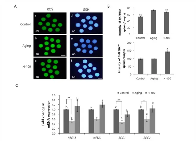
3.3. Hesperetin prevents aberrant spindle organization and chromosome misalignment in aging porcine oocytes in vitro
To further investigate the effect of hesperetin on spindle organization, spindles without abnormalities were classified as normal, whereas those in which chromosomes failed to align at the metaphase plate were classified as abnormal (Lenie, Cortvrindt, Eichenlaub-Ritter, & Smitz, 2008) (Fig. 2A). The percentage of oocytes with normal meiotic spindles was significantly higher (p<0.01) in the H-100 group than in the aging group, and was similar in the control and H-100 groups (control, 72.1±3.5%; aging, 57.8±0.4%; and H-100, 71.2±2.3%; Fig. 2B).
15
Fig. 2. Effect of hesperetin on spindle morphology in porcine oocytes during aging in vitro. (A)
Images of oocytes with normal (a) and abnormal (b) spindles. (B) Percentages of oocytes with normal and abnormal spindles. Data were derived from five independent replicates per group(*p<0.01).
16
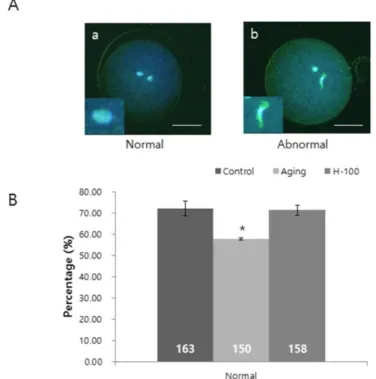
3.4. Hesperetin increases expression of cytoplasmic maturation markers in aging porcine oocytes in vitro
To investigate the effect of hesperetin on cytoplasmic maturation of aging oocytes, we examined mitogen-activated protein kinase (MAPK) activity and maternal gene expression (Fig. 3). Active phospho-p44/42 MAPK migrated as a doublet in lysates of maturing porcine oocytes by western blotting. The relative ratio of phospho-p44/42 MAPK activity in total p44/42 MAPK was significantly higher (p<0.05) in the H-100 group than in the aging group, and was similar in the control and H-100 groups (control, 98.2±0.01%; aging, 86.9±0.04%; and H-100, 98.0±0.02%; Fig. 3A).
Expression of the cytoplasmic maturation marker genes growth differentiation factor-9 (GDF9), cyclin B1 (CCNB1), bone morphogenetic protein 15 (BMP15), and MOS proto-oncogene, serine/threonine kinase (MOS) was analyzed by real-time RT-PCR (Fig. 3B). mRNA expression of
GDF9, CCNB1, and BMP15 was significantly higher (p<0.05) in the H-100 group than in the aging
group, and was similar in the control and H-100 groups. mRNA expression of MOS was significantly higher (p<0.01) and lower (p<0.001) in the H-100 group than in the aging and control groups, respectively (Fig. 3B).
17
Fig. 3. Effect of hesperetin on MAPK activity and maternal gene expression in porcine oocytes during
aging in vitro. (A) MAPK protein activity. (B) Maternal gene expression. Levels were normalized against that in the control group. Data were derived from four independent replicates per group (a–
b
18
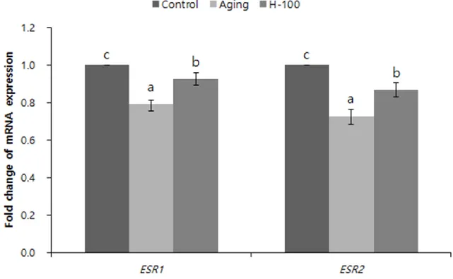
3.5. Hesperetin increases expression of estrogen receptor genes in aging porcine oocytes in vitro
The effects of hesperetin on mRNA expression of estrogen receptor 1 (ESR1) and 2 (ESR2) were analyzed by real-time RT-PCR to determine if hesperetin activates these receptors.mRNA expression of these genes was significantly higher (p<0.05) and lower (p<0.05) in the H-100 group than in the aging and control groups, respectively (Fig. 4).
19
Fig. 4. Effect of hesperetin on expression of estrogen receptor genes in porcine oocytes during aging
in vitro. Expression was normalized against that in the control group. Data were derived from two
20
3.6. Hesperetin improves the developmental capacity and quality of embryos derived from aging porcine oocytes in vitro
To investigate whether hesperetin treatment during IVM of oocytes influences subsequent embryo development and quality, oocytes were matured in the presence or absence of 100 μM hesperetin and then parthenogenetically activated. The blastocyst formation rates were calculated at 7 days after PA (Fig. 5). The total number of cells per blastocyst was significantly higher (p<0.01) in the control and
H-100 groups than in the aging group (control, 81.7±6.1; aging, 54.3±4.5; and H-100, 72.9±4.8; Fig. 5B). Genomic DNA fragmentation was assessed by terminal deoxynucleotidyl transferase dUTP nick-end labeling to detect apoptotic cells in blastocysts. The percentage of apoptotic cells was significantly higher (p<0.05) in the aging group than in the control and H-100 groups (control, 5.0±0.4%; aging, 7.9±0.9%; and H-100, 5.6±0.5%; Fig. 5C).
21
Fig. 5. Effect of hesperetin treatment during aging of porcine oocytes in vitro on subsequent embryo
development and quality. (A) Cleavage rate. (B) Blastocyst formation rate. (C) Total cell number per blastocyst. (D) Percentage of apoptotic cells in blastocysts. Data were derived from four independent replicates per group (a–cp<0.05, *p<0.01, **p<0.001).
22
4. DISSCUSTION
The mechanism by which oocytes are protected against aging in vitro is unknown. This study investigated the effects of the antioxidant hesperetin on aging of porcine oocytes in vitro. Treatment with 100 μM hesperetin during 24 h of aging significantly decreased the level of ROS and significantly increased expression of antioxidant genes (PRDX5, NFE2L2, SOD1, and SOD2), expression of maternal genes (GDF9, CCNB1, BMP15, and MOS), and the percentage of oocytes with normal spindles. In addition, embryos derived from these hesperetin-treated oocytes exhibited an improved developmental capacity and quality. This study demonstrates that hesperetin protects oocytes against oxidative stress during aging in vitro.
Oxidative stress arises in postovulatory aging oocytes, in which ROS production is increased and antioxidant protection is concomitantly decreased (Lord & Aitken, 2013). Intracellular GSH protects oocytes against oxidative damage; however, its level gradually decreases during aging (Boerjan & de Boer, 1990). The level of ROS was lower and higher in the H-100 group than in the aging and control groups, respectively. Moreover, the level of GSH was significantly higher in the H-100 group than in the control and aging groups, but was similar in the control and aging groups (Fig. 1A–B). Expression of antioxidant genes was higher in the H-100 group than in the aging group, and was similar in the control and H-100 groups (Fig. 1C). PRDX5 mediates ROS-based redox signaling and is involved in defense against a variety of cellular stresses. SOD1 catalyzes the conversion of superoxide radicals into molecular oxygen and hydrogen peroxide (McCord & Fridovich, 1969). SOD2 is the first line of defense against superoxide produced as a byproduct of oxidative phosphorylation (Y. Li et al., 1995). The transcription factor NFE2L2 is activated by antioxidants and chemopreventive agents (Cho, Reddy, & Kleeberger, 2006). Our results indicate that hesperetin protects oocytes against oxidative stress.
23
Chromosome condensation is the first visible event in meiotic maturation and is important for the formation of chromosomes and their proper segregation (Jelinkova & Kubelka, 2006). Postovulatory aging changes the levels of spindle-associated proteins (Cecconi et al., 2014) and induces acetylation of α-tubulin (A. R. Lee et al., 2013). The percentage of oocytes with normal spindles was lower in the aging group than in the control group, but was similar in the H-100 and control groups (Fig. 2B). These results demonstrate that treatment with hesperetin during aging of oocytes in vitro protects chromosomes and spindles at MII.
MAPK activity gradually decreases during aging of porcine oocytes (Larbi et al., 2005). The ratio of phospho-p44/42 MAPK to total p44/42 MAPK was higher in the H-100 group than in the aging group, and was similar in the control and H-100 groups (Fig. 3A). Expression of the maternal genes
CCNB1, MOS, BMP15, and GDF9 was higher in the H-100 group than in the aging group (Fig. 3B). GDF9 plays multifunctional roles in communication of oocyte granulosa cells and regulation of the
differentiation and function of follicular cells (Elvin, Clark, Wang, Wolfman, & Matzuk, 1999).
BMP15 prevents apoptosis of cumulus cells (Hussein, Froiland, Amato, Thompson, & Gilchrist, 2005). BMP15 and GDF9 interact and thereby elicit synergistic effects (Liu et al., 2018). CCNB1 forms a
complex with cyclin-dependent kinase 1 that permits transition from G2 to M phase of the cell (Robert, Hue, McGraw, Gagne, & Sirard, 2002; Zhang et al., 2011). MOS participates in MII arrest and meiotic asymmetric division, and is thus vital for oocyte maturation (Evangelou et al., 2002). Consequently, the increases in expression of these genes upon hesperetin treatment mean elicit beneficial effects during aging of oocytes in vitro.
To investigate whether hesperetin enters oocytes via estrogen receptors, we determined the expression levels of ESR1 and ESR2. Estrogen receptor is working more often as classical channel but also it could be mediated by antiatherogenic effect. Expression of these genes was significantly higher and lower in the H-100 group than in the aging and control groups, respectively (Fig. 4). Hesperetin enters cells via estrogen receptors (Hwang & Yen, 2011) and protects oxidative damage (Zhu, Dong, Liu, Ren, & Cui, 2017). The two forms of estrogen receptor are ERα and ERβ, which are encoded by
24
ESR1 and ESR2, respectively. Alteration of pre-mRNA splicing and expression levels of ESR1
modulate the function of ERα (Ghali et al., 2018). ERβ is homologous to ERα, and these two proteins have similar, but not identical, tissue distributions (Mosselman, Polman, & Dijkema, 1996). Our results demonstrate that hesperetin enters porcine oocytes via estrogen receptors.
The easiest way to assess the quality of oocytes in vitro is to identify the developmental rate. Developmental rate determines the efficiency with which embryos are produced in vitro. Aging negatively affects oocyte competency and embryo development (Marshall & Rivera, 2018), and reduces the cleavage rate (W. J. Yang et al., 2015). The percentage of oocytes that became cleavage was similar in the control and H-100 groups, and was lower in the aging group than in the control group. Many factors increase the developmental rate of oocytes (R. Li, Liu, Pedersen, & Callesen, 2015; Park, So, & Hyun, 2017; Shin et al., 2018). Oocytes damaged by aging or oxidative stress reportedly have a reduced developmental potential (Chaube et al., 2005; Goud et al., 2008; Liang et al., 2017). To investigate blastocyst quality, we checked the numbers of total and apoptotic cells per blastocyst. The total cell number per blastocyst was higher in the H-100 group than in the aging group, and was similar in the control and H-100 groups. The percentage of apoptotic cells was significantly lower in the H-100 group than in the aging group, and was similar in the control and H-100 groups. Thus, the developmental rate was decreased in the aging group and this was prevented in the H-100 group, suggesting that treatment with 100 µM hesperetin protects oocytes against aging in vitro.
In conclusion, this study indicates that treatment with 100 μM hesperetin effectively protects oocytes against oxidative stress by reducing the level of ROS and thereby minimizes the deterioration in oocyte quality during aging in vitro. Therefore, hesperetin can be used to improve assisted reproductive technologies.
25
REFERENCE
Boerjan, M. L., & de Boer, P. (1990). First cell cycle of zygotes of the mouse derived from
oocytes aged postovulation in vivo and fertilized in vivo. Mol Reprod Dev, 25(2),
155-163. doi:10.1002/mrd.1080250208
Bomfim, M. M., Andrade, G. M., Del Collado, M., Sangalli, J. R., Fontes, P. K., Nogueira, M.
F. G., . . . Perecin, F. (2017). Antioxidant responses and deregulation of epigenetic
writers and erasers link oxidative stress and DNA methylation in bovine blastocysts.
Mol Reprod Dev, 84(12), 1296-1305. doi:10.1002/mrd.22929
Cecconi, S., Rossi, G., Deldar, H., Cellini, V., Patacchiola, F., Carta, G., . . . Canipari, R.
(2014). Post-ovulatory ageing of mouse oocytes affects the distribution of specific
spindle-associated proteins and Akt expression levels. Reprod Fertil Dev, 26(4),
562-569. doi:10.1071/RD13010
Chaube, S. K., Prasad, P. V., Thakur, S. C., & Shrivastav, T. G. (2005). Hydrogen peroxide
modulates meiotic cell cycle and induces morphological features characteristic of
apoptosis in rat oocytes cultured in vitro. Apoptosis, 10(4), 863-874.
doi:10.1007/s10495-005-0367-8
Cho, H. Y., Reddy, S. P., & Kleeberger, S. R. (2006). Nrf2 defends the lung from oxidative
stress. Antioxid Redox Signal, 8(1-2), 76-87. doi:10.1089/ars.2006.8.76
de Souza, V. T., de Franco, E. P., de Araujo, M. E., Messias, M. C., Priviero, F. B., Frankland
Sawaya, A. C., & de Oliveira Carvalho, P. (2016). Characterization of the antioxidant
activity of aglycone and glycosylated derivatives of hesperetin: an in vitro and in vivo
study. J Mol Recognit, 29(2), 80-87. doi:10.1002/jmr.2509
Eichenlaub-Ritter, U. (2012). Oocyte ageing and its cellular basis. Int J Dev Biol, 56(10-12),
841-852. doi:10.1387/ijdb.120141ue
Elvin, J. A., Clark, A. T., Wang, P., Wolfman, N. M., & Matzuk, M. M. (1999). Paracrine
actions of growth differentiation factor-9 in the mammalian ovary. Mol Endocrinol,
13(6), 1035-1048. doi:10.1210/mend.13.6.0310
Evangelou, K., Balaskas, C., Marinos, E., Dosios, T., Kittas, C., & Gorgoulis, V. G. (2002).
Immunohistochemical localization of c-mos at the light and electron microscope level
26
in non-small cell lung carcinomas. Biotech Histochem, 77(2), 85-91.
Finkel, T., & Holbrook, N. J. (2000). Oxidants, oxidative stress and the biology of ageing.
Nature, 408(6809), 239-247. doi:10.1038/35041687
Ghali, R. M., Al-Mutawa, M. A., Al-Ansari, A. K., Zaied, S., Bhiri, H., Mahjoub, T., &
Almawi, W. Y. (2018). Differential association of ESR1 and ESR2 gene variants with
the risk of breast cancer and associated features: A case-control study. Gene, 651,
194-199. doi:10.1016/j.gene.2018.02.011
Goto, Y., Noda, Y., Mori, T., & Nakano, M. (1993). Increased generation of reactive oxygen
species in embryos cultured in vitro. Free Radic Biol Med, 15(1), 69-75.
Goud, A. P., Goud, P. T., Diamond, M. P., Gonik, B., & Abu-Soud, H. M. (2008). Reactive
oxygen species and oocyte aging: role of superoxide, hydrogen peroxide, and
hypochlorous
acid.
Free
Radic
Biol
Med,
44(7),
1295-1304.
doi:10.1016/j.freeradbiomed.2007.11.014
Hirata, A., Murakami, Y., Shoji, M., Kadoma, Y., & Fujisawa, S. (2005). Kinetics of
radical-scavenging activity of hesperetin and hesperidin and their inhibitory activity on
COX-2 expression. Anticancer Res, COX-25(5), 3367-3374.
Hussein, T. S., Froiland, D. A., Amato, F., Thompson, J. G., & Gilchrist, R. B. (2005).
Oocytes prevent cumulus cell apoptosis by maintaining a morphogenic paracrine
gradient of bone morphogenetic proteins. J Cell Sci, 118(Pt 22), 5257-5268.
doi:10.1242/jcs.02644
Hwang, S. L., Lin, J. A., Shih, P. H., Yeh, C. T., & Yen, G. C. (2012). Pro-cellular survival
and neuroprotection of citrus flavonoid: the actions of hesperetin in PC12 cells. Food
Funct, 3(10), 1082-1090. doi:10.1039/c2fo30100h
Hwang, S. L., & Yen, G. C. (2011). Effect of hesperetin against oxidative stress via ER- and
TrkA-mediated actions in PC12 cells. J Agric Food Chem, 59(10), 5779-5785.
doi:10.1021/jf104632a
Jelinkova, L., & Kubelka, M. (2006). Neither Aurora B activity nor histone H3
phosphorylation is essential for chromosome condensation during meiotic maturation
of porcine oocytes. Biol Reprod, 74(5), 905-912. doi:10.1095/biolreprod.105.047886
Larbi, A., Douziech, N., Fortin, C., Linteau, A., Dupuis, G., & Fulop, T., Jr. (2005). The role
27
with aging. Immun Ageing, 2(1), 6. doi:10.1186/1742-4933-2-6
Lee, A. R., Kishigami, S., Amano, T., Matsumoto, K., Wakayama, T., & Hosoi, Y. (2013).
Nicotinamide: a class III HDACi delays in vitro aging of mouse oocytes. J Reprod
Dev, 59(3), 238-244.
Lee, S. E., Sun, S. C., Choi, H. Y., Uhm, S. J., & Kim, N. H. (2012). mTOR is required for
asymmetric division through small GTPases in mouse oocytes. Mol Reprod Dev,
79(5), 356-366. doi:10.1002/mrd.22035
Lenie, S., Cortvrindt, R., Eichenlaub-Ritter, U., & Smitz, J. (2008). Continuous exposure to
bisphenol A during in vitro follicular development induces meiotic abnormalities.
Mutat Res, 651(1-2), 71-81. doi:10.1016/j.mrgentox.2007.10.017
Li, R., Liu, Y., Pedersen, H. S., & Callesen, H. (2015). Effect of ambient light exposure of
media and embryos on development and quality of porcine parthenogenetically
activated embryos. Zygote, 23(3), 378-383. doi:10.1017/S096719941300066X
Li, Y., Huang, T. T., Carlson, E. J., Melov, S., Ursell, P. C., Olson, J. L., . . . Epstein, C. J.
(1995). Dilated cardiomyopathy and neonatal lethality in mutant mice lacking
manganese superoxide dismutase. Nat Genet, 11(4), 376-381.
doi:10.1038/ng1295-376
Liang, S., Nie, Z. W., Zhao, M., Niu, Y. J., Shin, K. T., & Cui, X. S. (2017). Sodium fluoride
exposure exerts toxic effects on porcine oocyte maturation. Sci Rep, 7(1), 17082.
doi:10.1038/s41598-017-17357-3
Lim, J., & Luderer, U. (2011). Oxidative damage increases and antioxidant gene expression
decreases with aging in the mouse ovary. Biol Reprod, 84(4), 775-782.
doi:10.1095/biolreprod.110.088583
Liu, C., Yuan, B., Chen, H., Xu, M., Sun, X., Xu, J., . . . Zhang, J. (2018). Effects of
MiR-375-BMPR2 as a Key Factor Downstream of BMP15/GDF9 on the Smad1/5/8 and
Smad2/3
Signaling
Pathways.
Cell
Physiol
Biochem,
46(1),
213-225.
doi:10.1159/000488424
Lord, T., & Aitken, R. J. (2013). Oxidative stress and ageing of the post-ovulatory oocyte.
Reproduction, 146(6), R217-227. doi:10.1530/REP-13-0111
Marshall, K. L., & Rivera, R. M. (2018). The effects of superovulation and reproductive
aging on the epigenome of the oocyte and embryo. Mol Reprod Dev, 85(2), 90-105.
28
doi:10.1002/mrd.22951
McCord, J. M., & Fridovich, I. (1969). Superoxide dismutase. An enzymic function for
erythrocuprein (hemocuprein). J Biol Chem, 244(22), 6049-6055.
Miao, Y. L., Kikuchi, K., Sun, Q. Y., & Schatten, H. (2009). Oocyte aging: cellular and
molecular changes, developmental potential and reversal possibility. Hum Reprod
Update, 15(5), 573-585. doi:10.1093/humupd/dmp014
Mosselman, S., Polman, J., & Dijkema, R. (1996). ER beta: identification and
characterization of a novel human estrogen receptor. FEBS Lett, 392(1), 49-53.
Orrenius, S., Gogvadze, V., & Zhivotovsky, B. (2007). Mitochondrial oxidative stress:
implications for cell death. Annu Rev Pharmacol Toxicol, 47, 143-183.
doi:10.1146/annurev.pharmtox.47.120505.105122
Park, S. J., So, K. H., & Hyun, S. H. (2017). Effect of zeaxanthin on porcine embryonic
development during in vitro maturation. J Biomed Res, 31(2), 154-161.
doi:10.7555/JBR.31.20160079
Patra, R. C., Rautray, A. K., & Swarup, D. (2011). Oxidative stress in lead and cadmium
toxicity and its amelioration. Vet Med Int, 2011, 457327. doi:10.4061/2011/457327
Rajani, S., Chattopadhyay, R., Goswami, S. K., Ghosh, S., Sharma, S., & Chakravarty, B.
(2012). Assessment of oocyte quality in polycystic ovarian syndrome and
endometriosis by spindle imaging and reactive oxygen species levels in follicular
fluid and its relationship with IVF-ET outcome. J Hum Reprod Sci, 5(2), 187-193.
doi:10.4103/0974-1208.101020
Robert, C., Hue, I., McGraw, S., Gagne, D., & Sirard, M. A. (2002). Quantification of cyclin
B1 and p34(cdc2) in bovine cumulus-oocyte complexes and expression mapping of
genes involved in the cell cycle by complementary DNA macroarrays. Biol Reprod,
67(5), 1456-1464.
Shin, M. Y., Lee, S. E., Son, Y. J., Park, Y. G., Jeong, S. G., Kim, E. Y., & Park, S. P. (2018).
Lysophosphatidic acid accelerates development of porcine embryos by activating
formation of the blastocoel. Mol Reprod Dev, 85(1), 62-71. doi:10.1002/mrd.22938
Takeo, S., Kimura, K., Shirasuna, K., Kuwayama, T., & Iwata, H. (2017). Age-associated
deterioration in follicular fluid induces a decline in bovine oocyte quality. Reprod
Fertil Dev, 29(4), 759-767. doi:10.1071/RD15228
29
Wang, J., Zhu, H., Yang, Z., & Liu, Z. (2013). Antioxidative effects of hesperetin against lead
acetate-induced oxidative stress in rats. Indian J Pharmacol, 45(4), 395-398.
doi:10.4103/0253-7613.115015
Wang, Q., & Sun, Q. Y. (2007). Evaluation of oocyte quality: morphological, cellular and
molecular predictors. Reprod Fertil Dev, 19(1), 1-12.
Yang, H. W., Hwang, K. J., Kwon, H. C., Kim, H. S., Choi, K. W., & Oh, K. S. (1998).
Detection of reactive oxygen species (ROS) and apoptosis in human fragmented
embryos. Hum Reprod, 13(4), 998-1002.
Yang, W. J., Hwang, Y. C., Lin, C. S., Hwu, Y. M., Lee, R. K., & Hsiao, S. Y. (2015).
Embryonic early-cleavage rate is decreased with aging in GnRH agonist but not
inantagonist protocols. J Assist Reprod Genet, 32(5), 789-795.
doi:10.1007/s10815-015-0461-y
You, J., Kim, J., Lim, J., & Lee, E. (2010). Anthocyanin stimulates in vitro development of
cloned pig embryos by increasing the intracellular glutathione level and inhibiting
reactive
oxygen
species.
Theriogenology,
74(5),
777-785.
doi:10.1016/j.theriogenology.2010.04.002
Zarebczan, B., Pinchot, S. N., Kunnimalaiyaan, M., & Chen, H. (2011). Hesperetin, a
potential therapy for carcinoid cancer. Am J Surg, 201(3), 329-332; discussion 333.
doi:10.1016/j.amjsurg.2010.08.018
Zhang, D. X., Park, W. J., Sun, S. C., Xu, Y. N., Li, Y. H., Cui, X. S., & Kim, N. H. (2011).
Regulation of maternal gene expression by MEK/MAPK and MPF signaling in
porcine oocytes during in vitro meiotic maturation. J Reprod Dev, 57(1), 49-56.
Zhu, C., Dong, Y., Liu, H., Ren, H. & Cui, Z. (2017). Hesperetin protects against
H2O2-riggered oxidative damage via upregulation of the Keap1-Nrf2/HO-1 signal pathway
in
ARPE-19
cells.
Biomed
Pharmacother,
88,
124-133.
30
ABSTRACT IN KOREAN
감귤류 플라보노이드 헤스페레틴은 항산화, 항염증 및 항암작용을 비롯한 다양한 약리학적 작용을 한다. 본 연구는 헤스페레틴이 체외에서 돼지 난자가 노화하는 동안 난자의 핵 및 세포질의 성숙과 ROS 수준을 조절하여 배아의 발육능력에 어떤 영향을 미치는지 조사하였다. 돼지의 난소로부터 회수된 미성숙 난자는 44 시간 체외성숙 한 후 추가로 0, 1, 1,0 100, 250 μM 의 헤스페레틴을 첨가하여 24 시간 노화시켰다 (대조군, 노화군, H-1, H-10, H-100 와 H-250). 실험의 결과, 44 시간 체외성숙 난자와 추가로 24 시간 노화한 난자에서 차이가 없었지만 H-100 에서 배아형성률이 다른 노화군 보다 높고 대조군과 비슷한 수준을 보였다 (p<0.01). 그리고 H-100 군은 노화군에 비해 활성산소종의 수준이 현저하게 낮고 글루타티온 수준이 유의적으로 높게 나왔으며 항산화 유전자 PRDX5, NFE2L, SOD-1 와 SOD-2 의 발현도 유의하게 높게 나왔다 (p<0.01). 또한 H-100 군은 방추사의 비정상과 염색체 정렬의 불량을 억제하고 MAPK 의 수준 감소를 막고 세포질 성숙인자 유전자 GDF9, CCNB1, BMP15 와 MOS 의 발현을 증가시켰다. 배아에서는 H-100 군이 노화군에 비해 배반포 당 총 세포수가 증가하고 세포사멸은 감소하는 것이 대조군과 유사하였다. 이 결과는 헤스페레틴이 체외에서 노화하는 동안 산화스트레스로부터 돼지의 난자를 보호함으로써 돼지 난자의 질을 향상 시킨다는 것을 시사한다.31