저작자표시-비영리-변경금지 2.0 대한민국 이용자는 아래의 조건을 따르는 경우에 한하여 자유롭게 l 이 저작물을 복제, 배포, 전송, 전시, 공연 및 방송할 수 있습니다. 다음과 같은 조건을 따라야 합니다: l 귀하는, 이 저작물의 재이용이나 배포의 경우, 이 저작물에 적용된 이용허락조건 을 명확하게 나타내어야 합니다. l 저작권자로부터 별도의 허가를 받으면 이러한 조건들은 적용되지 않습니다. 저작권법에 따른 이용자의 권리는 위의 내용에 의하여 영향을 받지 않습니다. 이것은 이용허락규약(Legal Code)을 이해하기 쉽게 요약한 것입니다. Disclaimer 저작자표시. 귀하는 원저작자를 표시하여야 합니다. 비영리. 귀하는 이 저작물을 영리 목적으로 이용할 수 없습니다. 변경금지. 귀하는 이 저작물을 개작, 변형 또는 가공할 수 없습니다.
A Thesis for the Degree of Master of Science
The Novel Effects of
5-(3',4’-Dihydroxyphenyl)-γ-valerolactone, a major Metabolite of Procyanidins,
on C2C12 Murine Myoblasts Myogenesis
DHPV 의 노화성 근감소 억제 및 근육분화 촉진 효능
Feburary, 2018
By Seunghee Yang
A Thesis for the Degree of Master of Science
The Novel Effects of
5-(3',4’-Dihydroxyphenyl)-γ-valerolactone, a major Metabolite of Procyanidins,
on C2C12 Murine Myoblasts Myogenesis
DHPV 의 노화성 근감소 억제 및 근육분화 촉진 효능
Feburary, 2018
By Seunghee Yang
Department of Agricultural Biotechnology
Seoul National University
ABSTRACT
Procyanidins have many health benefits. After intake, procyanidins
are degraded into small metabolites by gastrointestinal microorganisms.
According to a previous study, 5-(3',4’-Dihydroxyphenyl)-γ-valerolactone
is the most abundant among these metabolites. The object of this study is to
determine the effects of DHPV on muscle and whether DHPV could recover
aged muscle to normal. TGF-β is connected to muscle aging and attenuates
the muscle differentiation and inhibits myogenesis.
In this study, I measured myoD, myogenin, and the myosin heavy
chain expression level to determine whether DHPV improved the myogenesis
the DHPV treated group in comparison to TGF-β only treated. In addition,
DHPV modulated the TGF-β signaling pathway and C2C12 myogenesis by
regulating p-smad2/3, p-JNK, p-ERK and p-p38 expression. Taken
together, it may be suggested that DHPV is an ideal therapeutic candidate for
recovering myogenesis and muscle differentiation evoked by aging.
Keywords: DHPV; myogenesis; procyanidins; TGF-
β; sarcopenia
CONTENTS
ABSTRACT
... iii
I. INTRODUCTION
... 1
II. MATERLIALS AND METHODS
... 4
1.
Chemicals and reagents
... 4
2.
Cell culture and treatments
... 5
3.
Sulforhodamine B assay
... 6
4.
Hematoxylin and Eosin staining (H&E staining)
... 6
5.
Western blot assay
... 7
6.
Real-time quantitative PCR
... 8
7.
Statistical analysis
... 10
III. RESULTS
... 11
1.
DHPV promotes myotube formation and has no toxic effect
on C2C12 murine myoblasts ... 11
2.
DHPV improves the myoD expression compared to TGF-
βtreatment ... 15
3.
DHPV up-regulates the expression of myogenin in spite of
TGF-
βtreatment on C2C12 myoblasts ... 19
4.
DHPV treatment counteracts the inhibition of myosin heavy
chain expression induced by TGF-
β... 22
5.
DHPV donw-regulates p-smad2/3 expressions even in
TGF-βtreated group ... 26
6.
The summary of the myogenic role of DHPV on C2C12
myogenesis against
TGF-
β... 29
IV. DISCUSSION
... 30
V. REFERENCES
... 34
I.
Introduction
The aging population is growing rapidly worldwide. According to the
United Nations, the number of people over 60 will rise to more than 2 billion
by 2050[1]. Decline of muscle mass and strength is one of the physical
changes associated with aging and gradually begins at age 40[2]. Sarcopenia,
decrease of muscle mass and loss of function, is an age-related senile disease.
In 2000, the estimated health care costs of sarcopenia in the U. S. was 18.5
billion dollars [3]. Since impaired physical function lowers the quality of life,
the importance of studying sarcopenia is growing[4].
Skeletal muscle occupies approximately half of the entire body weight
into skeletal muscle as well as to form myotubes and myofibers. [6, 7]. There
are several biomarkers verified during myogenesis, including myoD, myogenin
and myosin heavy chain [8].
Meanwhile, according to previous research, TGF-β is markedly
increased in the old muscle cells compared to young ones [9]. TGF-β is a
well-known muscle differentiation inhibitor [13-20], that mediates the
expression of myogenic biomarkers by regulating p-smad2/3 and non
smad2/3 pathways [10, 11]. During muscle differentiation, TGF-β signaling
activates smad2/3 by up-regulating p-smad2/3, which acts on muscle
regulatory factors [13, 14], and regulates non-smad pathways including the
ERK, JNK and p38 pathways [11].
of procyanidins, which are plentiful in the husks of Yak-Kong, and cocoa.
Previous study has shown that DHPV was the highest detected metabolite
after the uptake of cocoa polyphenol in human plasma [12]. According to the
kinase array conducted in our lab, DHPV attenuates the activity of TGF-β
receptor 1 by half. In our lab, we have studied DHPV’s beneficial effects such
as its anti-obesity effects and anti-oxidant effects in neurons, but there are
no studies of DHPV’s myogenic role. Based on the result of DHPV’s kinase
array and several health benefits, this study researches the muscle myogenic
effects of DHPV and investigates the effects of DHPV to indentify the
molecular mechanisms underlying the myogenic role of DHPV on C2C12
Ⅱ. Materials and methods
1. Chemical and reagents
Dulbecco’s modified eagle medium (DMEM), horse serum and fetal
bovine serum (FBS) were purchased from Thermo Fisher Scientific (Waltham,
MA, USA). 5-(3’,4’-Dihydroxyphenyl)-gamma-valerolactone (DHPV)
was purchased from Chemieliva pharmaceutical company (China). All-trans
retinoic acid was purchased from Sigma-Aldrich (St. Louis, MO, USA).
TGF-β was purchased from R&D Systems (Minneapolis, MN, USA). The
sulforhodamine B assay kit was purchased from Sigma-Aldrich (St. Louis,
MO, USA). The myosin heavy chain, myogenin, myoD,
Cruz Biotechnology (Santa Cruz, CA, USA). The anti-p-smad2 antibody was
purchased from Thermo Fisher Scientific (Waltham, MA, USA). The
anti-p-smad3 antibody was obtained from Ab cam (Bristol, UK).
2. Cell culture and treatments
C2C12 murine myoblasts (ATCC, Manassas, VA, USA) were
cultured in DMEM containing high glucose with FBS. Three days after being
seeded, the myoblasts were replenished to low glucose DMEM with horse
serum and each began differentiation. They were replenished everyday. 5
ng/ml TGF-β, 1 μM DHPV were used to lead to inhibit myogenesis and
promote myogenesis respectively. And All-trans retinoic acid was used as a
3. Sulforhodamine B assay (Cell viability)
C2C12 cells were cultured in the 96-well plate at a density of
2.5×10⁵ cells/ml in the presence of DMEM that contained low glucose
with horse serum for 24 h. They were then analyzed using a Sulforhodamine
B assay kit according to manufacturer’s instruction. This was done to measure
cell viability in the presence of TGF-β, DHPV and retinoic acid.
4. Hematoxylin and Eosin staining (H&Estaining)
After four days of differentiation, each sample was carefully washed
twice using PBS. To visualize myotubes and nuclei, hematoxylin and eosin
was for nucleus staining. These myotubes were examined at 100 times
magnification.
5. Western blot assay
Protein extracts were harvested by using RIPA buffer on ice, then
centrifuged at 13000 g for 10 min. Protein assay reagent kits (Bio-Rad
Laboratories, Hercules, CA, USA) were used to measure the protein
concentration, and then the proteins were electrically divided in
SDS-polyacrylamide gel. A transfer was conducted onto a Nitrocellulose blotting
membrane (GE Healthcare Life Science, Amersham, UK), which was blocked
in 5% skimmed milk for 1 h, followed by incubation with the primary
myogenin, myosin heavy chain, smad2/3, p-smad2, p-smad3, b-actin and
lamin B, as indicated. After incubation, the membranes were washed 3 times
with TBS-T for 10 min, and then incubated with secondary antibodies for 1
h in the presence of 5% skimmed milk. Following this, the membranes were
washed five times using TBS-T buffer for 8 min. The protein bands were
discovered through the use of an ECL detection kit (GE healthcare, St, Giles,
UK) in Chemidoc. Protein quantitative analysis was conducted by Image J
software (National Institutes of Health, Bethesda, MD, USA).
6. Real-time quantitative PCR
The RNA of C2C12 cells were collected and isolated in the presence of RNA
ND-2000 spectrophotometer (Thermo Fisher Scientific, MA, USA). In
addition, cDNA synthesis was conducted using a PrimeScript™ 1ststrand
cDNA Synthesis Kit (Takara, Kyoto, Japan). A real-time quantitative
polymerase chain reaction (RT-qPCR) was conducted using a Bio-Rad CFX
96real-time PCR detection system (Bio-Rad) with their respective primers
and SYBR Green Master Mix (Bio-Rad). The primers used were as follows:
mouse myoD forward (FW) 5’-GTG GCA GCG AGC ACT ACA GT-3’ and
mouse myoD reverse (RV) 5’-CTT GCA AAG GAA CTT GGG CTT-3’;
mouse myogenin forward (FW) 5’-GCA CTG GAG TTC GGT CCC AA-3’
and mouse myogenin reverse (RV) 5’-TAT CCT CCA CCG TGA TGC
TG-3’; mouse myosin heavy chain forward (FW) 5’-AAG CGA AGA GTA AGG
GAA CTT GGG CTT-3’; and mouse GAPDH forward (FW) 5’-CAA GGA
GTA AGA AAC CCT GGA CC-3’ and mouse GAPDH reverse (RV)
5’-CGA GTT GGG ATA GGG CCT CT-3’. The GAPDH was used as a
reference gene.
7. Statistical analysis
The data was specified as mean ± standard deviation. One-way
ANOVA test was used for comparative study between each groups, and
Student’ t-test was used to consider significant differences.
Ⅲ.
RESULTS
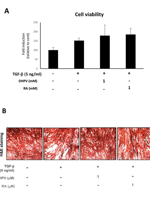
1. DHPV promotes myotube formation and has no toxic effect on
C2C12 murine myoblasts.
To identify the cytotoxicity of DHPV, TGF-β and Retinoic acid on
C2C12 murine myoblasts, the cell viability was tested using a Sulforhodamine
B assay. C2C12 myoblasts were treated with 1μM DHPV, 5 ng/ml TGF-β
and 1μM Retinoic acid for 24 h in a 96 well plate. The four groups were the
control, TGF-β only, DHPV with TGF-β and Retinoic acid with TGF-β.
The results showed that they are not toxic to C2C12 myoblasts at each
concentration (Fig. 1A). The cell viability was indicated as a relative ratio to
To visualize C2C12 myotube formation, I conducted hematoxylin
and eosin staining. TGF-β is a well-known myogenesis inhibitor [13-20],
that diminishes myotube formation in C2C12. According to this result,
TGF-β treatment inhibited cell differentiation and C2C12 myotube formation (Fig.
1B). A lower density of myotubes was observed in the TGF-β only group
compared with the control, DHPV and retinoic acid treated groups.
Figure 1. The cytotoxicity of DHPV and its effect on C2C12 myotube
acid and 5 ng/ml TGF-β have a cytotoxic effect on C2C12 cells (A). The cells
were incubated after treatment for 24 h. The cell viability of the C2C12 cells
was measured using a Sulforhodamine B assay, and marked according to the
relatively fold induction compared to the control. Each group was
triplicated and expressed as means ± standard deviation. (B) Representative
images of the C2C12 myoblasts which is differentiated in each sample for 96
h.
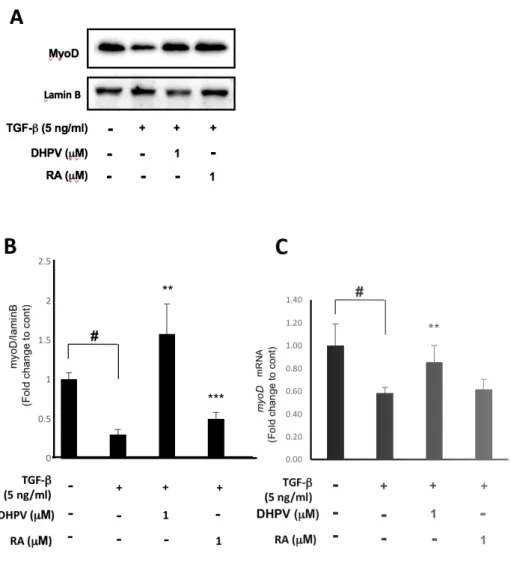
2. DHPV improves the myoD expression compared to TGF-
β
treatment.
The expression of the muscle differentiation biomarker myoD was
investigated to explore the role of DHPV on myogenesis to counteract
TGF-β. Since myoD usually manifests in the early stage of muscle differentiation,
the cells were incubated in a differentiation medium containing each sample
for 24 h. The results showed that TGF-β significantly decreased the myoD
protein expression level in comparison to the control (Fig. 2A, B). However,
DHPV and retinoic acid significantly enhanced myoD expression compared
to TGF-β (Fig. 2A, B). For further study, the mRNA expression level in each
group was investigated. 24 h after DM, RT-qPCR was conducted. As a result,
counteracted myoD expression (Fig. 2C). Retinoic acid made myoD
expression increase slightly, but not significantly (Fig. 2C). Each group was
triplicated and expressed as means ± SD. * p<0.05, ** p<0.01, *** p<0.001
Figure 2. DHPV treatment stimulates myoD expression against TGF-β. (A)
control. (B) The quantification of the protein bands from (A) (by Image J)
expressed relative to the control. (C) The myoD mRNA expression level in
C2C12 cells in each group after 24 h.
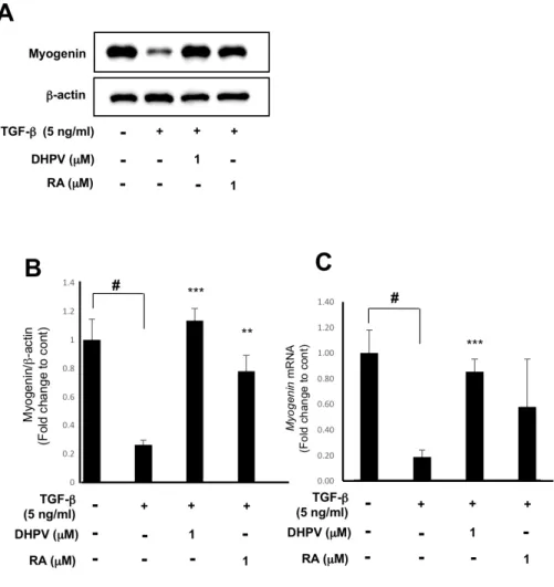
3. DHPV up-regulates the expression of myogenin in spite of
TGF-
βtreatment on C2C12 myoblasts.
Myogenin is usually expressed in the middle of muscle differentiation.
Thus, after each sample treatment was treated for 72 h, the myogenin
expression level was measured. It was found that TGF-β treatment
significantly attenuated myogenic protein expression in C2C12 myoblasts and
DHPV and Retinoic acid enhanced myogenin protein expression (Fig. 3A, B).
RT-qPCR was also conducted after 72 h of sample treatment, showing that
TGF-β reduced the myogenin mRNA level, and in contrast, DHPV enhanced
myogenin expression (Fig. 3C).
Figure 3. The inhibition of myogenin expression is counteracted by DHPV
loading control. (B) Quantification of myogenin protein expression by Image
J. (C) Myogenin expression in C2C12 myoblasts after 72 h of DHPV, Retinoic
acid and TGF-β treatment (n=7). * p<0.05, ** p<0.01, *** p<0.001
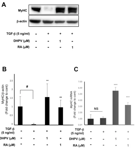
4. DHPV treatment couteracts the inhibition of myosin heavy chain
expression induced by
TGF-
β.
Myosin heavy chain is a biomarker that appears in the last stage of
muscle differentiation. To determine the myosin heavy chain expression level,
western blot analysis and RT-qPCR were conducted for each sample after 96
h of treatment. As with the previous two biomarkers, myosin heavy chain
protein expression was dramatically attenuated when treated with TGF-β
only (Fig. 4.A-C). According to the quantification of protein
bands, treatment with DHPV and Retinoic acid enhanced the myosin heavy
chain level to higher levels than in the control or similar, which was significant
(Fig. 4A, B). Even so, myosin heavy chain expression was slightly different
and showed no significance. However, the DHPV and Retinoic acid treatment
significantly elevated the myosin heavy chain level compared to the control
Figure 4. DHPV treatment elevates myosin heavy chain expression even in
was differentiated for 96 h in C2C12 myoblasts. (B) Quantification of the
western blot analysis from (A) (n=3). (C) Myosin heavy chain expression in
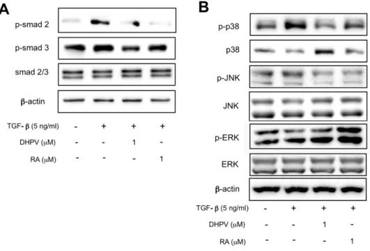
5.
DHPV promotes C2C12 myogenesis by regulating both smad 2/3
signaling and non-smad signaling pathways.
TGF-β induces the TGF-β signaling pathway and plays a key role in
inhibiting myogenesis in C2C12 myoblasts [13-20]. Therefore, I examined
the expression of phosphorylated smad2/3 which is a well-known
downstream protein of the TGF-β signaling pathway. The expression of
p-smad2/3 protein decreased when treated with DHPV in contradiction to
treatment with TGF-β only (Fig. 5A). MAPK singling pathway is also
related with TGF-β, which is consist of ERK, JNK and p38 signaling
pathways [28]. According to this data, DHPV down-regulated
phosphorylated-JNK and phosphorylated-p38 protein expression against the
treated compared to TGF-β treatment (Fig. 5B). From these results, it was
found that DHPV diminished myogenesis inhibition caused by TGF-β
signaling pathways.
Figure 5. DHPV mediates both smad2/3 signaling pathway and MAPK
signaling pathways. (A) DHPV decreased p-smad2/3 protein expression
which was increased by the TGF-β. β-actin was used as a loading control.
(B) The protein expression of three kinds of MAPK was examined. DHPV
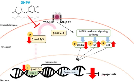
Figure 6. The summary of the myogenic role of DHPV on C2C12 myogenesis
Ⅳ. Discussion
There are several previous studies regarding the health benefits of
procyanidins such as their neuroprotective effects and anti-atherosclerotic
ability [23-26]. Furthermore, procyanidins were previously found to have
antioxidant effects and anti-obesity effects [21, 22]. At this point, the
question arises astowhatothernovel health benefits of DHPV may haveand
whattype ofprogresscouldbe madeby deactivating TGF-β receptor 1. As I
metioned earlier, according to the kinase array results, DHPV lowered the
TGF-β receptor 1 activity by half. Since TGF-β is deeply relevant to old
muscle [9], DHPV’s role muscle protection against TGF-β was studied.
control [27]. There are two types of pathways in TGF-β signaling pathway,
the smad2/3 pathway and non-smad pathways. The results show that DHPV
promotes the protein expression of three biomarkers of muscle differentiation
as well as RNA expression, and is usually more effective than retinoic acid.
DHPV was also confirmed to promote C2C12 myotubes formation more than
TGF-β inhibits it, showing a high density of differentiated muscle. In the
signaling pathway, the results show that DHPV regulates TGF-β signaling by
down-regulating smad2/3 phosphorylation as well as JNK and p38
phosphorylation and upregulating ERK phosphorylation. In conclusion,
DHPV promotes C2C12 myogenesis (Fig. 6).
This study, which is just the beginning of this researcher’s work
TGF-β because only two pathways were studied. As such, further study on other
TGF-β signaling pathways should be conducted. Since there are no previous
studies regarding DHPV or procyanidins and myogenesis promotion, these
results are novel in their discovery of new features of the metabolites of
natural substances.
It was observed that DHPV has myogenic effects on C2C12 murine
myoblasts, and as such, its precursor procyanidins have the possibility to have
similar effects as DHPV. Therefore, for further study, it is necessary that the
research into the effects of procyanidins on C2C12 myoblasts should be
conducted. Whether DHPV directly binds to TGF-β receptors or not should
Administrating DHPV treatment to C2C12 murine myoblasts
recovers the muscle differentiation suppressed by TGF-β to normal levels and
promotes myogenesis. This new approach suggests that DHPV may be used
Ⅴ. Reference
1. United Nations, Department of Economic and Social Affairs, Population
Division. World Population Aging 2013, United Nations, 2013.
2. McGregor, R., Cameron-Smith, D. & Poppitt, S. It is not just muscle mass:
a review of muscle quality, composition and metabolism during ageing as determinants of muscle function and mobility in later life. Longev. Healthspan, 2014, 3:9.
3. Janssen I, Shepard DS, Katzmarzyk PT, Roubenoff R., The Healthcare Costs
of Sarcopenia in the United States. Journal of the American Geriatric Society, 2004, 52:80–85.
4. RYAN D. NIPP, Sarcopenia Is Associated with Quality of Life and Depression in Patients with Advanced Cancer, the oncologist, 2018, 23(1):97-104.
5. GEORGE H. CARDINET III, Skeletal Muscle Function, CLINICAL BIOCHEMISTRY OF DOMESTIC ANIMALS, FIFTH EDITION, academic press, 407-440, 1997.
6. Anna Montesano, Resveratrol promotes myogenesis and hypertrophy in murine myoblasts Journal of Translational Medicine 2013, 11:310.
7. Ian C. Scott & Wendy Tomlinson & Andrew Walding & Beverley Isherwood & Iain G. Dougall, Large-scale isolation of human skeletal muscle satellite cells from post-mortem tissue and development of quantitative assays to evaluate modulators of myogenesis, J Cachexia Sarcopenia Muscle, 2013, 4:157–169.
8. M. G. Cusella-De Angelis, MyoD, Myogenin Independent Differentiation of Primordial Myoblasts in Mouse Somites, J cell biology, 1992, 116(5):1243-55.
9. Morgan E. Carlson, Imbalance between pSmad3 and Notch induces CDK inhibitors in old muscle stem cells, Nature, 2008, 454, 528–532
10. Dong Liu, TGF-beta inhibits muscle differentiation through functional repression of myogenic transcription factors by Smad3, Genes & Dev. 2001, 15:2950-2966.
11. Ying E Zhang, Non-Smad pathways in TGF-β signaling, Cell Res., 2009, 19(1): 128–139.
12. Urpi-Sarda M et al., Journal of Chromatography A, 2009, 1216(43): 7258-67
13. Liu D, Black BL, Derynck R. TGF-beta inhibits muscle differentiation through functional repression of myogenic transcription factors by Smad3. Genes Dev. 2001, 15:2950–66.
14. Liu D, Kang JS, Derynck R. TGF-beta-activated Smad3 represses MEF2-dependent transcription in myogenic differentiation. EMBO J. 2004, 23:1557–66.
15. Massague J, Cheifetz S, Endo T, Nadal-Ginard B. Type beta transforming growth factor is an inhibitor of myogenic differentiation. Proc Natl Acad Sci U S A. 1986, 83:8206–10.
16. Miyake T, Alli NS, McDermott JC. Nuclear function of Smad7 promotes myogenesis. Mol Cell Biol. 2010, 30:722–35.
17. Olson E, Sternberg E, Hu J, Spizz G, Wilcox C. Regulation of myogenic differentiation by type beta transforming growth factor, J Cell Biol, 1986,
18. Xu Q, Kopp JB. Retinoid and TGF-beta families: crosstalk in development, neoplasia, immunity, and tissue repair. Semin Nephrol, 2012, 32:287–94.
19. Yang X, Letterio J, Lechleider R, Chen L, Hayman R, Gu H, et al. Targeted disruption of SMAD3 results in impaired mucosal immunity and diminished T cell responsiveness to TGF-beta. EMBO J. 1999, 18:1280–91.
20. Zhu S, Goldschmidt-Clermont PJ, Dong C. Transforming growth factor-beta induced inhibition of myogenesis is mediated through Smad pathway and is modulated by microtubule dynamic stability, Circ Res., 2004, 94:617–25.
21. Wang XP, Advances of mechanism research on procyanidin in prevention and treatment of type 2 diabetes mellitus, zhongguo zhong yao za zhi, 2017, 42(20):3866-3872.
22. Kandhare AD, Ameliorative effects of type-A procyanidins polyphenols from cinnamon bark in compound 48/80-induced mast cell degranulation, Anat Cell boil, 2017, 50(4):275-283.
23. Kw Lee, Cocoa procyanidins inhibit expression and activation of MMP-2 in vascular smooth muscle cells by direct inhibition of MEK and MT1-MMP activities, Cardiovasc Res, 2008, 79(1):34-41.
24. Kw Lee, Cocoa has more phenolic phytochemicals and a higher antioxidant capacity than teas and red wine, J Agric Food chem, 51(25):7292-5 .
25. Cho ES, Cocoa procyanidins attenuate 4-hydroxynonenal-induced apoptosis of PC12 cells by directly inhibiting mitogen-activated protein kinase kinase 4 activity, Free radic biol med, 2009, 46(10):1319-2.
26. Lee CC, 5-(3',4'-Dihydroxyphenyl-γ-valerolactone), a Major Microbial Metabolite of Proanthocyanidin, Attenuates THP-1 Monocyte-Endothelial
27. Émilie Lamarche, Retinoic acid promotes myogenesis in myoblasts by antagonizing transforming growth factor-beta signaling via C/EBPβ, Skeletal Muscle, 2015, 5:8.
28. Zhiqing Huang, Signal Proteins Involved in Myogenic Stem Cells Differentiation, Current Protein and Peptide Science, 2017, 18: 571-578.
Ⅵ. 국문초록
프로시아니딘은 약콩 껍질에 풍부한 폴리페놀류의 물질로, 건강에 유익한 효능이 많이 밝혀져있다. 섭취 후, 프로시아니딘은 장내 균총에 의해 작은 대사체들로 분해되는데, 선행 연구에 따르면 5-(3',4’-Dihydroxyphenyl)-γ-valerolactone 은 프로시아니딘에 의해 생성된 대사체들 중 가장 풍부한 양을 차지한다. 또한 DHPV 는 노인성 근감소증을 일으키는 주요한 원인으로 꼽히는 TGF-β 의 리셉터의 활성을 절반가량으로 떨어뜨렸는데, 이러한 kinase array 결과에 기반하여 이 연구가 시작되었다. 이 연구의 목적은 DHPV 의 TGF-beta 에 의해 분화 억제된 근육세포에서 분화 회복여부를 관찰히고, DHPV 가 노인성 근 감소증을 진행시키는 분자적 매커니즘을 조절하여 노인성 근 감소증을 억제하는 것을 밝혀내는데에 있다.
이 연구는 TGF-β에 의해 분화억제된 C2C12 cell에서 DHPV가
근육분화의 바이오마커인 myoD, myogenic, myosin heavy chain의 발현을
정상상태만큼 증가시키는 것을 확인하였다. 더 나아가 TGF-β의 하위
시그널의 단백질의 발현을 조절하며 억제된 myogenesis를 회복하는 것을
확인하였다.
본 연구를 통해, 프로시아니딘 대사체 DHPV 가 노화성 근감소를