*Corresponding author. Tel: +82-51-510-8097; Fax: +82-55-382- 8090; E-mail: immunpym@pusan.ac.kr
http://dx.doi.org/10.5483/BMBRep.2012.45.2.79 Received 16 January 2012, Revised 16 January 2012, Accepted 17 January 2012
Keyword: Asthma, GATA-3, Rhamnogalacturonan II (RG-II), T helper 2 (TH2), T-bet
RG-II from Panax ginseng C.A. Meyer suppresses asthmatic
reaction
In Duk Jung
1,2, Hye Young Kim
3, Jin Wook Park
1, Chang-Min Lee
1, Kyung Tae Noh
1, Hyun Kyu Kang
1, Deok Rim Heo
1,
Su Jung Lee
1, Kwang Hee Son
1, Hee-ju Park
3, Sung Jae Shin
4, Jong-Hwan Park
5, Seung-Wook Ryu
6& Yeong-Min Park
1,2,*
1Department of Microbiology and Immunology, School of Medicine, Pusan National University, 2Research Institute of Convergence of
Biomedical Science and Technology, Pusan National University Yangsan Hospital, Yangsan 626-770, 3Department of Pediatrics, Pusan
National University Hospital, Pusan 602-739, 4Department of Microbiology, College of Medicine, Chungnam National University, Daejeon
301-747, 5Department of Biochemistry, College of Medicine, Konyang University, Daejeon 302-711, 6Cell Signaling and Bioimaging
Laboratory, Department of Bio and Brain Engineering, KAIST, Daejeon 305-701, Korea
In asthma, T helper 2 (TH2)-type cytokines such as interleukin
(IL)-4, IL-5, and IL-13 are produced by activated CD4+ T cells.
Dendritic cells played an important role in determining the fate of naïve T cells into either TH1 or TH2 cells. We
determined whether RG-II regulates the TH1/TH2 immune
response by using an ovalbumin-induced murine model of asthma. RG-II reduced IL-4 production but increased inter-feron-gamma production, and inhibited GATA-3 gene expression. RG-II also inhibited asthmatic reactions including an increase in the number of eosinophils in bronchoalveolar lavage fluid, an increase in inflammatory cell infiltration in lung tissues, airway luminal narrowing, and airway hyperres-ponsiveness. This study provides evidence that RG-II plays a critical role in ameliorating the pathogenic process of asthmatic inflammation in mice. These findings provide new insights into the immunotherapeutic role of RG-II in terms of its effects in a murine model of asthma. [BMB reports 2012; 45(2): 79-84]
INTRODUCTION
Asthma is a chronic inflammatory disease affecting the airways that is characterized by recurring symptoms, including rever-sible airflow obstruction and bronchospasm (1). Asthma epi-sodes are thought to be caused by a combination of genetic and environmental factors such as allergens, tobacco smoke, and emotional stress (2, 3). As a model, ovalbumin (OVA)-in-duced asthma is characterized by airway hyperresponsiveness
(AHR) and airway inflammation (4), and is closely associated with the accumulation of eosinophils, neutrophils, and lym-phocytes in the bronchial lumen and lung tissues (4). These cellular infiltrates release various chemical mediators capable of inducing AHR (5, 6). Additionally, recruitment of these in-flammatory cells from the blood to sites of inflammation is re-garded as a central event in the development and prolongation of airway inflammation (7).
The roots of P. ginseng are precious since the plant requires 4-6 years to harvest, whereas the leaves can be harvested ev-ery year. If the leaves of P. ginseng had similar pharmaco-logical activity as the roots, much more of the therapeutic compounds could be available for clinical use. Previous stud-ies have reported that polysaccharides from the leaves of P.
ginseng possess potent anti-complementary (8) and anti-ulcer
activities (9), indicating the potential clinical value of the leaves.
Antigen-activated CD4+ T cells are able to differentiate into different types of effector cells, each with distinct functional properties conferred by cytokines (10, 11). The T helper 2 (TH2)-type cytokines interleukin-4 (IL)-4, IL-5, and IL-13, all of which are expressed by activated CD4+ T cells, have critical roles in the pathogenesis of asthma by controlling immuno-globulin E (IgE) production, mast cell growth, as well as differ-entiation and activation of mast cells and eosinophils (12, 13). In contrast, TH1 cytokines such as interferon-gamma (IFN-γ) and IL-12, which downregulate the TH2 response, inhibit the development of allergic lung inflammation (14, 15). Therefore, therapeutic interventions that simultaneously inhibit TH2 cyto-kine production while enhancing TH1 cytokine production may be useful in treating allergic asthma (16).
GATA-3, a member of the GATA family of transcription fac-tors, is a transcription factor that binds to the T cell re-ceptor-alpha (TCR-α) gene enhancer (17). Specifically, GATA- 3 is induced through the action of the STAT6 protein upon binding of IL-4 to its receptor and plays a critical role in regu-lating TH1 and TH2 cell differentiation. GATA-3 specifically regulates TH2 cytokine expression at the transcriptional level
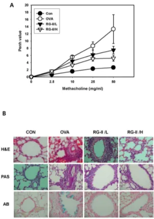
Fig. 1. Effect of RG-II on airway responsiveness, lung inflamma-tion, and inflammatory cell infiltration in OVA-treated mice. (A) Airway responsiveness was measured 24 hours after the final challenge in saline-inhaled mice administered PBS (CON), OVA-sensitized mice administered saline (OVA), OVA-sensitized mice administered a low dose of RG-II (20 mg/kg, RG-II/L), and OVA-sensitized mice administered a high dose of RG-II (100 mg/kg, RG-II/H). Airway responsiveness to aerosolized methacho-line was measured in unrestrained and conscious mice. Mice were placed into the main chamber and nebulized first with PBS, followed by increasing doses (2.5 to 50 mg/ml) of methacholine for 15 min for each nebulization. Readings of breathing parame-ters were taken for 3 minutes after each nebulization, during which Penh values were determined. (B) Mice were sensitized and challenged as described above. Sections were obtained from the lungs of mice receiving control (CON), OVA (OVA), OVA plus a low dose of RG-II (RG-II/L), and OVA plus a high dose of RG-II (RG-II/H). Lungs were removed 24 hours after the final air-way challenge. Sections were stained by haematoxylin and eosin, PAS, and Alcian blue (400×).
by binding directly to the promoters of IL-5 and IL-13, as well as by affecting chromatin remodeling, resulting in opening of the IL-4 locus (18). The T-box transcription factor T-bet (Tbx21) has emerged as a key regulator of dendritic cells as well as the type 1 immune response, playing an essential role in establish-ing effector cell fate in T and B lymphocytes (19). T-bet ex-pression is induced in TH1 cell,s but not TH2 cells, upon signal transduction, and acts as a potent transactivator of the IFN-γ gene in TH1, NK, and B cells (20). Therefore, we investigated the effects of RG-II on T-bet and GATA-3 expression in a mur-ine model of asthma.
In this study, administration of RG-II before the final airway OVA challenge resulted in significant inhibition of asthmatic reactions, suggesting that RG-II could play a critical role in the improvement of the pathogenic processes of asthma in mice.
RESULTS
RG-II inhibits AHR, lung inflammation, and inflammatory cell infiltration
Airway responsiveness was measured as a Penh value in re-sponse to increasing doses of methacholine. In OVA-sensitized and -challenged mice, the dose-response curve of the Penh value was shifted to the left compared to that of control mice (Fig. 1A). In addition, the Penh value produced by methacho-line administration (at doses ranging from 2.5-50 mg/ml) was significantly higher in the OVA-sensitized and -challenged mice compared to controls. In OVA-sensitized and -challenged mice treated with RG-II, the dose-response curve of the Penh value was shifted to the right compared to that of untreated OVA-sensitized and -challenged mice. Moreover, the shift was dose-dependent.
Histological analyses revealed the typical pathological fea-tures of asthma in OVA-exposed mice compared to control mice, with the OVA-exposed mice displaying numerous in-flammatory cells, including infiltrated eosinophils around the bronchioles (Fig. 1B). Mice treated with RG-II showed a marked decrease in inflammatory cell infiltration in the peri-bronchiolar and perivascular regions (Fig. 1B). Therefore, the increases in total lung inflammation and cell infiltration were significantly inhibited by administration of RG-II. These results suggest that RG-II inhibits OVA-induced airway hyper-responsiveness and antigen-induced inflammation in the lungs, including the influx of eosinophils.
RG-II reduces TH2 cytokine levels in lung tissues of
OVA-se-nsitized and -challenged mice
BAL fluids were obtained 24 hours after the final airway challenge. In the BAL fluids of mice subjected to airway chal-lenge with OVA, the levels of IL-4, IL-5, and IL-13 were sig-nificantly higher than those in control mice. Administration of RG-II reduced the secretion of IL-4 (Fig. 2A), IL-5 (Fig. 2B), and IL-13 (Fig. 2C). However, the levels of the TH2 cytokines IL-4, IL-5, and IL-13, as well as those of the TH1 cytokines IFN-γ
(Fig. 2D) and IL-12 (Fig. 2E) were higher in OVA-sensitized and OVA-challenged mice compared to cytokine levels in sal-ine-sensitized and -challenged control mice. These results in-dicate that RG-II treatment reduces TH2 cytokine levels, such as IL-4, IL-5, and IL-13, in BAL fluids.
RG-II decreases IgE levels in the serum and the number of inflammatory cells in BAL fluid
Fig. 2. Effect of RG-II on TH1 and TH2 cytokine production.
OVA-sensitized mice were treated as described in Materials and methods. BAL fluid was obtained 7 days after the final airway challenge as described by the manufacturer. IL-4 (A), IL-5 (B), IL-13 (C), IFN-γ (D), and IL-12 (E) levels in the BAL fluid was measured by ELISA.
Fig. 3. Effect of RG-II on IgE and IgG2a levels in serum of OVA-treated mice and the number of total cells and different cell types in BAL fluids of OVA-treated mice. Blood was collected by cardiac puncture, and serum IgE (A) and IgG2a (B) levels were measured. IgE and IgG2a levels were analyzed by using ELISA (n=5). (C) Mice were sensitized with OVA on days 0 and 14 by i.p. injection of OVA emulsified in 1 mg of aluminum hydroxide. Three days later, mice were treated with vehicle (CON), OVA (OVA), OVA plus a low dose of RG-II (20 mg/kg, RG-II/L), or OVA plus a high dose of RG-II (100 mg/kg, RG-II/H). The mice were challenged for 30 minutes with a 5% (w/v) OVA aerosol in saline (or saline alone as a control) using an ultrasonic nebulizer. The BAL cells were collected 24 hours after OVA challenge. The results shown are from a single representative experiment of the total five experiments performed.
Fig. 4. Effect of RG-II on expression of GATA-3 and T-bet in the lungs of OVA-treated mice. Samples were obtained 7 days after challenge from mice treated with control (CON), OVA (OVA), OVA plus a low dose of RG-II (20 mg/kg, RG-II/L), or OVA plus a high dose of RG-II (100 mg/kg, RG-II/H) and then subjected to Western blot analysis with α-GATA-3 (A) and α-T-bet (B) antibody.
by increasing IgE levels, which in turn favors the TH2 in-flammatory pathway, we measured how much RG-II modu-lates serum IgE levels in OVA-induced mice. As shown in Fig. 3, serum IgE levels in OVA-induced mice were significantly higher compared to those in control mice. RG-II significantly decreased serum IgE levels (Fig. 3A), but not serum IgG2a lev-els (Fig. 3B). These data indicate that RG-II modulates the TH1/TH2 balance towards TH1 in an OVA-induced asthma model. The numbers of total cells, eosinophils, lymphocytes, and macrophages in BAL fluid increased significantly 24 hours after OVA challenge compared to the numbers of cells after saline inhalation (Fig. 3C). These increases in the numbers of inflammatory cells were significantly inhibited by RG-II administration.
RG-II reduces GATA-3 expression in lung tissues of OVA-sen-sitized and -challenged mice
Western blot analysis showed that GATA-3 and T-bet ex-pression was significantly increased in lung tissues 24 hours af-ter OVA-challenge compared to control tissues. Administration of RG-II significantly inhibited this increase in GATA-3 ex-pression (Fig. 4A). On the other hand, RG-II administration in-duced an increase in T-bet expression (Fig. 4B).
DISCUSSION
inflammation in a murine model of asthma. Notably, RG-II profoundly inhibited asthmatic reactions, including leukocyte recruitment into the airway and lung inflammation. We also observed that RG-II regulates the TH1/TH2 balance by media-ting the levels of T-bet and GATA3.
OVA-induced asthma is recognized as a disease caused by chronic airway inflammation, which is characteristically asso-ciated with the infiltration of lymphocytes, eosinophils, and neutrophils into the bronchial lumen. Presently, we observed that OVA-induced asthma increased eosinophil infiltration, thickness of the bronchial wall, and surface area of smooth muscle. However, these events were significantly inhibited by administration of RG-II. T-bet, a member of the T-box family of transcription factors, is a master determinant of the TH1 cell lineage (20, 21). Indeed, T-bet-deficient mice lack the TH1 im-mune response (22), which inhibits allergic responses (23, 24). Moreover, ectopic expression of T-bet in murine TH2 cells di-rects activation of IFN-γ as well as upregulation of IL-12Rβ (25, 26). On the other hand, GATA-3 belongs to the GATA family of transcription factors. Six members (GATA-1 to GATA-6) of this family have been identified in birds, and homologues have been found in both mammals and birds. Based on their expression profiles and structures, GATA proteins may be clas-sified as either hematopoietic (GATA-1 to GATA-3) (27) or non-hematopoietic (GATA-4 to GATA-6). Naïve CD4+ T cells express low levels of GATA-3. However, the expression of GATA-3 is dependent upon T cell lineage; it is markedly upre-gulated in cells differentiating along the TH2 pathway, whereas it is downregulated in cells differentiating along the TH1 path-way (28). These data demonstrate that RG-II inhibits the in-crease in GATA3 expression in OVA-sensitized and OVA-chal-lenged mice (Fig. 4). Therefore, we can conclude that RG-II ad-ministration is a novel, selective way to simultaneously sup-press GATA-3 and increase T-bet exsup-pression in asthmatic re-actions in vivo (29).
In addition, TH1 and TH2 cytokine production was examined in CD4+ T cells. RG-II inhibited the increase in the level of IL-4, a TH2 cytokine produced in OVA-sensitized and OVA-challenged mice. In contrast, RG-II increased the level of IFN-γ, a TH1 cytokine produced in mice administered RG-II. Taken together, these results strongly indicate that RG-II re-duces allergic airway inflammation and hyperresponsiveness through modulation of the TH1/TH2 balance by suppressing GATA-3 expression and increasing T-bet expression.
In conclusion, these data suggest that RG-II might offer a new therapeutic approach to the treatment of allergic airway diseases.
MATERIALS AND METHODS
MiceFemale, 6 to 8-week-old, BALB/c mice that were certified free of murine-specific pathogens were obtained from Charles River Laboratories (Yokohama, Japan). All experimental
ani-mals were maintained under a protocol approved by the Institutional Animal Care and Use Committee of Pusan National University Medical School.
Purification of RG-II
A crude polysaccharide fraction (GL-2) was prepared from the leaves of Panax ginseng by hot water extraction, ethanol pre-cipitation, and dialysis (8). GL-2 was fractionated by a Cetavlon (cetyltrimethylammonium bromide) precipitation, and a weakly acidic polysaccharide fraction (GL-4) was obtained. The Fc re-ceptor expression-enhancing polysaccharide (RG-II) was puri-fied from GL-4 by anion-exchange chromatography on DEAE Sepharose CL-6B as described previously (30). To remove the colored materials in the polysaccharide, RG-II was further puri-fied on a Q-Sepharose column (C1- form). For this, the column was washed with water and eluted sequentially with 0.1, 0.2, 0.3, 0.4, 0.5, and 1.0 M NaC1. The major fraction, which was eluted with 0.3 M NaC1, was further fractionated by gel filtra-tion on a Bio-Gel P-30 column to obtain the purified GL-4IIb2 (yield: 5.8 mg/kg dry leaves).
Experimental protocol
Mice were sensitized intraperitoneally (i.p.) with 20 μg of OVA (Sigma-Aldrich, St. Louis, MO, USA) emulsified in 25 μl of aluminum hydroxide (Pierce, Rockford, IL, USA) on days 0 and 14. Mice were challenged for 30 min with OVA (5%) via the airway from days 20 to 22. Brochoalveolar lavage (BAL) fluid was obtained 24 hours after the final challenge. At the time of lavage, the mice (five mice from each group) were kil-led using an overdose of ether. The chest cavity was exposed to allow expansion, after which the trachea was carefully in-cubated and the catheter secured with ligatures. Pre-warmed saline solution was then slowly infused into the lungs and withdrawn. The aliquots were pooled and stored at 4oC. A por-tion of each pooled aliquot was then centrifuged, and the su-pernatants were kept at −70oC until use.
Administration of RG-II
Female BALB/c mice were injected i.p. with 20 mg/kg/day or 100 mg/kg/day of RG-II (in a 200 μl volume) everyday from days 17 to 19.
Total cell counting
Total cell numbers were counted with a hemocytometer. Smears of BAL cells prepared with Cytospin II (Shandon, Runcorn, UK) were stained with Diff-Quick solution (Merck, Darmstadt, Germany) for differential cell counting. Two in-dependent and blinded investigators counted the cells using a microscope. Approximately 200 cells were counted in each of four random locations.
Immunohistochemistry
Twenty-four hours after the final challenge, lungs were moved from the mice following sacrifice. Prior to lung
re-moval, the lungs and trachea were filled by intratracheal ad-ministration of a fixative (4% paraformaldehyde) using a liga-ture around the trachea. Lung tissues were then fixed with 10% (v/v) paraformaldehyde. The specimens were dehydrated and embedded in paraffin. For histological examination, 4 μm-thick sections of fixed and embedded tissues were cut on a Leica model 2165 rotary microtome (Leica, Nussloch, Germany), placed on glass slides, deparaffinized, and sequen-tially stained with hematoxylin 2 and eosin-Y (Richard-Allan Scientific, Kalamazoo, MI, USA). To identify mucus-producing cells, lung sections were stained with periodic acid-Schiff (PAS) and Alcian blue (AB).
Determination of airway responsiveness to methacoline
Airway responsiveness in mice was measured 24 hours after the final challenge in an unrestrained conscious state, as pre-viously described (9). Mice were placed in a barometric ple-thysmographic chamber (All Medicus, Seoul, Korea), and base-line readings were taken for 3 minutes and then averaged. Aerosolized methacholine at various concentrations (2.5 to 50 mg/ml) was nebulized through an inlet of the main chamber. Enhanced pause (Penh), which was calculated according to the manufacturer’s protocol as (expiratory time/relaxation time-1) ×(peak expiratory flow/peak inspiratory flow), is a dimension-less value that is a function of the proportion of maximal ex-piratory to maximal insex-piratory box pressure signals and a function of the timing of expiration. In this study, Penh was used as a measure of airway responsiveness to methacholine. Results are expressed as the percent increase in Penh follow-ing challenge with each concentration of methacholine, and the baseline Penh (after saline challenge) was set to 100%. Penh values taken for 3 minutes following each nebulization were averaged and evaluated.
Measurement of cytokines
Cytokine levels in BAL fluid was determined by enzyme-linked immunosorbent assay (ELISA). ELISA kits from R&D Systems (Minneapolis, MN, USA) were employed for the measurement of IL-4, IL-5, IL-13, IL-12, and IFN-γ.
Western blot analysis
The lung tissues were homogenized, washed with phos-phate-buffered saline (PBS), and incubated in lysis buffer con-taining a protease inhibitor cocktail (Sigma-Aldrich) to obtain extracts of lung and spleen proteins. The samples were loaded onto 10% sodium dodecyl sulfate-polyacrylamide gel electro-phoresis gels and the separated proteins were electrotrans-ferred to polyvinylidene difluoride membranes. The blots were incubated with anti-GATA-3 antibody or anti-T-bet antibody overnight at 4°C. After washing, the blots were next incubated with horseradish peroxidase-conjugated secondary antibody. Following three washes with Tris-buffered saline containing Tween-20, immunoreactive bands were visualized using an enhanced chemiluminescence detection system.
Measurement of OVA-specific serum IgE levels
OVA-specific serum IgE levels were determined in samples collected 24 hours after the final OVA challenge using ELISA. Briefly, a 96-well microtiter plate was coated with OVA (10 mg/ml), followed by incubation with mouse sera and then bio-tin-conjugated rat anti-mouse IgE (Pharmingen, San Diego, CA, USA). Then, avidin-horseradish peroxidase solution was added to each well. Units are shown as optical density readings at 405 nm.
Acknowledgements
This study was financially supported by the [2010 Post-Doc. Development Program] of Pusan National University and by a grant of the Korean Health Technology R&D Project, Ministry of Health & Welfare, Republic of Korea (A111025-1101- 0000100).
REFERENCES
1. Franchini, M., Gilli, U., Akens, M. K., Fellenberg, R. V. and Bracher, V. (1998) The role of neutrophil chemotactic cytokines in the pathogenesis of equine chronic ob-structive pulmonary disease (COPD). Vet. Immunol.
Immunopathol. 66, 53-65.
2. Yawn, B. P. (2008) Factors accounting for asthma varia-bility: achieving optimal symptom control for individual patients. Prim Care Respir J. 17, 138-147.
3. Spicuzza, L., Bonfiglio, C. and Polosa, R. (2003) Research applications and implications of adenosine in diseased airways. Trends Pharmacol. Sci. 24, 409-413.
4. Mapp, C. E., Boschetto, P., Zocca, E., Milani, G. F., Pivirotto, F., Tegazzin, V. and Fabbri, L. M. (1987) Pathogenesis of late asthmatic reactions induced by ex-posure to isocyanates. Bull Eur. Physiopathol. Respir. 23, 583-586.
5. Bousquet, J., Chanez, P., Lacoste, J. Y., Barneon, G., Ghavanian, N., Enander, I., Venge, P., Ahlstedt, S., Simony-Lafontaine, J. and Godard, P. (1990) Eosinophilic inflammation in asthma. N. Engl. J. Med. 323, 1033-1039. 6. Kay, A. B. (1991) Asthma and inflammation. J. Allergy
Clin. Immunol. 87, 893-910.
7. Park, H. J., Lee, C. M., Jung, I. D., Lee, J. S., Jeong, Y. I., Chang, J. H., Chun, S. H., Kim, M. J., Choi, I. W., Ahn, S. C., Shin, Y. K., Yeom, S. R. and Park, Y. M. (2009) Quercetin regulates Th1/Th2 balance in a murine model of asthma. Int. Immunopharmacol. 9, 261-267.
8. Gao, Q. P., Kiyohara, H., Cyong, J. C. and Yamada, H. (1989) Chemical properties and anti-complementary activ-ities of polysaccharide fractions from roots and leaves of Panax ginseng. Planta. Med. 55, 9-12.
9. Sun, X. B., Matsumoto, T. and Yamada, H. (1992) Purifi-cation of an anti-ulcer polysaccharide from the leaves of Panax ginseng. Planta. Med. 58, 445-448.
10. Cher, D. J. and Mosmann, T. R. (1987) Two types of mur-ine helper T cell clone. II. Delayed-type hypersensitivity is mediated by TH1 clones. J. Immunol. 138, 3688-3694. 11. Kim, J., Woods, A., Becker-Dunn, E. and Bottomly, K.
Ia-re-stricted helper T cells. J. Exp. Med. 162, 188-201. 12. Corrigan, C. J. and Kay, A. B. (1992) T cells and
eosino-phils in the pathogenesis of asthma. Immunol. Today 13, 501-507.
13. Karp, M. and Oker-Blom, C. (1999) A streptavidin-lucifer-ase fusion protein: comparisons and applications. Biomol.
Eng. 16, 101-104.
14. Iwamoto, I., Nakajima, H., Endo, H. and Yoshida, S. (1993) Interferon gamma regulates antigen-induced eosino-phil recruitment into the mouse airways by inhibiting the infiltration of CD4+ T cells. J. Exp. Med. 177, 573-576. 15. Tanaka, H., Komai, M., Nagao, K., Ishizaki, M., Kajiwara,
D., Takatsu, K., Delespesse, G. and Nagai, H. (2004) Role of interleukin-5 and eosinophils in allergen-induced air-way remodeling in mice. Am. J. Respir. Cell. Mol. Biol. 31, 62-68.
16. Sur, S., Lam, J., Bouchard, P., Sigounas, A., Holbert, D. and Metzger, W. J. (1996) Immunomodulatory effects of IL-12 on allergic lung inflammation depend on timing of doses. J. Immunol. 157, 4173-4180.
17. Ho, I. C., Vorhees, P., Marin, N., Oakley, B. K., Tsai, S. F., Orkin, S. H. and Leiden, J. M. (1991) Human GATA-3: a lineage-restricted transcription factor that regulates the ex-pression of the T cell receptor alpha gene. Embo. J. 10, 1187-1192.
18. Zhou, M. and Ouyang, W. (2003) The function role of GATA-3 in Th1 and Th2 differentiation. Immunol. Res. 28, 25-37.
19. Renz, H., Saloga, J., Bradley, K. L., Loader, J. E., Greenstein, J. L., Larsen, G. and Gelfand, E. W. (1993) Specific V beta T cell subsets mediate the immediate hy-persensitivity response to ragweed allergen. J. Immunol. 151, 1907-1917.
20. Szabo, S. J., Kim, S. T., Costa, G. L., Zhang, X., Fathman, C. G. and Glimcher, L. H. (2000) A novel transcription factor, T-bet, directs Th1 lineage commitment. Cell 100, 655-669.
21. Montefort, S. and Holgate, S. T. (1991) Adhesion mole-cules and their role in inflammation. Respir. Med. 85, 91-99.
22. Szabo, S. J., Sullivan, B. M., Stemmann, C., Satoskar, A. R., Sleckman, B. P. and Glimcher, L. H. (2002) Distinct ef-fects of T-bet in TH1 lineage commitment and IFN-gamma production in CD4 and CD8 T cells. Science 295, 338-342.
23. Gavett, S. H., O'Hearn, D. J., Li, X., Huang, S. K., Finkelman, F. D. and Wills-Karp, M. (1995) Interleukin 12 inhibits antigen-induced airway hyperresponsiveness, in-flammation, and Th2 cytokine expression in mice. J. Exp.
Med. 182, 1527-1536.
24. Li, X. M., Chopra, R. K., Chou, T. Y., Schofield, B. H., Wills-Karp, M. and Huang, S. K. (1996) Mucosal IFN-gam-ma gene transfer inhibits pulmonary allergic responses in mice. J. Immunol. 157, 3216-3219.
25. Afkarian, M., Sedy, J. R., Yang, J., Jacobson, N. G., Cereb, N., Yang, S. Y., Murphy, T. L. and Murphy, K. M. (2002) T-bet is a STAT1-induced regulator of IL-12R expression in naive CD4+ T cells. Nat. Immunol. 3, 549-557.
26. Mullen, A. C., High, F. A., Hutchins, A. S., Lee, H. W., Villarino, A. V., Livingston, D. M., Kung, A. L., Cereb, N., Yao, T. P., Yang, S. Y. and Reiner, S. L. (2001) Role of T-bet in commitment of TH1 cells before IL-12-dependent selection. Science 292, 1907-1910.
27. Orkin, S. H. (1995) Hematopoiesis: how does it happen?
Curr. Opin. Cell. Biol. 7, 870-877.
28. Ouyang, W., Ranganath, S. H., Weindel, K., Bhattacharya, D., Murphy, T. L., Sha, W. C. and Murphy, K. M. (1998) Inhibition of Th1 development mediated by GATA-3 through an IL-4-independent mechanism. Immunity 9, 745-755.
29. Bian, T., Yin, K. S., Jin, S. X., Zhang, X. L., Zhou, J. Y., Ma, X. Q., Hu, J. J. and De, W. (2006) Treatment of allergic airway inflammation and hyperresponsiveness by imiqui-mod imiqui-modulating transcription factors T-bet and GATA-3.
Chin. Med. J. (Engl) 119, 640-648.
30. Shin, K., Lee, J. C., Choi, H. J., Jeong, J. M., Son, M., Lee, Y. J., Lee, E. B., Hong, S. H. and Song, Y. W. (2007) Radiation synovectomy using 188Re-tin colloid improves knee synovitis as shown by MRI in refractory rheumatoid arthritis. Nucl. Med. Commun 28, 239-244.