Volume 12, Number 2, December, 2009
Introduction
The discovery of two small RNAs, lin-4 and let-7 during the study of genes involved in timing of larval development of Caenohabditis elegans was the official introduction of the microRNAs (miRNA) to the world1,2)
. Only 17 to 23 nucelotides in length, these small RNAs were soon recognized as a key regulator of the devel-opment of C.elegans and interestingly
they were highly conserved across species. Following the initial discovery, numerous groups have began their own expedition onto the unknown territories of the world of miRNA, and have successfully identi-fied that not only were they crucial for the larval development but also in cell proliferation3) and apoptosis4) , immunore-sponse5) , protein expression6) and tissue development7)
as well as in various other biological processes. Currently, few
※ 통신저자: 이 진 우
서울특별시 서대문구 성산로 250 연세대학교 의과대학 정형외과학교실
TEL: 02) 2228-2190 FAX: 02) 393-8345 E-mail: ljwos@yuhs.ac 접수일: 2009년 11월 23일, 게재확정일 2009년 11월 27일
The Role of microRNAs Involved in
Mesenchymal Stem Cell Differentiation
Seungil Paik, Ho Sun Jung, Jin Woo Lee, M.D.*
Department of Orthopaedic Surgery, Yonsei University College of Medicine, Korea
= Abstract =
Over the past decade, microRNA has been emerging as a key regulator involved in various biological and physiological process including cell cycle, tissue development and pathogenesis. The effort to decipher the complicated machinery behind microRNA function is underway yet the complete understanding is far from being reached. However, various groups have made successful identification of the role of a specific microR-NA involved in a specific process. This article summarizes the microRmicroR-NAs identified in one such specific area, namely, differentiation of mesenchymal stem cell. Characterized by its unique potential to differentiate into three separate tissue cell types, adipocyte, chondrocyte and osteoblast, mesenchymal stem cells have been of immense interest for its possible applicability in regenerative medicine. Despite this interest, however, the study of microRNA function in MSCs differentiation remains rudimentary. The aim of this paper is to summa-rize the currently identified, experimentally validated and accepted by the scientific community as novel, microRNAs involved in MSC differentiation.
miRNA are clearly identified and assigned to specific function in mammals. miR-181
promotes B cell development in mice8)
, and it targets the homeobox protein Hox-A11 during mammalian myoblast differentia-tion9)
. miR-196 regulates several Hox genes while the brain-specific miR-134
regulates dendritic spine development10)
. miR-1, miR-133 and miR-206 are
specifi-cally induced during myogenesis11)
and miR-143 are known to regulate adiocytic
differentiation12)
. However, despite the recent achievements in identification of miRNAs involved in various processes, the studies on the miR function in one partic-ular field of physiology remain at primi-tive stage: the differentiation of mes-enchymal stromal cells.
Mesecnhymal stromal cell, also known as MSCs, are plastic-adherent, fibroblast-like cells first identified and described by
Friedenstein13)
. Following its initial discov-ery, various groups have identified the cells capacity to differentiate into multiple connective tissue cell types, including fat,
tendon, cartilage and bone14,15,16)
. Their wide abundance in body coupled with their mul-tipotent differentiation capacity has made the MSCs an ideal candidate for use in regenerative medicine, which led to the creation of its alias, mesenchymal stem cell, coined by Caplan. Despite this immense interest in dissecting the exact mechanism behind MSCs capacity to differ-entiate into multiple lineages, our under-standing of all the regulators involved in MSC differentiation remains elusive. Until recently, MSCs differentiation was thought to be solely regulated by various transcrip-tion and growth factors, hormones, and signaling pathways provided in an optimal condition. However, with the discovery of
miRNA, and considering its comprehensive impact on other biological processes, the regulatory role of miRNA on MSC differ-entiation seems convincing.
The functional mechanism of
microRNA
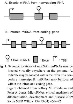
Before attempting to understand the complex regulatory functions miRNA may have on the differentiation process of MSCs, sound knowledge in how miRNA are synthesized and, more importantly how they induce inhibitory effect on its target gene must be established. Current-ly, 10883 hairpin precursor miRNAs, expressing 10581 mature miRNA products, in 115 species have been identified in the Sanger database version 14.0. Being either intronic or exonic, and often located in non-coding regions between genes
(inter-Fig. 1. Genomic locations of miRNAs. miRNAs may be
located virtually anywhere on the genome. A. miRNA may be located within the exon of a non-coding transcript B. miRNAs may be located within the intron of a coding gene.
Figure obtained from Jeffrey M. Friedman and Peter A. Jones, MicroRNAs: critical mediators of differentiation, development and disease 2009 Swiss MED WKLY 139(33-34):466-472
genic) and sometimes within the coding region, miRNA expression may be induced by its own promoter (intergenic) or by that of its host gene (Fig. 1). The tran-scription product quickly folds back onto itself, forming a hairpin structure which is then cleaved by Drosha within the nucleus17)
. Once cleaved, it is exported to
cytoplasm by exportin18)
, secondary cleav-age by Dicer before forming a functional complex called RNA-induced silencing
complex (RISC)19)
. This complex recognizes the complementary sequence of its miRNA on the target genes 3’UTR region, which does not require a perfect match, and induce inhibition of gene expression by either of the two mechanisms: mRNA
cleavage20)
or translational repression21)
. However, the complete identification of all regulators involved in miRNA expression and its functioning pathway remains to be fully identified and the data presented here only summarizes the knowledge established and accepted currently.
MSC differentiation
As previously mentioned, MSCs have trilineage differentiation potential to
dif-ferentiate into adipoblast, chondroblast and osteoblast resulting in formation of fat, cartilage and bone, respectively. Recent advances in transcriptomics and proteomics have allowed us to study tran-scriptional regulation involved in these stage in more detail and have yielded valuable information. In the following sections, the differentiation process into three separate lineages will be examined closely involving identified transcription factors, genes and miRNA.
I. Osteogenesis
The long and complicated process of osteogenesis is initiated with cellular pro-liferation followed by extracellular matu-ration (ECM) which ultimately leads to matrix mineralization. Bone morphogenic protein (BMP), Wnt and Hedgehog signal-ing are accepted as the key signalsignal-ing pathway involved during the process. These signaling pathways work symbioti-cally in a systematisymbioti-cally regulated path-way and exert their impact on down-stream transcription factors such as Cbfa1/Runx2, Msx2, Dlx5 and Osterix. Over the past decade, handfuls of
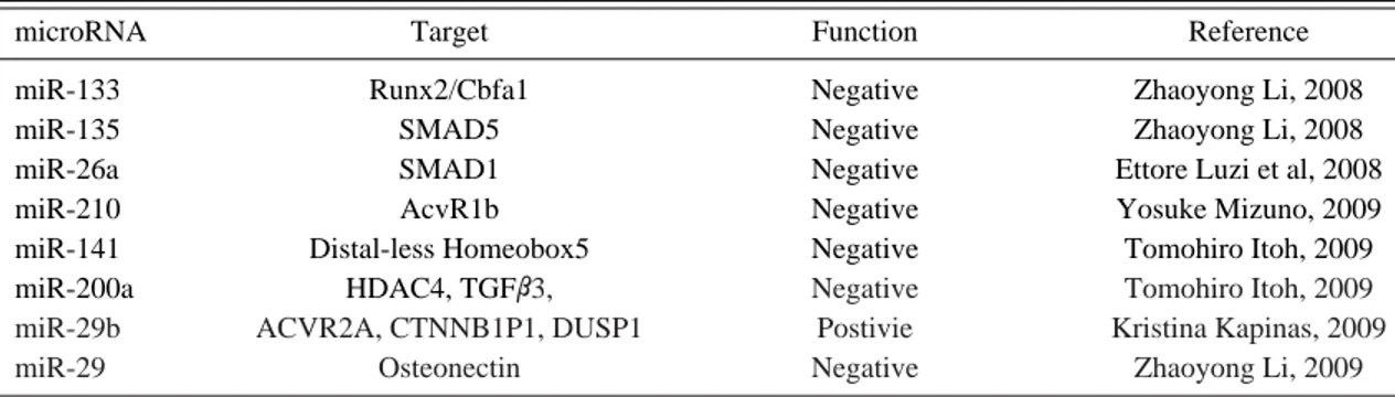
miR-Table 1. Identified microRNAs involved in osteogenesis and their targets
microRNA Target Function Reference
miR-133 Runx2/Cbfa1 Negative Zhaoyong Li, 2008
miR-135 SMAD5 Negative Zhaoyong Li, 2008
miR-26a SMAD1 Negative Ettore Luzi et al, 2008
miR-210 AcvR1b Negative Yosuke Mizuno, 2009
miR-141 Distal-less Homeobox5 Negative Tomohiro Itoh, 2009 miR-200a HDAC4, TGFβ3, Negative Tomohiro Itoh, 2009
miR-29b ACVR2A, CTNNB1P1, DUSP1 Postivie Kristina Kapinas, 2009
miR-29 Osteonectin Negative Zhaoyong Li, 2009
List of the published microRNAs targeting genes involved in osteogenesis. Under function column, the microRNAs shown to inhibit osteogenesis are labeled as “negative” and the microRNAs shown to promote osteogenesis are labeled “positive.”
NAs have been identified to inhibit or induce osteogenesis, which are summarized on Table 1. Specifically, Zhaoyong Li et al. have identified that miR-133 directly tar-gets Runx2/Cbfa1 while miR-135 tartar-gets Smad5, which is a key transducer of the BMP-2 osteogenic signal, both of which had a complement sequence to respective
miRNAs on their 3’UTR region22)
. SMAD1 was found to have two binding sites for
miR-26a on its 3’UTR region23)
while miR-210 was identified to promotes osteogenesis via targeting AcvR1b and that miR-141 and -200a targets Distal-less Homeobox 5, respectively 24,25)
. Kristina Kapinas et al. have discovered that miR-29 suppresses
osteonectin in osteoblast26)
. Although major-ity of the data have identified the miRNAs which had an inhibitory effect on osteogen-esis, some have also found the miR target-ing the inhibitors of osteogenesis, which in turn promoted the differentiation. One particular study was carried out by Zhaoy-ong Li, the same group which have suc-cessfully identified that miR-133 targets Runx2/Cbfa1, in which they have discov-ered that miR-29b targets multiple inhibitors of osteoblast differentiations,
HDAC4, TGFβ3, ACVR2A, CTNNBIP1 and
DUSP227)
. From these findings, one can understand that although the basic func-tioning mechanism of miR is to inhibit the expression of its target gene by binding to the 3’UTR region, based on which gene the miR targets, miR can either promote or inhibit the differentiation process. Although the majority of the data men-tioned in this review have used mouse osteoblast cell lines, such as MC3T3 and ST2, and human adipose derive stem cell (hADSC), the repeating theme here is that the miRNA is highly conserved across
species and as such the resulting data from these alternative cell lines are expected to be readily translated onto MSCs as well.
II. Chondrogenesis
Similar to osteogenesis, chondrogenesis also occur via multiple stages initiated by cell condensation driven by upregulated N-cadherin, neural cell adhesion moleu-cule (NCAM) and fibronectin, promoting cell-to-cell and cell-to-ECM interactions. Once the differentiating cells become com-mitted into chondroblast phase, the cells switch the major components of the ECM from Type I collagen, which is a charac-teristic of an undifferentiated MSCs, to more cartilaginous make up involving Type-II, Type-IX and Type -XI collagen, Cartilage oligomeric matrix protein-1 (COMP-1) and Aggrecan. Ultimately, the chondrocyte reaches the hypertrophic state where the cells start to express Type-X collagen, unique to the hypertrophic stage of chondrogenesis. Eventually, as part of the natural bone growth and develop-ment, the hypertrophic chondrocytes undergo apoptosis and are replaced by bone. This long and complicated process of chondrogenesis are directed by number of
signaling pathways involving TGFβ, FGF,
Wnt and Indian hedgehog (Ihh) signaling to name a few. In turn, the aforemen-tioned signaling pathways direct the expression of various transcription factors involved in chondrogenesis such as the SOX 5, SOX 6, SOX 9, also known as the SOX trio. Currently, handful of miRNAs have been experimentally validated as a key regulator in chondrogenesis. One study reported that miRNA18a targets CCN family protein 2/connective tissue
growth factor (CCN2/CTGF), which is a central player in endochondral bone
for-mation28)
. Additionally, miR-140 was found to inhibit the expression of histone
deacetylase 429)
, which may be involved in long bone development. miR-199a* had a profound inhibition on the early stages of chondrogenesis in pluripotent C3H10T1/2 stem cells grown on pellet culture. When treated with miR-199a*, the authors have observed significant reduction of key chondrogenic markers, namely; cartilage oligomeric matrix protein (COMP), sox 9 and Type II Collagen, whereas inhibition of miR-199a* resulted in increase of these genes30)
. Interestingly, unlike in studies which focused on the effect of miRNA during osteogenesis, several groups study-ing miRNA in chondrogenesis approached the subject with quite different aspect. Walter Dunn et al. have examined the miRNAs expressed on different areas of articular cartilage, specifically classified by the degree of weight-bearing. The authors have found that miRNA-221 and miR-222 were significantly up-regulated on higher weight-bearing region identified as M1, which is located on the anterior part of the cartilage, suggesting that this particular localization on anterior medial condyle may have a certain function on
weight-bearing capacity of the cartilage31)
. Another group, have observed that miRNA-146a were gradually reduced as osteoarthritis progressed and have also found out that miR-146a targets matrix
metalloproteinase 13 (MMP-13)32)
. III. Adipogenesis
Similar to the patterns shared by osteo-genesis and chondroosteo-genesis, adipoosteo-genesis
also occur through multiple steps initiated by commitment to pre-adipocytes. The signaling cascade is initiated by the co-expression of CCAAT/enhancer binding
protein β (C/EBPβ) along with C/EBPδ,
which leads to the activation of down-stream transcription factors C/EBPα and peroxisome proliferator-activated receptor
γ (PPARγ). By the terminal
differentia-tion stage, numerous genes are expressed which include hormone-sensitive lipase (HSL), fatty acid binding protein (FABPs) and glycerol-3-phosphate dehy-drogenase (GPDH). During this period, various adipokines are also secreted including leptin (LEP) and adiponectin (ADIPOQ). Michael Karbiener et al. suc-ceeded in identifying the miRNA target-ing the key transcription factor durtarget-ing
adipogenesis: PPARγ. By using human
multipotent adipose-derived stem
(hMADS) cells, the authors have found that when the cells were treated with miR-27b over-expressing vector, number of adipogenic marker genes showed notable decrease in expression, which
included PPARγ, FABP4 and LPL33)
. Christine Esau et al. have also observed the increase of miR-143 in differentiating adipocytes, and that when miR-143 was inhibited so was the adipogenesis while BP Lewis et al. have suggested the possible target of miR-143 as the extracellular
sig-nal-regulated kinase 5 (ERK5)34)
. In last two years, number of groups has also suc-cessfully identified miRNAs involved in adipogenesis, including the positive effect of miR-103 and miR-19-92 cluster had on
adipogenesis35,36)
and the negative effect
induced by let-7 and miR-2737,38)
. In a study using ST2 cells, J.A. Kennell et al. have identified multiple miRNAs which
promoted adipogenesis, which were
miR-200c/141 and miR-200b,a/42939)
. In C3H10T1/2 cells, F.Sun et al. have suc-cessfully identified that miR-24 enhanced adipogenesis while miR-31 enforced nega-tive effect40)
.
Conclusion
Despite being relatively recently identi-fied, miRNAs are proving to be a critical mediators in various biological processes including tissue development, immunore-sponse, protein expression, viral infection and cell proliferation, division, apoptosis and differentiation. However, despite the recent achievements in identifying the functioning pathway of miRNA and the impact of individual miRNA on a specific pathway, we are still at a primitive stage in understanding the whole picture that miRNA draws. One such aspect is our limited understanding of how miRNA actually recognizes its target and induce its effect. Although a computerized algo-rithm and simplified means of communi-cations have yielded useful database such as TargetScan and PicTar, which predicts the potential target of each identified miRNA based on sequence complementari-ty, significant portion of the predicted targets remained independent of miRNA treatment. Such result makes us speculate the existence of additional regulatory mechanism that exists between the target mRNAs and the miRNA. Regardless, var-ious groups are making progress in under-standing the complete pathway of miRNA and more importantly, succeeding in applying miRNA in the field of medicine. For example, Gilad S et al. have devel-oped a novel technique to isolate miRNAs
from bodily fluids such as urine41)
, which Mitchell et al. have utilized to identify miR-141 isolated from plasma as a novel
indicator of prostate cancer42)
. One report suggested that more than one third of all protein coding genes in human genome
may be regulated by miRNA43)
, and con-sidering the unparalleled advance in tech-nology as well as the continued global effort to gain complete understanding of miRNA, its use and applicability seems, truly, endless. It seems, paradoxically, the potential for a molecule with such a tiny name, microRNA, has an endless potential.
REFERENCE
01) Lee, R.C., Feinbaum, R.L., and Ambrox: V The C. elegans heterochronic gene lin-4 encodes small RNAs with antisense complementarity to lin-14. Cell 75, 843-854, 1993.
02) Reinhart, B.J., Slack, F.J., Basson, M. et al.: The 21-nucleotide let-7 RNA regulates develop-mental timing in Caenorhabditis elegans. Nature 403, 901-906, 2000.
03) Hutvagner G, Zamore P.D.A: microRNA in a multiple-turnover RNAi enzyme complex. Sci-ence 297: 2056-2060, 2002.
04) Chen, Y., Stallings, R.L: Differential patterns of microRNA expression in neuroblastoma are cor-related with prognosis, differentiation, and apop-tosis. Cancer Res 67: 976-983, 2007
05) Wu, H., Neilson, J.R., Kumar, P., Manocha, M.,
Shankar, P., et al.: miRNA profiling of na ve,
effector and memory CD8 T cells. PLoS ONE 2: e1020, 2007
06) Poy, M.N., Eliasson, L., Krutzfeldt, J.,
Kuwaji-ma, S., Ma, X., et al.: A pancreatic islet-specific
microRNA regulates insulin secretion. Nature 432:226-230, 2004
control-ling left/right neuronal asymmetry in Caenorhab-ditis elegans. Nature 426: 845-849, 2003
08) Chen CZ, Li L, Lodish HF, Bartel DP: MicroRNAs modulate hematopoietic lineage dif-ferentiation. Science 303:83-86, 2004..
09) Naguibneva I, Ameyar-Zazoua M, Polesskaya
et al.: The microRNA miR-181 targets the
homeobox protein Hox-A11 during mammalian myoblast differentiation. Nat Cell Biol 8:278-284, 2006
10) Yekta S, Shih IH, Bartel DP: MicroRNA-direct-ed cleavage of HOXB8 mRNA. Science 304:594-596, 2004
11) Rao PK, Kumar RM, Farkhondeh M,
Baskerville S, Lodish HF: Myogenic factors
that regulate expression of musclespecific microRNAs. Proc Natl Acad Sci USA 6:8721-8726, 2006
12) Esau C, Kang X, Peralta E et al.: MicroRNA-143 regulates adipocyte differentiation. J Biol Chem 279:52361-5236, 2004
13) Friedenstein AJ: Marrow stromal fibroblasts. Calcif Tissue Int 1995; 56(suppl 1):S17.
14) Dennis JE, Merriam A, Awadallah A et al.: A quadripotential mesenchymal progenitor cell iso-lated from the marrow of an adult mouse. J Bone Miner Res;14:700-709, 1999.
15) Pittenger MF, Mackay AM, Beck SC et al.: Multilineage potential of adult human mesenchy-mal stem cells. Science;284:143-147, 1999. 16) Haynesworth SE, Baber MA, Caplan AI: Cell
surface antigens on human marrow-derived mes-enchymal cells are detected by monoclonal anti-bodies. Bone,13:69-80, 1992.
17) Lee Y, Ahn C, Han J et al.: The nuclear RNase III Drosha initiates microRNA processing. Nature;425:415, 2003
18) Yi R, Qin Y, Macara IG et al.: Exportin-5 mediates the nuclear export of pre-microRNAs and short hairpin RNAs. Genes Dev;17:3011, 2003
19) Hutvagner G, McLachlan J, Pasquinelli AE et
al.: A cellular function for the RNA-interference
enzyme Dicer in the maturation of the let-7 small temporal RNA. Science;293:834, 2001.
20) Aukerman MJ, Sakai H: Regulation of flower-ing time and floral organ identity by a MicroR-NA and its APETALA2-like target genes. Plant Cell15:2730-2741. 2003.
21) Zhang L, Huang J, Yang N et al.: microRNAs exhibit high frequency genomic alterations in human cancer. Proc Natl Acad Sci U S A103:9136 ?9141, 2006.
22) Zhaoyong Li, Mohammad Q. Hassan, Stefano
Volinia et al.: A microRNA signature for a
BMP2-induced osteoblast lineage commitment program. PNAS. 105;13906-13911, 2008. 23) Ettore Luzi, Francesca Marini, Silvia
Car-bonell Sala et al.: Osteogenic Differentiation of
Human Adipose Tissue-Derived Stem Cells Is Modulated by the miR-26a Targeting of the SMAD1 Transcription Factor. Journal of Bone and Mineral Research. 23; 287-295, 2008. 24) Yosuke Mizuno, Yoshimi Tokuzawa, Yuichi
Ninomiya et al.: miR-210 promotes osteoblastic
differentiation through inhibition of AcvR1b. FEBS Letts. 583; 2263-2268, 2009.
25) Tomohiro Itoh, Yoshinori Nozawa, and
Yuki-hiro Akao: MicroRNA-141 and -200a Are
Involved in Bone Morphogenetic Protein -2-induced Mouse Pre-Osteoblas Differentiation by Targeting Distal-less Homoeobox 5. J Biol Chem. 284; 19272-19279, 2009.
26) Kristina Kapinas, Catherin B. Kessler, and
Anne M. Delany: miR-29 Suppression of
Osteonectin in Osteoblasts: Regulation During Differentiation and by Canonical Wnt Signaling. J Cell Biochem.; 216-224, 2009
27) Zhaoyong Li, Mohammad Q. Hassan et al.: Biological Functions of miR-29b Contributes to Positive Regulation of Osteoblast Differentia-tion. J Biol Chem. 284; 15676-1568, 2009 28) Toshihiro Ohgawara, Satoshi Kubota et al.:
RNA 18a: Involvement of Ccn2/Ctgf as a major target gene. FEBS letts.. 583; 1006-1010, 2009. 29) Shigeru Miyaki, Tomoyuki Naksa et al.:
MicroRNA-140 is expressed in differentiated human articular chondrocytes and modulates interleukin-1 response. Arthritis Rheum. 60; 2723-2730, 2009
30) Edward A. Lin, Li Kong, Xiao-Hui Bai, Yi
Luan, and Chuan-ju Liu: miR-199a*, a Bone
Morphogenic Protein 2-responsive microRNA, regulates chondrogenesis via direct targeting to Smad1. J Biol Chem.. 284; 11326-1133, 2009 31) Walter Dunn, Grayson DuRaine, and A.Hari
Reddi: Profiling microRNA expression in
bovine articular cartilage and implications for mechanotransduction. Arthritis Rheum. 60;2333-2339, 2009
32) Keiichiro Yamasaki, Tomoyuki Nakasa et al.: Expression of MicroRNA-146a in Osteoarthritis Cartilage. Arthritis Rheum. 60; 1035-1041, 2009. 33) Michael Karbiener, Christoph Fischer,
Susanne Nowitsch et al.: microRNa miR-27b
impairs human adipocyte differentiation and tar-gets PPARγ. Biochemcial and Biophysical Research Communications. 390; 247-251, 2009 34) C. Esau, X. Kang, E. Peralta, E et al.:
MicroR-NA-143 regulates adipocyte differentiation, J. Biol. Chem. 279 52361-52365, 2004.
35) H. Xie, B. Lim, H.F: Lodish, MicroRNAs induced during adipogenesis that accelerate fat cell development are downregulated in obesity, Diabetes 58 (2009) 1050-1057.
36) Q. Wang, Y.C. Li, J. Wang, J. Kong, Y. Qi,
R.J: Quigg, X. Li, miR-17-92 cluster accelerates adipocyte differentiation by negatively regulating tumor-suppressor Rb2/p130, Proc. Natl. Acad. Sci. USA 10; 2889-2894, 2008.
37) T. Sun, M. Fu, A.L. Bookout, S.A. Kliewer, D.J: Mangelsdorf, MicroRNA let-7 regulates 3T 3-L1 adipogenesis, Mol. Endocrinol. 23; 925-931, 2009
38) Q. Lin, Z. Gao, R.M. Alarcon, J. Ye, Z. Yun: A role of miR-27 in the regulation of adipogene-sis, FEBS J. 276 ;2348-2358, 2009.
39) J.A. Kennell, I. Gerin, O.A. MacDougald, K.M: Cadigan, The microRNA miR-8 is a con-served negative regulator of Wnt signaling, Proc. Natl. Acad. Sci. USA 105 ; 15417-15422, 2008. 40) F. Sun, J. Wang, Q. Pan, Y. Yu, Y. Zhang, Y.
Wan, J. Wang, X. Li, A: Hong, Characteriza-tion of funcCharacteriza-tion and regulaCharacteriza-tion of miR-24-1 and miR-31, Biochem. Biophys. Res. Commun. 380; 660-665, 2008.
41) Gilad S, Meiri E, Yogev Y, Benjamin S, Lebanony D, Yerushalmi N, et al.: Serum microRNAs are promising novel biomarkers. PLoSONE;3(9):e3148, 2008.
42) Mitchell PS, Parkin RK, Kroh EM et al.: Cir-culating microRNAs as stable blood-based mark-ers for cancer detection. Proc Natl Acad Sci U S A;105(30):10513-8, 2008.
43) Berezikov E, Guryev V, van de Belt J, Wien-holds E, Plasterk RH, Cuppen E: Phyolgenetic shadowing and computation identification of human micro RNA genes. Cell. 120;21-24, 2005.