Cytochrome P450 Monooxygenase-Mediated Metabolic
Utilization of Benzo[a]Pyrene by Aspergillus Species
Erin M. Ostrem Loss,aMi-Kyung Lee,bMing-Yueh Wu,c* Julia Martien,cWanping Chen,dDaniel Amador-Noguez,c Colin Jefcoate,aChristina Remucal,a,eSeunho Jung,fSun-Chang Kim,g Jae-Hyuk Yua,c,f
aMolecular and Environmental Toxicology Center, University of Wisconsin—Madison, Madison, Wisconsin, USA
bKorea Research Institute of Bioscience and Biotechnology (KRIBB), Daejon, Republic of Korea
cDepartment of Bacteriology, University of Wisconsin—Madison, Madison, Wisconsin, USA
dCollege of Food Science and Technology, Huazhong Agricultural University, Wuhan, People’s Republic of China
eDepartment of Civil and Environmental Engineering, University of Wisconsin—Madison, Madison, Wisconsin, USA
fDepartment of Systems Biotechnology, Konkuk University, Seoul, Republic of Korea
gDepartment of Biological Sciences, Korea Advanced Institute of Science and Technology, Daejon, Republic of Korea
ABSTRACT Soil-dwelling fungal species possess the versatile metabolic capability to degrade complex organic compounds that are toxic to humans, yet the mechanisms they employ remain largely unknown. Benzo[a]pyrene (BaP) is a pervasive carcino-genic contaminant, posing a significant concern for human health. Here, we report that several Aspergillus species are capable of degrading BaP. Exposing Aspergillus
ni-dulans cells to BaP results in transcriptomic and metabolic changes associated with
cellular growth and energy generation, implying that the fungus utilizes BaP as a growth substrate. Importantly, we identify and characterize the conserved bapA gene encoding a cytochrome P450 monooxygenase that is necessary for the meta-bolic utilization of BaP in Aspergillus. We further demonstrate that the fungal NF- B-type velvet regulators VeA and VelB are required for proper expression of bapA in re-sponse to nutrient limitation and BaP degradation in A. nidulans. Our study illuminates fundamental knowledge of fungal BaP metabolism and provides novel insights into enhancing bioremediation potential.
IMPORTANCE We are increasingly exposed to environmental pollutants, including the carcinogen benzo[a]pyrene (BaP), which has prompted extensive research into human metabolism of toxicants. However, little is known about metabolic mecha-nisms employed by fungi that are able to use some toxic pollutants as the sub-strates for growth, leaving innocuous by-products. This study systemically demon-strates that a common soil-dwelling fungus is able to use benzo[a]pyrene as food, which results in expression and metabolic changes associated with growth and en-ergy generation. Importantly, this study reveals key components of the metabolic utilization of BaP, notably a cytochrome P450 monooxygenase and the fungal NF-B-type transcriptional regulators. Our study advances fundamental knowledge of fungal BaP metabolism and provides novel insight into designing and implementing enhanced bioremediation strategies.
KEYWORDS Aspergillus, benzo[a]pyrene, catabolic enzyme system, cytochrome P450
monooxygenase, polycyclic aromatic hydrocarbons, velvet regulators, bioremediation, genome-wide expression, high-performance liquid chromatography, metabolomics, molecular genetics
P
olycyclic aromatic hydrocarbons (PAHs) are major soil pollutants that are formed by the partial combustion of organic matter and the five-ring PAH benzo[a]pyrene (BaP) poses a significant risk to human health (1). The increased use of hydrocarbons forCitation Ostrem Loss EM, Lee M-K, Wu M-Y,
Martien J, Chen W, Amador-Noguez D, Jefcoate C, Remucal C, Jung S, Kim S-C, Yu J-H. 2019. Cytochrome P450 monooxygenase-mediated metabolic utilization of benzo[a]pyrene by Aspergillus species. mBio 10:e00558-19.https://
doi.org/10.1128/mBio.00558-19.
Editor Reinhard Fischer, Karlsruhe Institute of
Technology (KIT)
Copyright © 2019 Ostrem Loss et al. This is an
open-access article distributed under the terms of theCreative Commons Attribution 4.0
International license.
Address correspondence to Jae-Hyuk Yu, jyu1@wisc.edu.
* Present address: Ming-Yueh Wu, Ginkgo Bioworks, Boston, Massachusetts, USA.
Received 4 March 2019 Accepted 18 April 2019 Published 28 May 2019
crossm
®
on December 1, 2019 at KOREA ADVANCE INST SCI & TECH
http://mbio.asm.org/
energy during the past century has consequently increased the deposition of BaP, making it an abundant pollutant found in the environment (2).
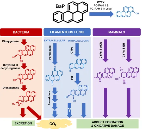
Organisms have various ways of metabolizing BaP, depending on their ecological niche (Fig. 1). Saprophytic bacteria create BaP ring cleavage products, leading to usable nontoxic fragments (3). Humans are equipped with cytochrome P450 monooxygenases (CYPs) to transform and excrete BaP, but this process results in the creation of reactive intermediates, which cause adduct formation and oxidative stress in cells (4, 5). This makes BaP an especially harmful compound, resulting in cancer and immune dysregu-lation (2). In addition, its chemical properties make BaP stable in the environment and resistant to abiotic degradation (1).
Fungi are one of nature’s most resourceful organisms, accounting for up to 75% of the soil microbial biomass (6). Aspergillus, the most common genus of soil-dwelling fungi, frequently prevails in contaminated sites and can metabolize certain PAHs (7).
Aspergillus species harbor abundant and diverse enzymatic systems, which allow them
to metabolically utilize complex organic molecules that are highly toxic to animals (8, 9). However, specific genes involved in metabolic utilization of BaP in fungi remain to be revealed.
Part of the metabolic armory harbored by Aspergillus species is over 100 CYPs encoded in the genome (10). These enzymes participate in a variety of physiological
BaP
CO2 Dioxygenase Dihydrodiol dehydrogenase Dioxygenase OH HO HO HO O O O OBACTERIA FILAMENTOUS FUNGI
EXCRETION OH CY P s & E H CY P s & A K R MAMMALS P e ro xidase CYPs: PC-PAH 1 & PC-PAH 3 in yeast P e ro xidase O O OH HO O OH HO INTRACELLULAR EXTRACELLULAR ADDUCT FORMATION & OXIDATIVE DAMAGE
?
CY P s O EH OH HOFIG 1 Schematic presentation of BaP transformation in mammals, fungi, and bacteria. Bacteria and fungi are both
able to mineralize BaP; however, the initial enzymatic transformation steps differ drastically in that bacteria utilize dioxygenase enzymes, whereas fungi utilize extracellular peroxidase enzymes (46). Mammalian and intracellular fungal pathways overlap in their utilization of CYP enzymes yielding similar metabolites; however, far less is known about the specific CYPs and metabolites produced by fungi. The CYPs Pc-PAH1 and Pc-PAH3 in P. chrysosporium, which have the ability to convert BaP to 3-hydroxybenzo[a]pyrene when expressed in P. pastoris, are shown. Mammals utilize other enzymes in partnership with CYPs, such as epoxide hydrolases (EH) and aldo-keto reductases (AKR), but because the limited studies on fungal CYPs were done using heterologous expression, it is unknown whether other enzymes are involved in BaP metabolism and what the final metabolic products are.
on December 1, 2019 at KOREA ADVANCE INST SCI & TECH
http://mbio.asm.org/
activities that allow the fungi to adapt to new ecological niches. Soil is a hostile and competitive environment, so these CYPs play a role in the synthesis and degradation of various toxic compounds. Aspergillus nidulans contains 119 predicted CYPs, for which the functions of 13 have been determined experimentally, and 32 are positioned near key secondary metabolite synthases, suggesting their potential biosynthetic role (11). Therefore, a large number of CYPs have no known or predicted function.
The white rot fungus Phanerochaete chrysosporium has an outstanding capability for degrading and/or mineralizing high-molecular-weight PAHs and contains an extraor-dinarily large repertoire (over 150) of CYPs in its genome (12). An excellent study by Syed and colleagues identified and characterized six CYPs in P. chrysosporium (Pc-PAH1 to Pc-PAH6) capable of oxidizing different PAHs (13). These CYPs were inducible by naphthalene, phenanthrene, pyrene, and BaP. Expression of each of the six Pc-PAH CYPs in the yeast Pichia pastoris in conjunction with the homologous P450 oxidoreduc-tase led to identification of Pc-PAH1 and Pc-PAH3 as CYPs with the ability to oxidize BaP to 3-hydroxybenzo[a]pyrene (13) (Fig. 1). This was the first report to identify a set of specific fungal CYPs having catalytic activity toward BaP. However, the functions of these CYPs have not been studied in vivo due to the limited ability of genetic manipulation in this organism, and hence further metabolism and the resulting prod-ucts remain a mystery. Likewise, many reports about BaP-degrading fungal species isolated from contaminated sites lack systematic study due to limited genetic tools (7). As Aspergillus species fill a similar saprophytic niche and have diverse metabolic capabilities, we hypothesize that they can metabolize BaP using a specific CYP-mediated pathway. We show that many, if not all, Aspergillus species can degrade BaP and uncover key aspects of cellular degradation of BaP by A. nidulans, using compre-hensive genetic, genomic, and biochemical approaches. Importantly, we identify a gene (bapA [AN1884]) predicted to encode CYP617D1 and show that bapA is necessary for degradation of BaP in vivo in two Aspergillus species. These critical findings further allow us to investigate the velvet regulators associated with BaP metabolic degradation. Our study illuminates fundamental knowledge of fungal BaP metabolism and provides novel insight into designing and implementing enhanced bioremediation strategies. RESULTS
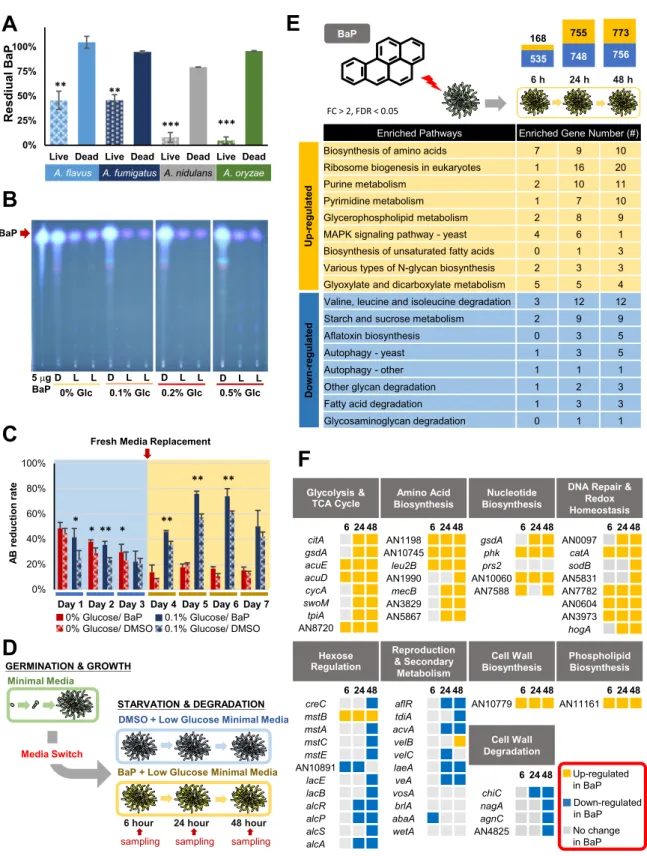
Aspergillus species can degrade BaP effectively. To test our initial hypothesis that
Aspergillus species are able to degrade BaP, we employed A. nidulans, A. flavus, A. oryzae, and A. fumigatus. These fungal species are distantly related to each other,
covering a broad range of Aspergillus (14). In all four species, the amount of BaP recovered from 7-day-old cultures was significantly lower in the living cells than that of dead cells (Fig. 2A), indicating that BaP was degraded or transformed in all species tested. The chromatogram showed no additional fluorescent peaks, suggesting that the degraded products were water soluble and/or not fluorescent at the same wavelengths as BaP (see Fig. S1 in the supplemental material). A. nidulans and A. oryzae were able to remove 92%⫾ 4.9% and 95% ⫾ 3.5% of the added 200M BaP, respectively. As A.
nidulans is a well-studied genetic model, we used it to further uncover the genetic and
biochemical mechanisms of BaP degradation. Thin-layer chromatography (TLC) analy-ses of residual BaP in different concentrations of glucose in minimal medium (MM) have revealed that MM with 0.1% glucose led to the most effective degradation of BaP by A.
nidulans (Fig. 2B).
BaP increases fungal cell viability. To test whether A. nidulans uses BaP as a substrate for growth, we measured changes in cell viability. The alamarBlue assay showed that cells exposed to BaP had significantly higher viability than the dimethyl sulfoxide (DMSO) controls, including time points after the addition of fresh medium to supply cells with the additional nutrients for the continuous proliferation (Fig. 2C). As found in TLC data, the prolonged viability of the cells with BaP was observable with the presence of a small amount of glucose in the medium (0.1%), whereas BaP addition alone was able to increase the cell viability at 2 days postexposure (Fig. 2C). This suggests that BaP can be used as a carbon source, and such effects can be enhanced
on December 1, 2019 at KOREA ADVANCE INST SCI & TECH
http://mbio.asm.org/
535 748 756 168 755 773 0% 25% 50% 75% 100%
Live Dead Live Dead Live Dead Live Dead
A. flavus A. fumigatus A. nidulans A. oryzae
*** *** ** ** l a ui d s e RB a P 0% 20% 40% 60% 80% 100% et ar n oi t c u d er B A
Fresh Media Replacement
0% Glucose/ BaP 0% Glucose/ DMSO
0.1% Glucose/ BaP 0.1% Glucose/ DMSO Day 1 Day 2 Day 3 Day 4 Day 5 Day 6 Day 7
*
* ** * **
** **
Minimal Media
GERMINATION & GROWTH
STARVATION & DEGRADATION DMSO + Low Glucose Minimal Media
BaP + Low Glucose Minimal Media Media Switch
6 hour 24 hour 48 hour sampling sampling sampling citA gsdA acuE acuD cycA swoM tpiA AN8720 6 24 48 AN1198 AN10745 leu2B AN1990 mecB AN3829 AN5867 creC mstB mstA mstC AN10891 mstE lacE lacB alcR alcP alcS alcA gsdA phk prs2 AN10060 AN7588 AN10779 AN11161 aflR tdiA acvA velB velC laeA veA vosA brlA abaA wetA chiC nagA agnC AN4825 AN0097 catA sodB AN5831 AN7782 AN0604 AN3973 hogA Glycolysis & TCA Cycle Amino Acid Biosynthesis Nucleotide Biosynthesis Phospholipid Biosynthesis Hexose Regulation Reproduction & Secondary Metabolism Cell Wall Degradation
DNA Repair & Redox Homeostasis Cell Wall Biosynthesis Up-regulated in BaP Down-regulated in BaP No change in BaP 6 24 48 6 24 48 6 24 48 6 24 48 6 24 48 6 24 48 6 24 48 6 24 48 24 h 6 h 48 h BaP
Enriched Pathways Enriched Gene Number (#)
Biosynthesis of amino acids 7 9 10 Ribosome biogenesis in eukaryotes 1 16 20
Purine metabolism 2 10 11
Pyrimidine metabolism 1 7 10 Glycerophospholipid metabolism 2 8 9 MAPK signaling pathway - yeast 4 6 1 Biosynthesis of unsaturated fatty acids 0 1 3 Various types of N-glycan biosynthesis 2 3 3 Glyoxylate and dicarboxylate metabolism 5 5 4 Valine, leucine and isoleucine degradation 3 12 12 Starch and sucrose metabolism 2 9 9 Aflatoxin biosynthesis 0 3 5
Autophagy - yeast 1 3 5
Autophagy - other 1 1 1
Other glycan degradation 1 2 3 Fatty acid degradation 1 3 3 Glycosaminoglycan degradation 0 1 1 U p -r e g u lat ed D o w n -r e g u lat ed FC > 2, FDR < 0.05
A
B
C
D
E
F
BaP 5 Pg BaP D L L D L L D L L D L L 0% Glc 0.1% Glc 0.2% Glc 0.5% GlcFIG 2 Degradation and utilization of BaP by Aspergillus. (A) Percentage of BaP remaining after 7-day cultures of each species. Three
separate experiments were performed in triplicate. Mean values are plotted with standard errors of the mean (SEM) throughout all the
graphs in the paper.**, P⬍ 0.01; ***, P ⬍ 0.001. (B) Thin-layer chromatograms of residual BaP after 7-day cultures of A. nidulans in MM
with various glucose concentrations. For reference, the 5-g BaP standard is shown. Note the differences of the BaP intensity in dead cells
(D) versus live cells (L). MM with 0.1% glucose with 200M BaP consistently resulted in the most effective degradation of BaP in multiple
experiments. (C) alamarBlue reduction of A. nidulans cells treated with BaP and DMSO with (blue) or without (red) 0.1% glucose.
Experiments were performed in triplicate.*, P⬍ 0.05; **, P ⬍ 0.01. (D) Experimental overview for RNA sequencing. Samples were collected
in triplicate at each time point. (E) KEGG pathways with upregulated (yellow) and downregulated (blue) DEGs at each time point. The bar
chart indicates the total number of DEGs (log2fold change [FC] of⬎1 or ⬍⫺1 and P value of ⬍0.05) at each time point. (F) Categories
of transcriptional responses previously seen in carbon-starved A. nidulans cells and relative expression of specific genes in BaP- versus control (DMSO)-treated cells.
on December 1, 2019 at KOREA ADVANCE INST SCI & TECH
http://mbio.asm.org/
with the supplementation of a small amount of glucose, which may provide additional resources for energy and enzyme production.
BaP leads to upregulation of cell growth-associated genes. To further test the hypothesis that the fungus uses BaP as a growth substrate, we investigated the genome-wide expression responses of A. nidulans to BaP via transcriptome sequencing (RNA-seq). Transcript levels were measured in BaP-treated cells relative to controls (DMSO) at 6, 24, and 48 h postexposure by KEGG analysis (Fig. 2D). The number of differentially expressed genes (DEGs) was low at 6 h (703 DEGs); this increased to 1,503 and 1,529 DEGs at 24 and 48 h, respectively (Fig. 2E). KEGG pathway analysis indicated that, as time progresses with BaP exposure, genes associated with cell growth, such as ribosome biogenesis, biosynthesis of amino acids, nucleotide metabolism, biosynthesis of unsaturated fatty acids, N-glycan biosynthesis (cell wall), and glycerophospholipid (membrane), were upregulated (Fig. 2E). Conversely, genes categorized into the path-ways indicative of cell starvation and stress, including amino acid degradation, au-tophagy, aflatoxin (sterigmatocystin) biosynthesis, and starch metabolism, were down-regulated in BaP-treated cells (Fig. 2E). These results indicate that BaP-treated cells are actively growing compared to controls and that the fungus is able to use BaP to sustain growth.
The transcriptomic response to carbon starvation in A. nidulans shows upregulation of genes involved in programmed cell death, macroautophagy, cell wall component degradation, asexual reproduction, and secondary metabolite production (15). Down-regulation of genes involved in glycolysis and oxidative phosphorylation, cell wall component synthesis, and nitrogen and lipid anabolic pathways was also seen in the starving cells (15). We carried out an integrated analysis of the differentially expressed genes by BaP treatment with the carbon starvation stress response and found that the BaP-treated cells showed upregulation of the following genes: (i) citA, gsdA, acuE, acuD, and cycA, involved in the tricarboxylic acid (TCA) cycle, replenishment of TCA cycle intermediates, and oxidative phosphorylation; (ii) AN11161, involved in phospholipid biosynthesis; (iii) AN10779, involved in-1,6-glucan biosynthesis; and (iv) several genes involved in amino acid biosynthesis pathways (Fig. 2F). BaP-treated cells show down-regulation of aflR and other sterigmatocystin biosynthesis genes, abaA, involved in conidiation, and agnC, nagA, chiC, and AN4825, involved in cell wall component hydrolase enzymes (Fig. 2F). Taken together, the data demonstrate that BaP enables the cells to grow more actively than control cells and provide some evidence that BaP is metabolized and shuttled into carbon utilization pathways.
Finally, in an attempt to address whether BaP metabolism causes oxidative stress and/or DNA damage responses in A. nidulans as in mammalian cells, we compared our RNA-seq data with those representing responses to cells treated with other known oxidizing compounds. Some redox-balancing genes were upregulated in BaP-treated cells, including catA and sodB (Fig. 2F). Expression of some DNA repair genes was also induced by BaP, including AN0604 and AN0097 (Fig. 2F). These results suggest that BaP may cause DNA damage in A. nidulans as in mammalian cells, although it is shown that fungi have additional capacity to prevent extensive DNA damage (16).
Identification of CYP necessary for metabolic utilization of BaP. Initial oxidation of BaP in mammalian cells is mediated by a CYP, adding a single molecular oxygen, leading to the formation of BaP epoxide intermediates, which can be further converted into hydroxylated products (4, 17–19).
With the hypothesis that A. nidulans employs a CYP to degrade BaP, we first examined our RNA-seq data to search for the CYP genes upregulated by BaP treatment and found that no specific CYPs were clearly induced by BaP. We then used the CYPs of P. chrysosporium, Pc-PAH1 and Pc-PAH3, which when expressed in the yeast cells, converted BaP to 3-hydroxybenzo[a]pyrene (13), to search for closely related CYPs in the A. nidulans genome. Despite the 723 million years of divergence between the two fungi (20), several CYPs similar to Pc-PAH1and Pc-PAH3 were identified (Fig. 3A; see Table S1 in the supplemental material). Among these, mRNA levels of AN1884 in
on December 1, 2019 at KOREA ADVANCE INST SCI & TECH
http://mbio.asm.org/
A
B
C
D
E
OH CYP
PC-PAH1 & PC-PAH3
PC-PAH 1 AN11142 AN1884 AN7399 PC-PAH 3 AN1601 AN0338 AN8615 AN3917 PC-PAH3 like PC-PAH1 like 0.2 0% 20% 40% 60% 80% 100% 0 4 8 12 16 20 4 10 24 48 168 BaP Degradation bapA Expression B a P D e g ra dat ion bapA Log 0 ot e vi t al er C Fh r Hours ΔbapA 0% 20% 40% 60% 80% 100% 120%
Live Dead Live Dead Live Dead
R esi d u a l B aP WT C’bapA p < 0.0001 p < 0.0001 -2.2 -1.8 -1.4 -1 -0.6 -0.2 0.2 0.6 1 1.4 1.8 2.2 ) O S M D/ P a B( 2 g ol WT ΔbapA C'bapA
Day 1 Day 2 Day 3 Day 4 Day 5 Day 6 Fresh Media Replacement
* ** * * ** ** * ** ** ** * * * WT ∆bapA Alanine * Arginine Asparagine Aspartate * Glutamate * Glutamine * Histidine Isoleucine Leucine Lysine * Methionine Ornithine * Phenylalanine Proline Serine Threonine Tryptophan Tyrosine Valine WT ∆bapA Aconitate α-ketoglutarate * Citrate Glycerate Malate Fumarate Gluconate NAD+ Pyruvate WT ∆bapA Guanine Guanosine Uridine AMP ATP dGMP Amino Acids -0.5 0 0.5 log2FC(BaP/DMSO) Primary Metabolites
FIG 3 Identification and characterization of the CYP BapA (AN1884) necessary for metabolic utilization of BaP. (A) Phylogenetic tree
representing A. nidulans CYPs similar to Pc-PAH1 and Pc-PAH3. (B) Correlation of bapA (AN1884) mRNA levels and BaP degradation. Time point 0 indicates exponential growth (18 h postgermination) in regular MM (1% glucose), and each time point represents hours after the switch to 0.1% glucose MM. The mean is plotted as a bar, and error bars represent standard error. BaP degradation (blue bar) was
determined by measuring residual BaP compared to the initial 200M BaP. (C) Residual BaP in each indicated strain. Three separate
experiments were performed in triplicate. Note the lack of BaP degradation in the ΔbapA mutant compared to WT and complemented
(C=) strains (P⬍ 0.001). *, P ⬍ 0.05; **, P ⬍ 0.01; and ***, P ⬍ 0.001. (D) Comparison of the log2values of the alamarBlue reduction levels
in WT, ΔbapA, and C= strains in cells treated with BaP relative to DMSO only (DMSO) at days 1 to⬃6. “Fresh media replacement” indicates
collection and washing of cells and addition of fresh medium. All treatments were performed in triplicate, and Student’s t test was used
(*, P⬍ 0.05; **, P ⬍ 0.01). (E) Relative quantification of cellular components in the WT and the ΔbapA mutant. Each colored square
represents log2fold change (FC) of the mean quantity of the metabolites in BaP versus DMSO. Each sample was prepared using biological
triplicates.*, P⬍ 0.05.
on December 1, 2019 at KOREA ADVANCE INST SCI & TECH
http://mbio.asm.org/
glucose-limited medium and kinetics of BaP degradation were closely aligned, indicat-ing that this CYP may be associated with metabolism of BaP (Fig. 3B; Fig. S2B).
To test its function in BaP metabolism in vivo, we generated independent deletion (Δ) strains of AN1884 in A. nidulans and found that all three independent null mutant strains lost the ability to degrade BaP, with no distinct growth and developmental phenotypic changes (Fig. S3). Reintroducing an AN1884 coding region into null mutant strains restored the BaP degradation ability to that of the wild type (WT) (Fig. 3C), supporting the hypothesis that AN1884 is responsible for the breakdown of BaP, so it was named bapA (benzo[a]pyrene metabolism locus A). Measuring the cell viability of the WT, the ΔbapA mutant cells, and complemented (C=bapA) cells treated with BaP relative to those treated with DMSO revealed that the ΔbapA mutant cells were not only less viable than WT and C=bapA cells treated with BaP, but less viable than ΔbapA mutant cells treated with DMSO (Fig. 3D). This indicates BaP may be toxic to cells unable to metabolize it. To our knowledge, this is the first report providing evidence of
in vivo function of a CYP in cellular degradation of BaP.
We attempted to identify the BaP metabolite(s) formed by BapA. We isolated microsome-containing fractions from the WT, ΔbapA, and C=bapA, cells and incubated them with BaP. The WT and C=bapA, but not ΔbapA, chromatograms showed a small fluorescent peak with a shorter retention time than BaP (Fig. S4A). The retention time of this unknown metabolite was compared to those of known BaP metabolites and matched that of benzo[a]pyrene-3,6-dione (Fig. S4A). However, the fluorescence spec-tra revealed that sample peaks do not show the same fluorescing as benzo[a]pyrene-3,6-dione (Fig. S4B). No other known metabolite standards matched the retention time of this peak (Fig. S4C). Although we were not able to reveal the exact identity of the product formed within the microsome fraction of cells expressing BapA, we were able to rule out many BaP metabolites formed by other organisms. This may indicate that BapA is involved in forming a unique metabolite not previously reported in other organisms.
The availability of the ΔbapA mutant allows us to further investigate the down-stream metabolomic outcomes of BaP. Since BaP treatment caused upregulation of genes associated with amino acid biosynthesis, which is an easily measured endpoint for carbon utilization, we quantified free amino acids in WT and ΔbapA cells treated with BaP relative to the control (DMSO). In agreement with alamarBlue data, only BaP-treated WT cells showed significant accumulation of glutamate, aspartate, glu-tamine, lysine, and the intermediate amino acid ornithine (Fig. 3E). The ΔbapA cells showed significant accumulation of alanine and ␣-ketoglutarate in control (DMSO) compared to BaP treatment (Fig. 3E). This provides additional evidence that loss of BaP metabolism causes lack of cell growth upon exposure to BaP and alludes to toxicity caused by the lack of BaP-degrading ability. While insignificant due to large deviations among samples, accumulation of several primary metabolites involved in energy metabolism, such as citrate and malate, and nucleotides involved DNA/RNA synthesis or signaling, such as cyclic AMP (cAMP), appeared to be affected by BaP (Fig. 3E).
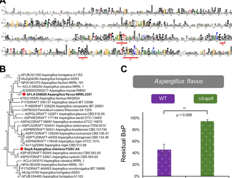
BapA is widely distributed in ascomycota and is functionally conserved in A. flavus. The structural analysis showed that the four widely recognized consensus regions (a to d) (Fig. 4A) contributing to the core function of P450s (10) are highly conserved in the CYP617 family. Interestingly, the conserved motif a (AGHETT) of the CYP617 family is very specific and is highly similar to those of archaea and bacteria (10). We performed a phylogenic analysis to determine how widely distributed BapA is in fungi. We found 64 CYP617 family members in the fungal kingdom, covering dothideo-mycetes, eurotiodothideo-mycetes, leotiodothideo-mycetes, and sordariodothideo-mycetes, with the number rang-ing from one to several per species (see Fig. S5 and Table S3 in the supplemental material). CYP617 members are limited to ascomycetes. The BapA subfamily CYP617D1 was mostly distributed in the genus Aspergillus (Fig. 4B), suggesting a conserved role within the genus.
To examine a potential conservation of its function, we identified a likely orthologue of BapA in A. flavus (AFLA_036020) and generated three individual null mutant strains.
on December 1, 2019 at KOREA ADVANCE INST SCI & TECH
http://mbio.asm.org/
All ΔAFLA_036020 strains lost the ability to degrade BaP, corroborating the conserved and essential role of BapA in degradation of BaP in Aspergillus species (Fig. 4C) under glucose-limiting conditions.
Requirement of fungal NF-B-type regulators in BaP degradation. Identification of bapA allowed us to further investigate its upstream regulatory components. The
velvet proteins are a family of global transcription factors (TFs) involved in diverse
aspects of fungal biology (21). They contain a DNA binding domain structurally similar to that of the human TF complex nuclear factor kappaB (NF-B) (22) p50, which governs cell survival upon exposure to BaP (23–26).
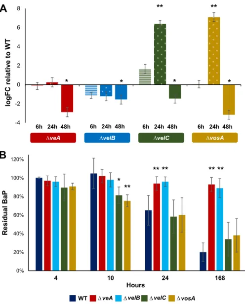
Due to this structural similarity and diverse regulatory functions between human NF-B and the fungal velvet complex, we hypothesized that velvet proteins might play a role in BaP degradation. To address this hypothesis, we first tested mRNA levels of
bapA in each velvet deletion mutant (ΔveA, ΔvelB, ΔvelC, and ΔvosA) and found that the
two regulators VeA and VelB were necessary for proper expression of bapA (Fig. 5A). Interestingly, the other two regulators, VosA and VelC, seem to play a repressive role at 10 h post-glucose starvation and an activating role at later time points (Fig. 5A). To
FIG 4 The BapA CYP signature motifs, distribution of BapA in Aspergillus species, and the role of BapA in BaP degradation in A. flavus. (A) Conserved domains
of the CYP617 family proteins queried from FungiDB (42) showing greater than 40% identity against the A. nidulans BapA protein (listed in Fig. S5). (B) Phylogenetic tree of the CYP617D members in Aspergillus having greater than 55% identity against the A. nidulans BapA protein. These proteins can be classified into the same subfamily, CYP617D, based on the rules of the International P450 Nomenclature Committee. For the expanded information, see Fig. S5 and Table S3. (C) Residual BaP in WT and the ΔbapA mutant of A. flavus. All experiments were performed in triplicate, and three independent ΔbapA strains were
tested.**, P⫽ 0.008.
on December 1, 2019 at KOREA ADVANCE INST SCI & TECH
http://mbio.asm.org/
verify that expression of bapA translates into BaP degradation, we tested the degra-dation ability of each deletion mutant and found that the ΔveA and ΔvelB mutants were unable to degrade BaP. On the contrary, the ΔvosA and ΔvelC mutants were able to degrade the same amount of BaP, yet faster than the WT (Fig. 5B). These results indicate that VeA and VelB play a key role in metabolic utilization of BaP, in part by controlling proper expression of bapA, whereas VosA and VelC may indirectly play a role in coordinating the proper timing of BapA expression in response to glucose limitation. DISCUSSION
BaP is a contaminant of significant concern because of its ubiquity and toxicity. As a result of its stability, biologically driven degradation remains the predominant form of removal from the environment (27). Thus, understanding how saprophytic bacteria and fungi effectively metabolize BaP is critical for the effective removal of BaP.
This is the first comprehensive study showing that Aspergillus species can effectively degrade BaP, resulting in cell survival and growth during carbon starvation (Fig. 6A). We
A
B
-4 -2 0 2 4 6 8 logFC re la tiv e to W T'veA velB velC vosA
6h 24h 48h
*
**
**
6h 24h 48h 6h 24h 48h 6h 24h 48h*
*
*
0% 20% 40% 60% 80% 100% 120% 4 10 24 168 l a u di s e RB a P HoursveA velB velC vosA
WT
*
**
** **
** **
' ' ' ' ' ' 'FIG 5 Fungal NF-B-type velvet regulators are required for proper bapA expression and BaP degradation.
(A) Levels of bapA mRNA were measured using 2⫺ΔΔCTrelative to actA (␥-actin) (ΔC
T) and then relative to
the WT (ΔΔCT). Time points indicate hours after the switch to 0.1% glucose medium. Technical and
biological triplicates were used for each time point and treatment.*, P⬍ 0.05; **, P ⬍ 0.01. (B) Residual BaP
remaining after being cultured for 7 days with each strain. Note the lack of BaP degradation in ΔveA and
ΔvelB strains. Three separate experiments were performed in triplicate.*, P⬍ 0.05; **, P ⬍ 0.01.
on December 1, 2019 at KOREA ADVANCE INST SCI & TECH
http://mbio.asm.org/
were unable to identify specific BaP intermediates in this study, so it is unclear which pathways are involved in further metabolism of BaP. The CYP-mediated metabolism of BaP in human cells has been well characterized, so we attempted to use BaP metabolite standards to identify the potential metabolite peak using high-performance liquid
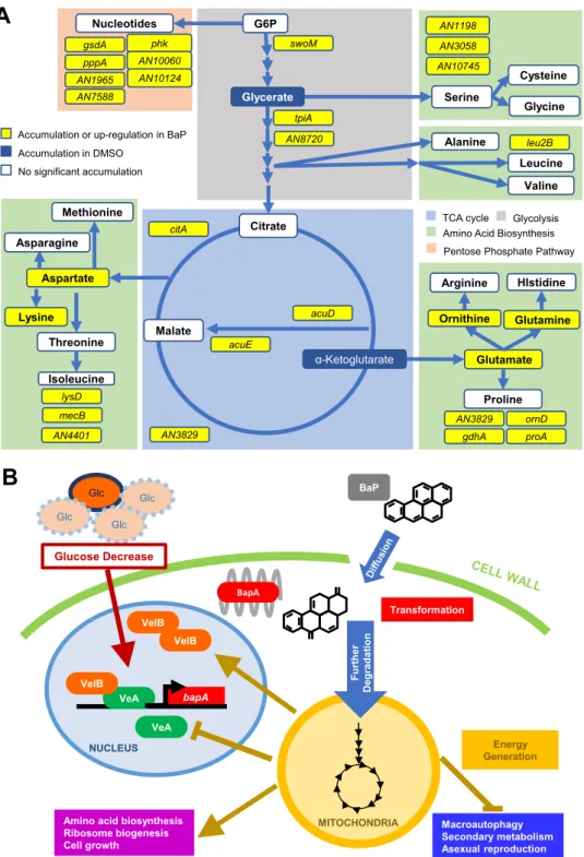
A
B
Glutamate Glutamine Arginine Proline HIstidine gdhA proA ornD AN3829 Ornithine Malate citA acuE acuD G6P Cysteine Serine Glycine AN10745 AN3058 AN1198 AN3829 Alanine Leucine Valine leu2B swoM tpiA AN8720TCA cycle Glycolysis Amino Acid Biosynthesis Pentose Phosphate Pathway
Nucleotides gsdA AN7588 phk pppA AN1965 AN10060 AN10124 Glycerate Citrate
Accumulation or up-regulation in BaP Accumulation in DMSO No significant accumulation α-Ketoglutarate Aspartate Threonine Methionine Asparagine Isoleucine Lysine lysD mecB AN4401 bapA Glc Glc Glc Glc Glucose Decrease NUCLEUS VeA VelB BapA BaP MITOCHONDRIA Energy Generation
Amino acid biosynthesis Ribosome biogenesis Cell growth Macroautophagy Secondary metabolism Asexual reproduction VelB VelB Furt her De g ra d a ti o n VeA Transformation
FIG 6 Models for BaP metabolic utilization in A. nidulans. (A) BaP confers changes in energy generation pathways
indicated by colored blocks. Upregulated DEGs are shown in yellow. Amino acids and other compounds found in greater quantity as a result of BaP treatment in WT but not ΔbapA strains are shown in yellow and white boxes,
where yellow indicates statistical significance (P⬍ 0.05). Blue boxes represent cellular components that
signifi-cantly decrease as a result of BaP treatment in WT but not in ΔbapA strains (P⬍ 0.05). (B) Proposed genetic pathway
for the metabolic utilization of BaP in A. nidulans. Limited glucose and the VeA-VelB complex are associated with increased expression of bapA.
on December 1, 2019 at KOREA ADVANCE INST SCI & TECH
http://mbio.asm.org/
chromatography (HPLC). None of the standards we tried matched the retention time or absorbance spectrum of the peak. Additionally, CYP metabolism of BaP in mammalian cells causes mutagenic and cytotoxic effects (28), whereas we observed an increase in viability of A. nidulans cells exposed to BaP. Together this leads us to conclude that BaP metabolism in Aspergillus sp. involves the unique CYP BapA, and further degradation of BaP may occur via metabolic pathways not found in mammalian cells. Further study is needed to understand the full metabolic pathway(s) of BaP degradation.
Our study does, however, identify a necessary CYP617D1 enzyme that not only provides information that can help effectively implement bioremediation strategies, but also gives us a unique insight into evolution of the fungal CYPs and their biocatalytic activity. We propose a model in which VeA and VelB activate expression of
bapA in response to nutrient limitation and BapA oxidizes BaP (Fig. 6B). We hypothesize
that oxidized BaP is further enzymatically fragmented, and the carbon is shuttled into energy-generating pathways, which in turn represses further expression of bapA (Fig. 6B).
Filamentous fungi harbor many more CYPs relative to their genome size than animals and bacteria, yet the functions of many remain unknown. The diversity of CYPs in fungi could be due to their need to metabolize many different carbon sources found in soils, including large cyclic compounds like lignin and plant polymers. It is also feasible that fungi, like animals, may need detoxification systems reliant on CYP activity to avoid toxic compounds produced by competing microbes and plants. Our results demonstrate that the regulation of bapA is governed by response to carbon starvation, rather than exposure to the toxicant BaP.
The A. nidulans, A. flavus, and A. fumigatus genomes each contain over 100 encoded CYPs, with 90, 93, and 57 family types, respectively, yet only 45 types are shared (10). Despite this diversity, BapA (CYP617D1) is found in all three distantly related Aspergillus and Penicillium species, and in A. nidulans and A. flavus it plays the same functional role of degrading BaP. Because CYPs demonstrate substrate promiscuity, it is likely that BapA oxidizes other compounds, such as other PAHs and/or large planar endogenous compounds. The deletion of bapA showed no obvious growth and developmental changes, suggesting that BapA does not likely play a major housekeeping role.
Regulation of bapA also demonstrates a novel understanding of how Aspergillus species respond to organic contaminants like BaP. Humans and fungi have evolved different strategies to deal with exposure to xenobiotics, yet both employ CYPs. Humans do not invest energy into utilizing carbon sources more complex than various sugars and a few of their polymers, so CYP transformation of BaP yields more polar metabolites that can then be excreted. Regulation of encoded BaP-metabolizing CYPs is predominantly governed by the aryl hydrocarbon receptor (29), yet BaP and its metabolites also activate NF-B (23–25). NF-B is a protein heterodimer consisting of p50 and RelA, which upon activation by many types of cellular stress, from microbial and viral proteins to ionizing radiation, promotes cell survival (26). Filamentous fungi, on the other hand, act more as ecological scavengers and are capable of utilizing large carbon-containing compounds, such as plant cell wall polymers. These fungi have evolved with the global regulators called the velvet proteins with a DNA binding domain structurally similar to that of NF-B p50 (22). The velvet regulators in Aspergillus species govern environmental sensing, orchestration of cell growth, reproduction, stress response, spore viability, and biosynthesis of various secondary metabolites, which similarly helps the fungal cells to survive environmental stressors (21, 30).
In this study, we have shown that CYP-mediated degradation of BaP requires functions of the velvet family proteins VeA and VelB. These regulatory proteins control expression of bapA in response to stress resulting from carbon insufficiency, as opposed to exposure to xenobiotics. As this CYP is functionally conserved across distantly related fungi, it may play the same role in many ascomycete fungi. Further investigation of substrates metabolized by BapA would reveal its activity on other environmental contaminants as well as give insight into a possible endogenous function.
on December 1, 2019 at KOREA ADVANCE INST SCI & TECH
http://mbio.asm.org/
MATERIALS AND METHODS
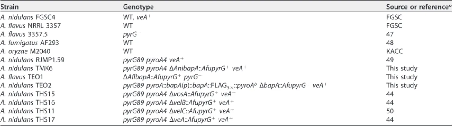
Strains, media, and culture conditions. The Aspergillus strains used in this study are listed in
Table 1. Initially, 106spores/ml were added to 400 ml minimal medium (MM) (31) with 1% glucose in
2-liter flasks and incubated for 18 h at 220 rpm at 37°C. The mycelial aggregates were then collected on sterile Miracloth (Sigma-Aldrich), rinsed, and transferred to 100 ml MM with 0.1% glucose in 250-ml Erlenmeyer flasks. Control dead cells were autoclaved on a liquid cycle at 121°C for 20 min to account for any nonmetabolic sources of loss of BaP. A 100 mM stock solution of BaP (Sigma-Aldrich) in dimethyl
sulfoxide (DMSO) was added to the cultures to a final concentration of 200M; the same volume of
DMSO was added to controls. All flasks were further incubated at 220 rpm at 37°C for the designated
time. Escherichia coli DH5␣ cells were grown in Luria-Bertani medium with ampicillin (100 g/ml) for
plasmid amplification.
Extraction and HPLC analysis. Extraction of BaP was optimized to recover all BaP adhered to and
taken up by cells, but not biotransformed. Individual fungal cell cultures were extracted using 100 ml 1:1
hexane-ethyl acetate with pyrene (Sigma-Aldrich [final concentration, 200M]) as an internal standard
to correct for extraction efficiency. The entire mixture was sonicated using a Sonic dismembrator model 100 (Fisher Scientific) with a 1/2-in. probe on full power for 6 min to ensure disintegration of hyphal pellets. Solvent (1 ml) was removed and centrifuged to remove particulate matter and diluted 100-fold in 1:1 solvent A (30 mM acetate buffer at pH 4.7, 10% acetonitrile)-solvent B (acetonitrile). BaP and pyrene were quantified by high-performance liquid chromatography (HPLC) using an Agilent 1260 system
equipped with a 3- by 50-mm Poroshell 120 EC-C182.7-m column (Fig. S1). A linear gradient that
ramped from 55% B to 90% B over 10 min at a flow rate of 0.75 ml/min was used, followed by
fluorescence detection (FLD [excitation⫽ 248 nm andemission⫽ 465 nm]). All standard curves were linear,
and the detection limits wereⱕ0.1M for pyrene and BaP.
TLC analysis of residual BaP. To further verify the degradation of BaP by A. nidulans at different
glucose concentrations (Fig. 2B), we also carried out TLC analyses 10 times and obtained a high degree of reproducibility. BaP extraction was performed by adding 0.5% (vol/vol) 6 N HCl (to stop all metabolic activity) to the fungal cultures (100 ml). The mycelium was collected through Miracloth and squeezed to maximize collection of the supernatant, which was transferred to a fresh 250-ml flask and mixed with 100 ml of ethyl acetate (1:1 ratio). Both liquids were then transferred to a new 250-ml separatory funnel. After shaking vigorously for 2 min, the organic phase was transferred in a new flask. Fresh solvent was added to the separatory funnel, and shaking and collecting were repeated two additional times. The resulting solvent was allowed to evaporate in the fume hood, and each dried sample was resuspended with ethyl acetate (1 ml) for TLC analysis. Ten microliters of each sample was applied to a TLC silica plate, including a fluorescence indicator (Kiesel gel 60, 0.25 mm thick; Merck). Authentic BaP standard was loaded as a control. The TLC plate was then developed with toluene-acetone-hexane (1:1:1 [vol/vol/vol]),
where the Rfvalue of BaP was 0.9. The TLC plate was exposed to UV at A320for 30 s, and images were
captured using a Canon EOS camera. To quantify the residual BaP shown in Fig. S3B, the density of each BaP spot on TLC was determined using ImageJ (NIH): the relative amount of BaP in live cells to the dead
cell control (⫽100%) is presented.
alamarBlue reduction assay. Cell viability was determined by percentage of alamarBlue (Bio-Rad)
reduction as described previously (32), with the following exceptions. Cells were prepared as described for BaP degradation with solvent (DMSO) only as a control, and 0.45 ml of cells was added to 0.45 ml fresh
MM with 0.1% glucose and 100l alamarBlue and incubated for 2 h at 37°C.
RNA preparation and qRT-PCR. Fungal cells from submerged cultures were collected at designated
time points, squeeze-dried, flash frozen in liquid N2, and stored at ⫺80°C until subjected to RNA
preparation. Total RNA isolation was done using TRIzol as described previously (33). cDNA was prepared using an avian myeloblastosis virus (AMV) reverse transcriptase kit (NewEngland Biolabs) with oligo(dT). Reverse transcriptase quantitative PCR (RT-qPCR) was performed with iTaq universal SYBR green super-mix (Bio-Rad) on a Bio-Rad CFX96 real-time PCR detection system. mRNA was normalized using threshold
cycle (2⫺ΔΔCT) method (34). Levels of bapA mRNA were determined using 2⫺ΔΔCT, in which bapA
TABLE 1 Aspergillus strains used in this study
Strain Genotype Source or referencea
A. nidulans FGSC4 WT, veA⫹ FGSC
A. flavus NRRL 3357 WT FGSC
A. flavus 3357.5 pyrG⫺ 47
A. fumigatus AF293 WT 48
A. oryzae M2040 WT KACC
A. nidulans RJMP1.59 pyrG89 pyroA4 veA⫹ 49
A. nidulans TMK6 pyrG89 pyroA4 ΔAnibapA::AfupyrG⫹veA⫹ This study
A. flavus TEO1 ΔAflbapA::AfupyrG⫹pyrG⫺ This study
A. nidulans TEO2 pyrG89 pyroA::bapA(p)::bapA::FLAG3⫻::pyroAbΔbapA::AfupyrG⫹veA⫹ This study
A. nidulans THS15 pyrG89 pyroA4 ΔvosA::AfupyrG⫹veA⫹ 44
A. nidulans THS16 pyrG89 pyroA4 ΔvelB::AfupyrG⫹veA⫹ 44
A. nidulans THS11 pyrG89 pyroA4 ΔvelC::AfupyrG⫹veA⫹ 50
A. nidulans THS17 pyrG89 pyroA4⌬veA::AfupyrG⫹veA⫹ 44
aFGSC, Fungal Genetic Stock Center; KACC, Korean Agricultural Culture Collection. bThe 3/4 pyroA marker causes targeted integration at the pyroA locus.
on December 1, 2019 at KOREA ADVANCE INST SCI & TECH
http://mbio.asm.org/
expression (CT) was found relative to the reference gene actA (␥-actin) (ΔCT) and then relative to time
point 0 (ΔΔCT). Time point 0 indicates exponential growth (18 h postgermination) in regular MM (1%
glucose), and each time point represents hours after the switch to MM with 0.1% glucose. Each experiment was performed using technical triplicates for RT-qPCR accuracy, and three biological tripli-cates were used for each time point. The oligonucleotides used are listed in Table S1. Total RNA was extracted and submitted to ProteinCT Biotechnologies (Madison, WI) for library preparation and RNA sequencing.
RNA sequencing. RNA sequencing was done as described previously (35). The library was
con-structed and purified and sequenced (SE100bp) using the Illumina HiSeq2500, and over 20 million high-quality reads per sample were achieved.
Data QC and analysis. Verification of the quality of reads (quality control [QC]), alignments, gene
annotation, and differential expression analysis were performed as described previously (35).
Functional enrichment analysis (KEGG). The KEGG pathway database was used to search against
A. nidulans KEGG pathway maps in order to identify A. nidulans metabolic pathways with the differentially
expressed genes (DEGs) after exposure to BaP on 20 February 2018 (36).
Metabolomics of amino acid and primary metabolites. Fungal cells prepared as described for BaP
degradation with the DMSO control were subject to extraction of cellular components as described previously (37), with the following exceptions. Hyphal mats were filtered and squeeze-dried, noting the mass after removing liquid, 1 day after transfer to BaP-containing medium. Tissue was flash frozen in
liquid N2and stored at⫺80°C. Two milliliters of extraction solvent (37) was added, and samples were
sonicated using a 1/4-in. probe for 3 min and centrifuged to remove cell debris. Additional sample prep and analysis were performed as described previously (37).
Protein alignment. CYP sequences similar to those of Pc-PAH1 and Pc-PAH3 in Aspergillus sp. were
identified using blastp (38). Protein sequences were found using NCBI, and protein alignment was calculated using Clustal Omega at EMBL-EBI output ClustalW with character counts (39). A phylogenetic tree was created using Jalview nearest neighbor joining (40).
Analysis of BapA families. BapA (AN1884) was assigned to CYP617D1 (11) and analyzed according
to the rules of the International P450 Nomenclature Committee (41). BapA protein sequence was used to query FungiDB (42). CYP617 members were aligned, and the phylogenetic tree was constructed as previously described (43).
Generation of⌬bapA and complemented strains. Double-joint PCR was used to generate the
deletion constructs of A. nidulans bapA (AN1884) and A. flavus bapA (AFLA_036020) (33). Briefly, the deletion construct containing the A. fumigatus pyrG marker with 5= and 3= flanking regions of bapA was introduced into the recipient strain RMJP1.59 (A. nidulans) or NRRL 3357.5 (A. flavus). Three independent ΔbapA strains each in A. nidulans (TMK6-1, -35, and -47) and A. flavus (TEO1 2, 8, and 9) were confirmed
and analyzed. To generate complemented strains of the ΔbapA mutant in A. nidulans, a bapA⫹gene
region, including its upstream 2-kb region, was introduced to pHS13 (44) and introduced into E. coli
DH5␣ for transformation. Upon sequence verification of the insert, the purified plasmid was introduced
into the recipient ΔbapA A. nidulans strain (TMK6). Three independent complemented strains (C=bapA 12, 16, and 17) were verified and analyzed.
Microsome isolation and BaP metabolic activity. Cells were prepared as described without BaP
treatment to capture peak bapA mRNA levels (Fig. S2A and B). After 1 day of incubation, cells were
filtered, washed, squeeze-dried, and flash frozen in liquid N2. Frozen tissue was ground to a fine powder
in liquid N2with the addition of glass beads in a mortar and pestle. The powder was resuspended in
30 ml homogenization buffer (0.1 M KPO4 at pH 7.25, 0.1 M KCl, 10 mM EDTA at pH 8, 0.25 mM
phenylmethylsulfonyl fluoride [PMSF], 0.1 mM dithiothreitol [DTT]) and kept in ice. A sonication probe (1/8 in.) was used on full power for 30 s to homogenize cells and form microsomal structures. Large
debris was filtered using Miracloth, and the supernatant was centrifuged for 20 min at 20,000⫻ g. The
supernatant was then transferred and centrifuged for 60 min at 105,000⫻ g. The resulting supernatant
was discarded, and the pellets were resuspended in 200l dilution buffer (0.25 M KPO4at pH 7.25, 20%
[vol/vol] glycerol, 10 mM EDTA at pH 8.0, 0.25 mM PMSF, 0.1 mM DTT), flash frozen in liquid N2, and
stored at ⫺80°C for a maximum of 1 week. The protein concentration was determined with the
Coomassie protein assay (Thermo Fisher). BaP metabolism was measured by incubating 2 mg/ml
microsomal protein, 1M BaP, and 1 M NADPH in up to 1 ml 50 mM phosphate buffer at pH 7.5 at 37°C
for 1 h. As a control, protein was denatured by boiling for 20 min prior to incubation. Metabolites were extracted by adding 2 ml of 2:1 acetone-ethyl acetate and vortexing for 2 min. Solvent was removed and
centrifuged at 13,000⫻ g for 10 min and dried under N2. Samples were resuspended in 50l 1:1 HPLC
solvent A-solvent B and analyzed by HPLC.
Statistics. Statistical significance was determined using Student’s t test with a two-tailed distribution
and two-sample unequal variance.
Data availability. All RNA-seq data files are available from the NCBI Gene Expression Omnibus
database (45) under accession no.GSE116804.
SUPPLEMENTAL MATERIAL
Supplemental material for this article may be found athttps://doi.org/10.1128/mBio .00558-19.
FIG S1, PDF file, 0.1 MB. FIG S2, PDF file, 0.2 MB. FIG S3, PDF file, 0.1 MB.
on December 1, 2019 at KOREA ADVANCE INST SCI & TECH
http://mbio.asm.org/
FIG S4, PDF file, 0.2 MB. FIG S5, PDF file, 0.3 MB. TABLE S1, DOCX file, 0.1 MB. TABLE S2, DOCX file, 0.1 MB. TABLE S3, XLSX file, 0.1 MB. ACKNOWLEDGMENTS
We thank the United States Environmental Protection Agency Students to Achieve Results (US EPA STAR) Fellowship for supporting E.O.L. This work was supported by EPA STAR Fellowship FP-91779101-1 and the Intelligent Synthetic Biology Center of Global Frontier Project funded by the Ministry of Education, Science and Technology of Korea (no. 2011-0031955).
We also thank Nancy Keller, Philipp Wiemann, Mengyao Niu, Andrew Maizel, Chris-topher Bradfield, Anna Shen, Susan Moran, and Trevor Penning for valuable help.
E.O.L., M.-K.L., J.M., W.C., C.R., C.J., D.A.-N., S.J., S.C.-K., and J.-H.Y. designed and performed the experiments and analyzed the data. E.O.L. and J.-H.Y. wrote the manu-script and designed the figures, M.-Y.W. designed the figures and manumanu-script structure. All authors contributed to revising the manuscript.
REFERENCES
1. Centers for Disease Control Agency for Toxic Substances and Disease Registry (ATSDR), Division of Toxicology and Human Health Sciences.
2013. 2013 ATSDR substance priority list.https://www.atsdr.cdc.gov/spl/
resources/2013_atsdr_substance_priority_list.html.
2. EPA. 2017. IRIS toxicological review of benzo[a]pyrene (final report).
https://cfpub.epa.gov/ncea/iris_drafts/recordisplay.cfm?deid⫽329750. 3. Cerniglia C. 1992. Biodegradation of polycyclic aromatic hydrocarbons.
Biodegradation 3:351–368.https://doi.org/10.1007/BF00129093.
4. Kim JH, Stansbury KH, Walker NJ, Trush MA, Strickland PT, Sutter TR. 1998. Metabolism of benzo(a)pyrene and benzo(a)pyrene-7,8-diol by
human cytochrome P450 1B1. Carcinogenesis 19:1847–1853.https://doi
.org/10.1093/carcin/19.10.1847.
5. Tsuji G, Takahara M, Uchi H, Takeuchi S, Mitoma C, Moroi Y, Furue M. 2011. An environmental contaminant, benzo(a)pyrene, induces oxida-tive stress-mediated interleukin-8 production in human keratinocytes via the aryl hydrocarbon receptor signaling pathway. J Dermatol Sci
62:42– 49.https://doi.org/10.1016/j.jdermsci.2010.10.017.
6. Harms H, Schlosser D, Wick LY. 2011. Untapped potential: exploiting fungi in bioremediation of hazardous chemicals. Nat Rev Microbiol
9:177–192.https://doi.org/10.1038/nrmicro2519.
7. Cerniglia CE, Sutherland JB. 2010. Degradation of polycyclic aromatic hydrocarbons by fungi, p 2079 –2110. In Timmis KN (ed), Handbook of hydrocarbon and lipid microbiology. Springer, Berlin, Germany. 8. Klich MA. 2007. Aspergillus flavus: the major producer of aflatoxin.
Mol Plant Pathol 8:713–722.https://doi.org/10.1111/j.1364-3703.2007
.00436.x.
9. Mukherjee A. 2016. Role of Aspergillus in bioremediation process, p 209 –214. In Gupta V (ed), New and future developments in microbial biotechnology and bioengineering. Elsevier, Amsterdam, The Nether-lands.
10. Chen W, Lee M-K, Jefcoate C, Kim S-C, Chen F, Yu J-H. 2014. Fungal cytochrome P450 monooxygenases: their distribution, structure, func-tions, family expansion, and evolutionary origin. Genome Biol Evol
6:1620 –1634.https://doi.org/10.1093/gbe/evu132.
11. Kelly DE, Kraševec N, Mullins J, Nelson DR. 2009. The CYPome (cyto-chrome P450 complement) of Aspergillus nidulans. Fungal Genet Biol
46:S53–S61.https://doi.org/10.1016/j.fgb.2008.08.010.
12. Yadav JS, Doddapaneni H, Subramanian V. 2006. P450ome of the white rot fungus Phanerochaete chrysosporium: structure, evolution and reg-ulation of expression of genomic P450 clusters. Biochem Soc Trans
34:1165–1169.https://doi.org/10.1042/BST0341165.
13. Syed K, Doddapaneni H, Subramanian V, Lam YW, Yadav JS. 2010. Genome-to-function characterization of novel fungal P450 monooxy-genases oxidizing polycyclic aromatic hydrocarbons (PAHs). Biochem
Biophys Res Commun 399:492– 497.https://doi.org/10.1016/j.bbrc.2010
.07.094.
14. Samson RA, Visagie CM, Houbraken J, Hong S-B, Hubka V, Klaassen CHW, Perrone G, Seifert KA, Susca A, Tanney JB, Varga J, Kocsubé S, Szigeti G, Yaguchi T, Frisvad JC. 2014. Phylogeny, identification and nomenclature
of the genus Aspergillus. Stud Mycol 78:141–173. https://doi.org/10
.1016/j.simyco.2014.07.004.
15. Szilágyi M, Miskei M, Karányi Z, Lenkey B, Pócsi I, Emri T. 2013. Tran-scriptome changes initiated by carbon starvation in Aspergillus nidulans.
Microbiology 159:176 –190.https://doi.org/10.1099/mic.0.062935-0.
16. Keller NP. 2015. Translating biosynthetic gene clusters into fungal armor
and weaponry. Nat Chem Biol 11:671– 677. https://doi.org/10.1038/
nchembio.1897.
17. Shimada T, Gillam EM, Sutter TR, Strickland PT, Guengerich FP, Yamazaki H. 1997. Oxidation of xenobiotics by recombinant human cytochrome P450 1B1. Drug Metab Dispos 25:617– 622.
18. Gautier J-C, Lecoeur S, Cosme J, Perret A, Prban P, Beaune P, Pompon D. 1996. Contribution of human cytochrome P450 to benzo[a]pyrene and benzo[a]pyrene-7,8-dihydrodiol metabolism, as predicted from
heterol-ogous expression in yeast. Pharmacogenetics 6:489 – 499. https://doi
.org/10.1097/00008571-199612000-00002.
19. Bauer E, Guo Z, Ueng YF, Bell LC, Zeldin D, Guengerich FP. 1995. Oxidation of benzo[a]pyrene by recombinant human cytochrome
P450 enzymes. Chem Res Toxicol 8:136 –142.https://doi.org/10.1021/
tx00043a018.
20. Kumar S, Stecher G, Suleski M, Hedges SB. 2017. TimeTree: a resource for timelines, timetrees, and divergence times. Mol Biol Evol 34:1812–1819.
https://doi.org/10.1093/molbev/msx116.
21. Bayram Ö, Braus GH. 2012. Coordination of secondary metabolism and development in fungi: the velvet family of regulatory proteins.
FEMS Microbiol Rev 36:1–24. https://doi.org/10.1111/j.1574-6976
.2011.00285.x.
22. Ahmed YL, Gerke J, Park H-S, Bayram Ö, Neumann P, Ni M, Dickmanns A, Kim SC, Yu J-H, Braus GH, Ficner R. 2013. The velvet family of fungal
regulators contains a DNA-binding domain structurally similar to NF-B.
PLoS Biol 11:e1001750.https://doi.org/10.1371/journal.pbio.1001750.
23. Bolotina NA, Gasparian AV, Dubovaja TK, Evteev VA, Kobliakov VA. 2007. Benzo[a]pyrene-dependent activation of transcription factors NF-kappaB and AP-1 related to tumor promotion in hepatoma cell
cultures. Biochemistry (Mosc) 72:552–557. https://doi.org/10.1134/
S0006297907050124.
24. Weng M-W, Hsiao Y-M, Chen C-J, Wang J-P, Chen W-C, Ko J-L. 2004. Benzo[a]pyrene diol epoxide up-regulates COX-2 expression through
NF-kappaB in rat astrocytes. Toxicol Lett 151:345–355.https://doi.org/
10.1016/j.toxlet.2004.03.007.
25. Ji K, Xing C, Jiang F, Wang X, Guo H, Nan J, Qian LU, Yang P, Lin J, Li M, Li J, Liao L, Tang J. 2013. Benzo[a]pyrene induces oxidative stress and endothelial progenitor cell dysfunction via the activation of the
on December 1, 2019 at KOREA ADVANCE INST SCI & TECH
http://mbio.asm.org/
kappaB pathway. Int J Mol Med 31:922–930.https://doi.org/10.3892/ ijmm.2013.1288.
26. Pahl HL. 1999. Activators and target genes of Rel/NF-kappaB
tran-scription factors. Oncogene 18:6853– 6866.https://doi.org/10.1038/sj
.onc.1203239.
27. Haritash K, Kaushik C. 2009. Biodegradation aspects of polycyclic
aro-matic hydrocarbons (PAHs): a review. J Hazard Mater 169:1–15.https://
doi.org/10.1016/j.jhazmat.2009.03.137.
28. Bell AO, States JC, Quan T, Reiners JJ, Jr, Hong N. 1994. Cytotoxicity and
genotoxicity of (⫾)-benzo[a]pyrene-trans-7,8-dihydrodiol in
CYP1A1-expressing human fibroblasts quantitatively correlate with CYP1A1
ex-pression level. Carcinogenesis 15:1827–1832.https://doi.org/10.1093/
carcin/15.9.1827.
29. Shimada T, Fujii-Kuriyama Y. 2004. Metabolic activation of polycyclic aromatic hydrocarbons to carcinogens by cytochromes P450 1A1
and 1B1. Cancer Sci 95:1– 6.https://doi.org/10.1111/j.1349-7006.2004
.tb03162.x.
30. Park H-S, Yu J-H. 2012. Genetic control of asexual sporulation in
filamen-tous fungi. Curr Opin Microbiol 15:669 – 677.https://doi.org/10.1016/j
.mib.2012.09.006.
31. Kafer E. 1977. Meiotic and mitotic recombination in Aspergillus and
its chromosomal aberrations. Adv Genet 19:33–131.https://doi.org/
10.1016/S0065-2660(08)60245-X.
32. Lee M-K, Kwon N-J, Choi JM, Lee I-S, Jung S, Yu J-H. 2014. NsdD is a key repressor of asexual development in Aspergillus nidulans. Genetics 197:
159 –173.https://doi.org/10.1534/genetics.114.161430.
33. Yu J-H, Hamari Z, Han K-H, Seo J-A, Reyes-Domínguez Y, Scazzocchio C. 2004. Double-joint PCR: a PCR-based molecular tool for gene
manipu-lations in filamentous fungi. Fungal Genet Biol 41:973–981.https://doi
.org/10.1016/j.fgb.2004.08.001.
34. Livak KJ, Schmittgen TD. 2001. Analysis of relative gene expression data
using real-time quantitative PCR and the 2⫺ΔΔCT method. Methods
25:402– 408.https://doi.org/10.1006/meth.2001.1262.
35. Alkahyyat F, Ni M, Kim SC, Yu J-H. 2015. The WOPR domain protein OsaA orchestrates development in Aspergillus nidulans. PLoS One 10:
e0137554.https://doi.org/10.1371/journal.pone.0137554.
36. Kanehisa M, Sato Y, Kawashima M, Furumichi M, Tanabe M. 2016. KEGG as a reference resource for gene and protein annotation. Nucleic Acids
Res 44:D457–D462.https://doi.org/10.1093/nar/gkv1070.
37. Wang P-M, Choera T, Wiemann P, Pisithkul T, Amador-Noguez D, Keller NP. 2016. TrpE feedback mutants reveal roadblocks and conduits toward increasing secondary metabolism in Aspergillus fumigatus. Fungal
Genet Biol 89:102–113.https://doi.org/10.1016/j.fgb.2015.12.002.
38. Altschul SF, Madden TL, Schäffer AA, Zhang J, Zhang Z, Miller W, Lipman
DJ. 1997. Gapped BLAST and PSI-BLAST: a new generation of protein
database search programs. Nucleic Acids Res 25:3389 –3402.https://doi
.org/10.1093/nar/25.17.3389.
39. McWilliam H, Li W, Uludag M, Squizzato S, Park YM, Buso N, Cowley AP, Lopez R. 2013. Analysis Tool Web Services from the EMBL-EBI. Nucleic
Acids Res 41(Web Server issue):W597–W600.https://doi.org/10.1093/
nar/gkt376.
40. Waterhouse AM, Procter JB, Martin DMA, Clamp M, Barton GJ. 2009. Jalview Version 2—a multiple sequence alignment editor and analysis
workbench. Bioinformatics 25:1189 –1191. https://doi.org/10.1093/bio
informatics/btp033.
41. Nelson DR. 2006. Cytochrome P450 nomenclature, 2004. Methods Mol
Biol 320:1–10.https://doi.org/10.1385/1-59259-998-2:1.
42. Stajich JE, Harris T, Brunk BP, Brestelli J, Fischer S, Harb OS, Kissinger JC, Li W, Nayak V, Pinney DF, Stoeckert CJ, Roos DS. 2012. FungiDB: an integrated functional genomics database for fungi. Nucleic Acids Res
40:D675–D681.https://doi.org/10.1093/nar/gkr918.
43. Bamal HD, Chen W, Mashele SS, Nelson DR, Kappo AP, Mosa RA, Yu JH, Tuszynski JA, Syed K. 2018. Comparative analyses and structural insights of the novel cytochrome P450 fusion protein family CYP5619 in
oomy-cetes. Sci Rep 8:6597.https://doi.org/10.1038/s41598-018-25044-0.
44. Park H-S, Ni M, Jeong KC, Kim YH, Yu J-H. 2012. The role, interaction and regulation of the velvet regulator VelB in Aspergillus nidulans. PLoS One
7:e45935.https://doi.org/10.1371/journal.pone.0045935.
45. Edgar R, Domrachev M, Lash AE. 2002. Gene Expression Omnibus: NCBI gene expression and hybridization array data repository. Nucleic Acids
Res 30:207–210.https://doi.org/10.1093/nar/30.1.207.
46. Ostrem Loss EM, Yu J-H. 2018. Bioremediation and microbial metabolism
of benzo(a)pyrene. Mol Microbiol 109:433– 444.https://doi.org/10.1111/
mmi.14062.
47. He Z-M, Price MS, Obrian GR, Georgianna DR, Payne GA. 2007. Improved protocols for functional analysis in the pathogenic fungus Aspergillus
flavus. BMC Microbiol 7:104.https://doi.org/10.1186/1471-2180-7-104.
48. Brookman JL, Denning DW. 2000. Molecular genetics in Aspergillus
fumigatus. Curr Opin Microbiol 3:468 – 474. https://doi.org/10.1016/
S1369-5274(00)00124-7.
49. Shaaban MI, Bok JW, Lauer C, Keller NP. 2010. Suppressor mutagenesis identifies a Velvet complex remediator of Aspergillus nidulans
sec-ondary metabolism. Eukaryot Cell 9:1816 –1824. https://doi.org/10
.1128/EC.00189-10.
50. Park H-S, Nam T-Y, Han K-H, Kim SC, Yu J-H. 2014. VelC positively controls sexual development in Aspergillus nidulans. PLoS One
9:e89883.https://doi.org/10.1371/journal.pone.0089883.