INTRODUCTION
The prevalence of traumatic anterior shoulder dislocation in the elderly has increased due to recent changes in lifestyle and life expectancy [1]. Recurrent instability is the main problem after Clin Shoulder Elbow 2020;23(3):144-151
https://doi.org/10.5397/cise.2020.00227
Magnetic resonance imaging analysis of rotator cuff tear after
shoulder dislocation in a patient older than 40 years
Jung-Han Kim
1, Jin-Woo Park
2, Si-Young Heo
3, Young-Min Noh
31Department of Orthopedic Surgery, Inje University Busan Paik Hospital, Inje University College of Medicine, Busan, Korea 2Department of Orthopedic Surgery, Gimhae-Sarang Hospital, Gimhae, Korea
3Department of Orthopedic Surgery, Dong-A University Hospital, Dong-A University College of Medicine, Busan, Korea
Received: August 4, 2020 Revised: August 16, 2020 Accepted: August 19, 2020
Correspondence to: Young-Min Noh
Department of Orthopedic Surgery, Dong-A University Hospital, Dong-A University College of Medicine, 26 Daesingongwon-ro, Seo-gu, Busan 49201, Korea
Tel: +82-51-240-5166, Fax: +82-51-254-6757, E mail: thugdoc77@dau.ac.kr, ORCID: https://orcid.org/0000-0001-7149-7526
IRB approval: Inje University Busan Paik Hospital (No. 20-0054).
Financial support: None.
Conflict of interest: None.
Background: This study was designed to evaluate characters of the rotator cuff tear (RCT) recognized after primary shoulder dislocation in patients older than 40.
Methods: From 2008 to 2019, patients who visited two hospitals after dislocation were retrospectively reviewed. Inclusion criteria were pa-tients over 40 who had dislocation, with magnetic resonance imaging (MRI) undergone. Exclusion criteria were papa-tients who lost to fol-low-up, combined with any proximal humerus fracture, brachial plexus injury, and previous operation or dislocation history in the ipsilat-eral shoulder. Also patients who had only bankart or bony bakart lesion in MRI were excluded. We evaluated RCTs that were recognized by MRI after the primary shoulder dislocation with regard to tear size, degree, involved tendons, fatty degeneration, the age when the first dis-location occurred, and the duration until the MRI was evaluated after the disdis-location.
Results: Fifty-five RCTs were included. According to age groups, the tear size was increased in coronal and sagittal direction, the number of involved tendons was increased, and the degree of fatty degeneration was advanced in infraspinatus muscle. Thirty-two cases (58.2%) con-ducted MRI after 3 weeks from the first shoulder dislocation event. This group showed that the retraction size of the coronal plane was in-creased significantly and the fatty accumulation of the supraspinatus muscle had progressed significantly.
Conclusions: Age is also a strong factor to affect the feature of RCT after the shoulder dislocation in patients over 40. And the delay of the MRI may deteriorate the degree of tear size and fatty degeneration.
Keywords: Dislocation; Magnetic resonance imaging; Rotator cuff; Fatty degeneration; Prognosis
shoulder dislocation in young patients, while rotator cuff tear is more common in the elderly [2,3]. However, immediate diagno-sis of pathology is very difficult because most patients visit and present via the emergency room and may not undergo further evaluation. Revisiting the clinic after reduction is relatively
un-eISSN 2288-8721
Copyright© 2020 Korean Shoulder and Elbow Society. All Rights Reserved.
This is an Open Access article distributed under the terms of the Creative Commons Attribution Non-Commercial License (http://creativecommons.org/licenses/by-nc/4.0/) which permits unrestricted non-commercial use, distribution, and reproduction in any medium, provided the original work is properly cited.
common especially for elderly patients, and it is currently debat-ed whether magnetic resonance imaging (MRI) should be per-formed within in a short time period after the first dislocation [2,4-6].
It is unclear whether shoulder dislocation triggers rotator cuff tear or whether an asymptomatic, pre-existing rotator cuff tear induces imbalanced shoulder movement, resulting in shoulder dislocation with minor trauma [7,8]. In older patients, it is diffi-cult to distinguish the acute and chronic features of rotator cuff tear that are recognized after primary shoulder dislocation. It is also uncertain whether delay of the diagnosis contributes to ad-verse outcomes and prognosis [4,5,9,10]. MRI is regarded as the most accurate diagnostic modality to detect rotator cuff tear and for estimating tear degree and tissue quality [6,11,12].
About 60% of nontraumatic rotator cuff tears remain asymp-tomatic for years [13-15], while most cases develop the first symptoms after significant trauma. One study reported signifi-cantly higher prevalence of rotator cuff tear after trauma com-pared to a control group without trauma [16]. Thus, it must be determined whether shoulder dislocation truly causes rotator cuff tear or if patients with an asymptomatic rotator cuff tear are exacerbated by a single dislocation event [9,17]. Although some studies suggest a number of symptoms to distinguish between solely traumatic and purely degenerative rotator cuff tear, such as patient medical history, physical examination, and imaging mo-dalities [18,19], these factors are insufficient for complete differ-entiation and lack evidence-based data. This study was designed to evaluate characteristics of rotator cuff tear using MRI to rec-ognize incidents after primary shoulder dislocation in patients older than 40 years.
METHODS
Seventy-four patients older than 40 years who presented for treatment of traumatic anterior dislocation of the shoulder from 2008 to 2019 were retrospectively reviewed. Of these patients, three were lost to follow-up. Eight patients with proximal humer-us fracture, brachial plexhumer-us injury, previohumer-us shoulder operation, or dislocation history in the ipsilateral shoulder were excluded. Another eight patients were excluded because they had only Ban-kart or bony BanBan-kart lesion on MRI. Therefore, a total of 55 pa-tients was enrolled in this study.
Demographic data were obtained through chart review. All ro-tator cuff tears were confirmed by MRI after the dislocation event. MRI was performed at two imaging centers using a 1.5- or 3.0-T imaging unit (Sigma; GE Medical Systems, Milwaukee, WI, USA) equipped with a dedicated shoulder coil. A standardized
imaging protocol was used according to hospital parameters, but two of the protocols did not align. The following MRI sequences were included for review: fat-suppressed T1-weighted fast spin echo sequences in the axial and oblique coronal planes parallel to the long axis of the supraspinatus tendon and the oblique sagittal plane perpendicular to the long axis of the supraspinatus tendon. Images were acquired with a slice thickness of 3 mm and an in-terslice gap with a 1 mm field of view of 16×16 cm. Images were interpreted using a standard picture archiving and communica-tion system (PACS) workstacommunica-tion (Centricity, GE Medical Sys-tems). All MRI findings were interpreted by two board-certified orthopedic surgeons (JHK, YMN) who confirmed the degree of fatty infiltration and tear size. Two weeks later, the procedure was repeated for validation (Table 1).
A tear was defined as a discontinuity of tendon fibers with the gap showing a high T2 signal [20-22]. A full-thickness tear was defined as a high signal extending through the depth of the ten-don. The tear was measured in two planes (anterior to posterior [AP] and medial to lateral [ML]) on the full-thickness supraspi-natus or infraspisupraspi-natus muscle.
Fatty infiltration was graded on a scale from 0 to 4 as a modifi-cation of the classifimodifi-cation of Goutallier et al. [23]. It was adapted to MRI: grade 0 means no fatty deposits; grade 1, some fatty streaks; grade 2, more muscle than fat; grade 3, fat equal to mus-cle; and grade 4, more fat than muscle.
To evaluate the effect of delayed MRI after the first injury event, we divided patients into two groups based on time from injury to MRI evaluation. The first group (group I) waited less than 3 weeks after dislocation, while group II was evaluated more than 3 weeks after dislocation. Three weeks was used as the cut-off based on previous clinical reports [4,24].
Comparative statistics were performed using Student t-test, chi-square test, or Fisher’s exact test (IBM SPSS ver. 19; IBM
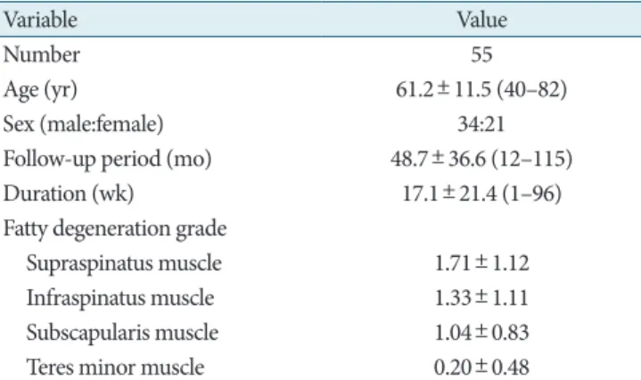
Table 1. Overall patient data
Variable Value
Number 55
Age (yr) 61.2±11.5 (40–82)
Sex (male:female) 34:21
Follow-up period (mo) 48.7±36.6 (12–115)
Duration (wk) 17.1±21.4 (1–96)
Fatty degeneration grade
Supraspinatus muscle 1.71±1.12
Infraspinatus muscle 1.33±1.11
Subscapularis muscle 1.04±0.83
Teres minor muscle 0.20±0.48
Values are presented as mean±standard deviation (range) or mean± standard deviation.
Corp., Armonk, NY, USA). The level of significance was set at P<0.05. Data are presented as mean±standard deviation.
RESULTS
Fifty-five rotator cuff tears were recognized on MRI after the first anterior dislocation of the shoulder. The average follow-up peri-od was 48.7±38.6 months (range, 12–115 months). The age of the patients at the time of primary shoulder dislocation was 61.2 ±11.5 years (range, 40–82 years). There were 21 women with a mean age of 64.5±10.2 years and 34 men with a mean age of 59.2±10.9 years. The mean duration between primary shoul-der dislocation and MRI evaluation was 17.1±21.4 weeks (range, 1–96 weeks). The mean degree of fatty degeneration was 1.71 in the supraspinatus muscle, 1.33 in the infraspinatus muscle, 1.04 in the subscapularis muscle, and 0.20 in the teres minor muscle (Table 1).
Of the 55 shoulders, 26 (47.3%) had isolated rotator cuff tear (R group) and 29 (52.7%) had combined Bankart lesion with rotator cuff tear (B+R group). The mean age was 69.12±8.84 years in the R group and 61.14±10.52 years in the B+R group, a signifi-cant difference. The mean tear size in the sagittal and coronal planes in the R group was 31.12±8.57 mm and 30.81±8.60 mm, respectively. The R group had significantly longer tear size in both directions compared with the B+R group (16.61±12.80 mm and 16.93±12.32 mm, respectively). The number of cases in the R group with more than two injured tendons was 26 (100%) and the number of cases that involved a long head of the biceps ten-don lesion was 22 (84.6%). The degree of fatty degeneration of
the rotator cuff was significantly different between the two groups, with exception of the teres minor muscle (Fig. 1). These findings are summarized in (Table 2).
Age and tear size in both directions were positively correlated (Pearson’s correlation coefficient 0.476 in the AP direction and 0.452 in the ML direction) (Fig. 2). The older age group showed significant increase in tear size, number of injured tendons, and long head biceps tendon lesions (Table 3). Interestingly, degree of fatty degeneration is advanced with age only in the infraspinatus muscle (Fig. 3).
Fig. 1. Distribution of degree of fatty degeneration of the supraspinatus muscle (A), infraspinatus muscle (B), and subscapularis muscle (C) be-tween the B+R and R groups. The results indicate significant differences for these muscles. B+R group: rotator cuff tear combined with labral tear group, R group: rotator cuff tear group, Grade (Gr)0: no fatty deposits, Gr1: some fatty streaks, Gr2: more muscle than fat, Gr3: fat equal to muscle.
Table 2. Comparison of age, tear size, number of injured tendons, and accompanying injuries between two groups
Variable B+R group R group P-value
Number 29 (52.7) 26 (47.3)
Age (yr) 61.12±10.52 69.12±8.84 0.000
Tear size (mm)
AP 16.61±12.80 31.12±8.57 0.000
ML 16.93±12.32 30.81±8.60 0.000
No. of injured tendons 0.005
1 (SS) 8 0
≥ 2 (SS+IS) 21 26
Accompanying injury
LHBT lesion 15 22 0.011
Labral tear 25 0 0.000
Hill Sachs lesion 22 20 0.926
Values are presented as number (%) or mean±standard deviation. B+R group: rotator cuff tear combined with labral tear group, R group: rota-tor cuff tear group.
AP: anterior to posterior, ML: medial to lateral, SS: supraspinatus, IS: infraspinatus, LHBT: long head of the biceps tendon.
30 25 20 15 10 5 0 30 25 20 15 10 5 0 30 25 20 15 10 5 0 Supraspinatus muscle P=0.027 B+R Group ■Gr0 ■Gr1 ■Gr2 ■Gr3 ■Gr0 ■Gr1 ■Gr2 ■Gr3 ■Gr0 ■Gr1 ■Gr2 ■Gr3 Group Group B+R B+R R R R P=0.002 P=0.814
Infraspinatus muscle Subscapularis muscle
B
A
C
Number Number Number
7 12 12 13 3 7 15 11 3 8 7 5 5 5 6 2 2 2 5 5 6 11 1 2
B
A
60 50 40 30 20 10 0 60 50 40 30 20 10 0 40 MM MM r = 0.476 r = 0.452 40 50 60 70 80 50 60 70 80 90 YEARS YEARSFig. 2. Distribution of tear size from anterior to posterior direction (A) and medial to posterior direction (B) as age increases. Both directions showed positive correlation.
Table 3. Comparison of tear size, number of injured tendons, and accompanying injuries according to age group
Variable 40–49 yr 50–59 yr 60–69 yr ≥70 yr P-value
Number 9 (16.4) 15 (27.3) 17 (30.9) 14 (25.4)
Tear size (mm)
AP 10.30±9.16 20.40±13.08 28.41±9.91 29.21±12.69 0.001
ML 10.78±10.28 21.93±13.15 28.06±8.18 27.79±13.29 0.003
No. of injured tendons 0.016
1 4 3 0 1
≥2 5 12 17 13
Accompanying injury
LHBT lesion 3 8 14 12 0.019
Labral tear 8 10 4 3 0.001
Hill Sachs lesion 8 12 10 12 0.215
Values are presented as number (%) or mean±standard deviation.
AP: anterior to posterior, ML: medial to lateral, LHBT: long head of the biceps tendon.
Fig. 3. Distribution of fatty degeneration grade in the supraspinatus muscle (A), infraspinatus muscle (B), and subscapularis muscle (C) ac-cording to age group. Degree of fatty degeneration showed a significant difference in the infraspinatus muscle. Grade (Gr)0: no fatty deposits, Gr1: some fatty streaks, Gr2: more muscle than fat, Gr3: fat equal to muscle.
■Gr0 ■Gr1 ■Gr2 ■Gr3 Age (yr) 40–49 50–59 60–69 ≥70 P=0.019 Infraspinatus muscle
B
20 15 10 5 0 Number 7 5 5 5 5 2 6 7 6 0 0 0 3 3 3 3 3 3 Supraspinatus muscle P=0.105 ■Gr0 ■Gr1 ■Gr2 ■Gr3 Age (yr) 40–49 50–59 60–69 ≥70A
20 15 10 5 0 Number 3 4 44 3 4 4 5 5 5 5 5 1 1 1 1 1 1 1 1 0 8 ■Gr0 ■Gr1 ■Gr2 ■Gr3 Age (yr) 40–49 50–59 60–69 ≥70 P=0.600 Subscapularis muscleC
20 15 10 5 0 Number 4 4 4 4 4 1 6 6When shoulders were divided into two groups by a 3-week du-ration between the first dislocation event and the MRI procedure, 23 (41.8%) were evaluated within 3 weeks after injury and 32 shoulders (58.2%) were evaluated after more than 3 weeks. There was no difference between the two groups in age, number of in-jured tendons, or associated lesions.
However, retraction size of the coronal plane was increased and fatty accumulation of the supraspinatus muscle was more advanced in group I (less than 3 weeks) than group II (more than 3 weeks) (Table 4, Fig. 4).
DISCUSSION
In this study, we focused on rotator cuff tear recognized on MRI after a dislocation event. As age progresses, the rotator cuff weak-ens and is more prone to tearing [25,26]. The results of this study indicate that age is a strong factor of tear size, number of involved tendons, and fatty degeneration of the infraspinatus muscle in primary shoulder dislocation in patients older than 40 years. It is apparent that fatty degeneration and tear progression progress in the AP direction (from supraspinatus to infraspinatus) because the degree of fatty degeneration was significantly different in the infraspinatus muscle in the older age group. Because the infraspi-natus muscle is the main depressor of the humeral head, dys-function results in upward migration of the humerus with sub-acromial impingement and loss of strength in external rotation and elevation [27,28]. We assumed that it is paramount to recog-nize rapid fatty infiltration of the Infraspinatus to avoid poor outcomes after cuff repair in patients older than 40 years,
partic-ularly for those with shoulder dislocation of fatty infiltration grade 2 or higher [29].
The rotator cuff significantly contributes to the stability of the glenohumeral joint, especially in elderly patients. It is possible that the higher prevalence of pre-existing rotator cuff disease in older patients may lead to abnormal glenohumeral motion and predispose an older individual to shoulder instability with low-energy trauma [7,30]. Hsu et al. [30] showed in a cadaver model that rotator cuff tear resulted in abnormal glenohumeral translation, and that larger tears had a greater tendency for direct
Fig. 4. Distribution of fatty degeneration grade in the supraspinatus muscle (A), infraspinatus muscle (B), and subscapularis muscle (C) ac-cording to duration from injury to magnetic resonance imaging. Degree of fatty degeneration showed a significant difference in the supraspi-natus muscle. Grade (Gr)0: no fatty deposits, Gr1: some fatty streaks, Gr2: more muscle than fat, Gr3: fat equal to muscle.
Table 4. Comparison of age, tear size, number of injured tendons, and accompanying injuries according to duration from injury to MRI
Variable Duration (wk) P-value
0–3 ≥ 3 Number 23 (41.8) 32 (58.2) Age (yr) 62.87±11.61 60.03±10.65 0.352 Tear size (mm) AP 20.52±13.79 25.58±12.42 0.160 ML 19.09±11.62 26.66±12.70 0.028
No. of injured tendons 0.120
1 1 7
≥2 22 25
Accompanying injury
LHBT lesion 14 23 0.391
Labral tear 12 13 0.396
Hill Sachs lesion 15 27 0.099
Values are presented as number (%) or mean±standard deviation. MRI: magnetic resonance imaging, AP: anterior to posterior, ML: me-dial to lateral, LHBT: long head of the biceps tendon.
B
A
C
■Gr0 ■Gr1 ■Gr2 ■Gr3 ■Gr0 ■Gr1 ■Gr2 ■Gr3 ■Gr0 ■Gr1 ■Gr2 ■Gr3
Age (yr) Age (yr) Age (yr)
0–3 wk ≥3 wk 0–3 wk ≥3 wk 0–3 wk ≥3 wk 35 30 25 20 15 10 5 0 35 30 25 20 15 10 5 0 35 30 25 20 15 10 5 0 Supraspinatus muscle P=0.027 P=0.164 P=0.479
Infraspinatus muscle Subscapularis muscle
Number Number Number
5 8 11 6 10 4 4 7 3 3 15 2 7 10 4 11 8 10 12 12 3 8 0 2
anterior translation. Pouliart and Gagey [31] showed that the hu-meral head dislocates in the presence of less extensive capsulolig-amentous lesions when rotator cuff lesions are present. Increased age, advanced fatty infiltration, and longer tear size might have weakened the posterior structures and resulted in anterior dislo-cation without anteroinferior labral lesion, such as Bankart lesion in the elderly. These characteristics are also shown in our results, which indicated that the rotator cuff tear group (R group) was statistically much older than the rotator cuff combined with labral tear group (B+R group). The R group cases all involved an infraspinatus tear, while infraspinatus tear involvement was pres-ent in about 72.4% of the BR group. These results suggest that shoulder dislocation without labral tear is strongly related with infraspinatus tear in elderly patients and is referred to as the pos-terior shoulder dislocation mechanism [7,30,31]. In our results, fatty degeneration of the subscapularis was not statistically differ-ent according to age or between the R and B+R groups.
Delayed diagnosis of rotator cuff tear after dislocation may de-crease the recovery potential. Previous studies [4,24] reported that patients who experienced acute injury with severe compro-mise of shoulder function that could be due to the rotator cuff tear should be diagnosed using further evaluations, and that sur-gical repair of rotator cuff tear should be performed within 3 weeks of injury to achieve the best surgical results. Failure to meet the conservative 3-week treatment window after primary dislocation in older patients and persistence of significant pain or weakness are indications for further investigation [32,33]. An-other study showed that infraspinatus fatty infiltration increased significantly when multiple tendons were torn, and that surgical repair should be performed before stage 2 fatty infiltration in older patients [34].
We hypothesized that rotator cuff tear identified by MRI after 3 weeks from dislocation may have features different from those of tear identified earlier. Based on our results, coronal tear size and degree of fatty infiltration in the supraspinatus muscle were increased significantly in the group with more than 3 weeks be-fore intervention (Table 4, Fig. 4). These results suggest that shoulder dislocation may worsen the course of degeneration even in the early period of post-dislocation in elderly patients. These results also provide a theoretical background for understanding why rapid rotator cuff repair might be considered in acute shoul-der dislocation due to the tendency of rapid fatty progression in elderly patients. Previous studies have reported inferior clinical results after delayed treatment of traumatic rotator cuff tear, which is likely due to loss of elasticity in tendons and the signifi-cantly increased tension of the repair. Increased tension is related to decreased viscoelastic properties of the tendons and poor
rota-tor cuff healing [24,35]. In a rat model, supraspinatus tendon de-tachment resulted in rapid loss of muscle mass by 1 week after injury [36,37]. In biomechanical studies, tension at the repaired tendon progressively increased with time from injury because of increase in retraction of the musculotendinous unit and in stiff-ness of the muscle and tendon [38,39]. Unfortunately, we could not evaluate clinical outcomes according to duration and so as-sumed that delayed diagnosis and treatment may have adverse effects on clinical outcomes based on our results and previous studies.
This study had several limitations; in particular, the relatively small number of cases and their retrospective enrollment. This study could not distinguish between acute lesion and chronic le-sion of the rotator cuff, and there were no clinical outcomes to evaluate function, satisfaction, or additional dislocation in pa-tients because of difficulty with long-term follow-up. We also did not have a uniform MRI protocol because the MRI studies were conducted at different institutions and during different time-frames from 2008 to 2019. However, we do not believe that such differences compromised our ability to analyze fatty infiltration.
We concluded that tear size of the rotator cuff and fatty infil-tration of the infraspinatus muscle are positively correlated with age in primary shoulder dislocation in patients older than 40 years. Combined Bankart lesion is more frequently observed in younger patients. Tear size (ML) of the rotator cuff and fatty in-filtration of the supraspinatus may advance faster after disloca-tion in this age group, and careful attendisloca-tion, diagnosis, and fol-low-up are important for optimizing patient outcomes.
ORCID
Jung-Han Kim https://orcid.org/0000-0002-6201-5895 Jin-Woo Park https://orcid.org/0000-0001-6506-3027 Si-Young Heo https://orcid.org/0000-0002-7452-4136 Young-Min Noh https://orcid.org/0000-0001-7149-7526
REFERENCES
1. Maier M, Geiger EV, Ilius C, Frank J, Marzi I. Midterm results after operatively stabilised shoulder dislocations in elderly pa-tients. Int Orthop 2009;33:719-23.
2. Hovelius L, Eriksson K, Fredin H, et al. Recurrences after initial dislocation of the shoulder: results of a prospective study of treatment. J Bone Joint Surg Am 1983;65:343-9.
3. Pevny T, Hunter RE, Freeman JR. Primary traumatic anterior shoulder dislocation in patients 40 years of age and older. Ar-throscopy 1998;14:289-94.
4. Bassett RW, Cofield RH. Acute tears of the rotator cuff: the tim-ing of surgical repair. Clin Orthop Relat Res 1983;(175):18-24. 5. Braune C, von Eisenhart-Rothe R, Welsch F, Teufel M, Jaeger A.
Mid-term results and quantitative comparison of postoperative shoulder function in traumatic and non-traumatic rotator cuff tears. Arch Orthop Trauma Surg 2003;123:419-24.
6. Dinnes J, Loveman E, McIntyre L, Waugh N. The effectiveness of diagnostic tests for the assessment of shoulder pain due to soft tissue disorders: a systematic review. In: NIHR health tech-nology assessment programme: executive summaries. South-ampton: NIHR Journals Library; 2003.
7. Loehr JF, Helmig P, Søjbjerg JO, Jung A. Shoulder instability caused by rotator cuff lesions: an in vitro study. Clin Orthop Relat Res 1994;(304):84-90.
8. Porcellini G, Paladini P, Campi F, Paganelli M. Shoulder insta-bility and related rotator cuff tears: arthroscopic findings and treatment in patients aged 40 to 60 years. Arthroscopy 2006; 22:270-6.
9. Neviaser RJ, Neviaser TJ, Neviaser JS. Anterior dislocation of the shoulder and rotator cuff rupture. Clin Orthop Relat Res 1993;(291):103-6.
10. Neviaser RJ, Neviaser TJ. Recurrent instability of the shoulder after age 40. J Shoulder Elbow Surg 1995;4:416-8.
11. Dwyer T, Razmjou H, Henry P, Gosselin-Fournier S, Holtby R. Association between pre-operative magnetic resonance imag-ing and reparability of large and massive rotator cuff tears. Knee Surg Sports Traumatol Arthrosc 2015;23:415-22.
12. Fuchs B, Weishaupt D, Zanetti M, Hodler J, Gerber C. Fatty de-generation of the muscles of the rotator cuff: assessment by computed tomography versus magnetic resonance imaging. J Shoulder Elbow Surg 1999;8:599-605.
13. Keener JD, Steger-May K, Stobbs G, Yamaguchi K. Asymptom-atic rotator cuff tears: patient demographics and baseline shoul-der function. J Shoulshoul-der Elbow Surg 2010;19:1191-8.
14. Minagawa H, Yamamoto N, Abe H, et al. Prevalence of symp-tomatic and asympsymp-tomatic rotator cuff tears in the general pop-ulation: from mass-screening in one village. J Orthop 2013;10:8-12.
15. Yamamoto A, Takagishi K, Kobayashi T, Shitara H, Osawa T. Factors involved in the presence of symptoms associated with rotator cuff tears: a comparison of asymptomatic and symp-tomatic rotator cuff tears in the general population. J Shoulder Elbow Surg 2011;20:1133-7.
16. Sørensen AK, Bak K, Krarup AL, et al. Acute rotator cuff tear: do we miss the early diagnosis?: a prospective study showing a high incidence of rotator cuff tears after shoulder trauma. J Shoulder Elbow Surg 2007;16:174-80.
17. Neviaser RJ, Neviaser TJ, Neviaser JS. Concurrent rupture of the rotator cuff and anterior dislocation of the shoulder in the older patient. J Bone Joint Surg Am 1988;70:1308-11.
18. Loew M, Porschke FB, Riedmann S, Magosch P, Lichtenberg S. Zur Unterscheidung zwischen traumatischer und degenerativer Rotatorenmanschettenruptur: eine klinische und radiologische Untersuchung. Obere Extremität 2014;9:209-14.
19. Loew M, Magosch P, Lichtenberg S, Habermeyer P, Porschke F. How to discriminate between acute traumatic and chronic de-generative rotator cuff lesions: an analysis of specific criteria on radiography and magnetic resonance imaging. J Shoulder El-bow Surg 2015;24:1685-93.
20. Balich SM, Sheley RC, Brown TR, Sauser DD, Quinn SF. MR imaging of the rotator cuff tendon: interobserver agreement and analysis of interpretive errors. Radiology 1997;204:191-4. 21. Owen RS, Iannotti JP, Kneeland JB, Dalinka MK, Deren JA,
Oleaga L. Shoulder after surgery: MR imaging with surgical validation. Radiology 1993;186:443-7.
22. Zanetti M, Jost B, Hodler J, Gerber C. MR imaging after rotator cuff repair: full-thickness defects and bursitis-like subacromial abnormalities in asymptomatic subjects. Skeletal Radiol 2000;29:314-9.
23. Goutallier D, Postel JM, Bernageau J, Lavau L, Voisin MC. Fatty muscle degeneration in cuff ruptures: pre- and postoperative evaluation by CT scan. Clin Orthop Relat Res 1994;(304):78-83. 24. Hantes ME, Karidakis GK, Vlychou M, Varitimidis S, Dailiana Z, Malizos KN. A comparison of early versus delayed repair of traumatic rotator cuff tears. Knee Surg Sports Traumatol Ar-throsc 2011;19:1766-70.
25. Yamaguchi K, Ditsios K, Middleton WD, Hildebolt CF, Galatz LM, Teefey SA. The demographic and morphological features of rotator cuff disease: a comparison of asymptomatic and symptomatic shoulders. J Bone Joint Surg Am 2006;88:1699-704.
26. Yamaguchi K, Tetro AM, Blam O, Evanoff BA, Teefey SA, Mid-dleton WD. Natural history of asymptomatic rotator cuff tears: a longitudinal analysis of asymptomatic tears detected sono-graphically. J Shoulder Elbow Surg 2001;10:199-203.
27. Inman VT, Saunders JB, Abbott LC. Observations on the func-tion of the shoulder joint. J Bone Joint Surg 1944;26:1-30. 28. Walch G, Edwards TB, Boulahia A, Nové-Josserand L, Neyton L,
Szabo I. Arthroscopic tenotomy of the long head of the biceps in the treatment of rotator cuff tears: clinical and radiographic results of 307 cases. J Shoulder Elbow Surg 2005;14:238-46. 29. Park JS, Park HJ, Kim SH, Oh JH. Prognostic factors affecting
rotator cuff healing after arthroscopic repair in small to medi-um-sized tears. Am J Sports Med 2015;43:2386-92.
30. Hsu HC, Luo ZP, Cofield RH, An KN. Influence of rotator cuff tearing on glenohumeral stability. J Shoulder Elbow Surg 1997;6:413-22.
31. Pouliart N, Gagey O. Concomitant rotator cuff and capsuloliga-mentous lesions of the shoulder: a cadaver study. Arthroscopy 2006;22:728-35.
32. Sonnabend DH. Treatment of primary anterior shoulder dislo-cation in patients older than 40 years of age: conservative versus operative. Clin Orthop Relat Res 1994;(304):74-7.
33. Simank HG, Dauer G, Schneider S, Loew M. Incidence of rota-tor cuff tears in shoulder dislocations and results of therapy in older patients. Arch Orthop Trauma Surg 2006;126:235-40. 34. Melis B, Wall B, Walch G. Natural history of infraspinatus fatty
infiltration in rotator cuff tears. J Shoulder Elbow Surg 2010; 19:757-63.
35. Gimbel JA, van Kleunen JP, Lake SP, Williams GR, Soslowsky
LJ. The role of repair tension on tendon to bone healing in an animal model of chronic rotator cuff tears. J Biomech 2007; 40:561-8.
36. Gimbel JA, van Kleunen JP, Mehta S, Perry SM, Williams GR, Soslowsky LJ. Supraspinatus tendon organizational and me-chanical properties in a chronic rotator cuff tear animal model. J Biomech 2004;37:739-49.
37. Barton ER, Gimbel JA, Williams GR, Soslowsky LJ. Rat supra-spinatus muscle atrophy after tendon detachment. J Orthop Res 2005;23:259-65.
38. Hersche O, Gerber C. Passive tension in the supraspinatus mus-culotendinous unit after long-standing rupture of its tendon: a preliminary report. J Shoulder Elbow Surg 1998;7:393-6. 39. Iannotti JP. The rotator cuff: current concepts and complex
problems. Rosemont, IL: American Academy of Orthopaedic Surgeons; 1998.