105
-Vol. 13, No. 2(December), 2016 The Journal of Medicine and Life Science
It is well known that F18-fluorodeoxyglucose (FDG) positron emission tomography/computed tomography (PET/CT) is valuable for evaluating recurrences of malignancies. It is limited occasionally in lower gastrointestinal, urologic and gynecologic cancers, because an interpretation of FDG PET/CT may be interrupted by a large amount of FDG which is excreted via urinary system. Diuretic or dual-time-point FDG PET/CT has been proposed to resolve these problems. Diuretic or dual-time-point FDG PET/CT improved the specificity and sensitivity of the cases of pelvic malignancies1-11). However, the present case shows that diuretic and dual-time-point FDG PET/CT mimicked a relapse due to an ureterovesical junction (UVJ) stricture in the patient who was suspicious of a recurrence of sigmoid colon cancer with elevated serum tumor marker levels.
In June 2010, a seventy-six year-old male had colectomy due to sigmoid colon cancer. Chemotherapy and radiation therapy was done due to metastatic lesions around right mid
ureter on 35 months after surgery. Thereafter follow-up radiologic studies and carcinoembryonic antigen (CEA) level are normal. FDG PET/CT was performed on 41 month after colectomy to evaluate a cause of elevated serum CEA level (14.62 ng/ml). To avoid the interruption of physiologic urine radioactivity in a bladder, 20 mg of furosemide was injected via intravascular line with FDG so that urine of the bladder was diluted. Diuretic FDG PET/CT showed focal radioactivity in right posterior wall area of bladder suggesting recur of colon cancer (Fig. 1A-D; empty arrows, maximum standardized uptake value (SUVmax); 7.9). As further evaluation magnetic resonance imaging (MRI) was performed and had no abnormal finding to suggest recur (Fig. 1E; T2-weighted image, F; T1-T2-weighted image). The oncologist decided a close observation without any treatment because of normal finding of MRI and history of percutaneous nephrostomy due to mid ureter obstruction occurred by external radiation therapy. Follow-up FDG PET/CT was performed on 9 months after first FDG PET/CT. This study did not use diuretics due to mistakes of technicians and showed normal image (Fig. 2A-D). To get more information, dual-time-point FDG PET/CT was acquired on 2 hours after FDG injection. The dual-time-point FDG PET/CT demonstrated focal radioactivity on the same region of the previous diuretic FDG PET/CT (Fig. 2E-G). Considering decreased serum CEA level (7.92 ng/ml) and the same pattern of the previous diuretic FDG PET/CT, the focal radioactivity was suspected urine radioactivity rather than a
Ureterovesical Junction Stricture on Diuretic and Dual-time-point FDG PET/CT
Mimicking Recur of Colon Cancer
Sanghoon Han
1, Heesung Song
2 Department of 1Internal Medicine and 2Nuclear Medicine,Jeju National University School of Medicine, Jeju, Republic of Koera
(Received December 2, 2016; Revised December 9, 2016; Accepted December 16, 2016)
FDG PET/CT is a useful modality to detect recurred lesions of many malignancies. Because a lot of FDG is excreted through urinary system, FDG PET/CT is limited especially in the cases of pelvic cavity. To overcome these restrictions diuretic or dual-time-point FDG PET/CT has been introduced and improved the specificity and sensitivity. Herein this report presents false-positive findings of diuretic and dual-time-point FDG PET/CT which mimicked a recurrence of sigmoid colon cancer due to an ureterovesical junction stricture. (J Med Life Sci 2016;12(2):105-107)
Key Words
: UVJ stricture, Diuretic, Dual-time-point, FDG, PET/CT, False-positiveIntroduction
Case report
Correspondence to : Heesung Song
Department of Nuclear Medicine, Jeju National University School of Medicine, 15, Aran 13gil, Jeju-si, Jeju Special self–governing province, 63241, Republic of Korea
E-mail : [email protected]
Abstract
Sanghoon Han and Heesung Song has no conflict of interest and no sources of funding.
106
-Sanghoon Han, Heesung Song
malignant lesion. Finally serum CEA returned to normal level on 3 months after follow-up FDG PET/CT without any treatment and the oncologist concluded clinically that the
focal radioactivity was false-positive finding due to the urine concentration of UVJ stricture strengthened by diuretics or delayed time.
FDG PET/CT is a useful tool for detecting recurrence of cancer. Physiologic urine radioactivity of bladder and ureter interrupts interpretations, especially in malignancy of lower abdomen and pelvic cavity, because FDG go through glomerular filtration comparable to glucose, but it is not reabsorbed in the tubules and is grandly excreted in urine, yielding high activity in the urinary tract1). The limitation can
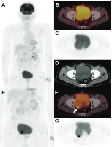
be overcome by diuretic or dual-time-point FDG PET/CT. In many cases diuretic or dual-time-point FDG PET/CT improves the sensitivity and specificity of FDG PET/CT. In the diuretic FDG PET/CT, FDG is injected with diuretics together so that urination increases and urine is diluted. Diluted urine can reveal pathologic lesions hidden by urine radioactivity in abdomen and pelvic cavity2-7). In the dual-time-point FDG PET/CT, first images are acquired 60 minutes after FDG injection and second images are obtained Figure 1. Diuretic FDG PET/CT was performed by
injection of 20 mg of furosemide via intravascular line with FDG. These images showed focal radioactivity in right posterior wall area of bladder suggesting recur of colon cancer (A-D; empty arrows, maximum standardized uptake value (SUVmax); 7.9). As further evaluation magnetic resonance imaging (MRI) was performed and had no abnormal finding to suggest recur (E; T2-weighted image, F; T1-weighted image).
Figure 2. Follow-up FDG PET/CT was performed on 9
months after first FDG PET/CT. This study did not use diuretics due to mistakes of technicians and showed normal image (A-D). To get more information, dual-time-point FDG PET/CT was acquired on 2 hours after FDG injection. The dual-time-point FDG PET/CT demonstrated focal radioactivity on the same region of the previous diuretic FDG PET/CT (2E-G).
Discussion
107
-Ureterovesical Junction Stricture on Diuretic and Dual-time-point FDG PET/CT Mimicking Recur of Colon Cancer
60-120 minutes after first images acquisition conventionally. As time goes by, non-specific urine retention and physiologic bowel radioactivity may vanish and true pathologic lesions can take in more FDG8-11). However, this case shows diuretic or dual-time-point FDG PET/CT may produce false-positive findings. First FDG PET/CT with furosemide shows intense FDG uptake in right posterior wall area of bladder. Actually this false positive finding was made by enhanced urine retention due to UVJ stricture, not recurred lesions. Follow-up dual-time-point FDG PET/CT without diuretics demonstrates normal finding on the standard images but intense FDG uptake in the same area of first FDG PET/CT on the delayed images. This false positive result was originated not by absorbing more FDG over time, but by urine retention due to UVJ stricture. To our best knowledge, there are no reports for false positive diuretic and dual-time-point FDG PET/CT due to urine retention of UVJ stricture. Because the present report shows that UVJ stricture can be a potential cause of false positive recurrence of cancer on diuretic or dual-time-point F-18 FDG PET/CT, physicians should interpret the images more carefully in similar cases.
1) Jadvar H, Parker JA. Clinical PET and PET/ CT. first ed. London: Springer; 2005. PET radiotracers; pp. 45– 67.
2) López-Gandul S1, Pérez-Moure G, García-Garzón JR, Soler-Peter M, Simó-Perdigó M, Lomeña F. Intravenous furosemide injection during FDG PET acquisition. J Nucl Med Technol. 2006;34:228–31.
3) Nair N, Basu S. Selected cases demonstrating the value of furosemide-primed 18F-FDG PET in identifying adrenal involvement. J Nucl Med Technol.
2005;33:166-71.
4) Anjos DA, Etchebehere EC, Ramos CD, Santos AO, Albertotti C, Camargo EE. 18F-FDG PET/CT delayed images after diuretic for restaging invasive bladder cancer. J Nucl Med. 2007;48:764-70.
5) Chen CJ, Chao TB, Shih DF. Recurrent colon cancer involving the urinary bladder identified with F-18 FDG PET/CT after forced diuresis. Clin Nucl Med. 2010;35:258-9.
6) Kamel EM1, Jichlinski P, Prior JO, Meuwly JY, Delaloye JF, Vaucher L, et al. Forced diuresis improves the diagnostic accuracy of 18F-FDG PET in abdominopelvic malignancies. J Nucl Med. 2006;47:1803–7.
7) Chen YW1, Huang MY, Hou PN, Chang CC, Lee CS, Lian SL. FDG PET/CT delayed diuretic imaging technique for differentiating invasive pelvic cancer. Clin Nucl Med. 2009;34:233–5.
8) Hustinx R, Smith RJ, Benard F, Rosenthal DI, Machtay M, Farber LA, et al. Dual time point fluorine-18 fluorodeoxyglucose positron emission tomography: a potential method to differentiate malignancy from inflammation and normal tissue in the head and neck. Eur J Nucl Med. 1999;26:1345–8.
9) Zhuang H, PourdehnadM, Lambright ES,YamamotoAJ, LanutiM, Li P, et al. Dual time point 18F-FDG PET imaging for differentiating malignant from inflammatory processes. J Nucl Med. 2001;42:1412–7.
10) Kubota K, Itoh M, Ozaki K, Ono S, Tashiro M, Yamaguchi K, et al. Advantage of delayed whole-body FDG-PET imaging for tumour detection. Eur J Nucl Med. 2001;28:696-703.
11) Cheng G, Torigian DA, Zhuang H, Alavi A. When should we recommend use of dual time-point and delayed time-point imaging techniques in FDG PET? Eur J Nucl Med Mol Imaging. 2013;40:779-87.