Korean J Anesthesiol Vol. 50, No. 4, April, 2006
서 론
건강한 성인1,2) 및 소아에서3,4) sevoflurane (SEVO)은 QTc 간격을 연장시키는 효과가 있으며,3-6) 한편 선천성 long QT syndrome이 있는 환자에서도 SEVO가 QT 간격을 더욱 연장 시키는 것이 보고되었다.5) 기니 픽 심실근 세포에서 Park 등6) 및 Shibata 등은7) SEVO가 활동전위 기간을 연장시키는 효과가 있음을 관찰하였다. 심전도상에서 QT 간격은 심실 근 세포의 활동전위 기간과 일치한다는 사실을 두고 볼 때 Park 등6) 및 Shibata 등의 결과는7) 임상에서의 심전도 결과 와 일치한다고 하겠다. 활동전위의 연장과 관련된 전기생리 학적인 연구로서 Park 등은6) 기니 픽 심실근 세포에서 SEVO 가 투여 농도에 비례하여 delayed outward K+ current (Ik)를 억제시키나, inwardly rectifying K+ current (IkI)에는 영향이 없는 점으로 보아 SEVO의 활동전위 연장 효과는 주로 Ik의 억제때문인 것으로 생각하였다. 동일 종의 심실근 세포에서 Stadnicka 등은8) SEVO가 IkI의 inward component를 감소시키 나(0.75 mM: ∼20%), outward component는 증가시킨다고 하 였으나(0.75 mM: ∼12%) 이 역시 그 정도가 크지 않음을Electrophysiologic Mechanisms of Sevoflurane on Prolongation of the QT Interval: K+ Currents in Rat
Ventricular Myocytes
Jee Eun Chae, B.S.1, Chong Hoon Kim, M.D.1,2, Kyung Tae Min, M.D.1,2, and Wyun Kon Park, M.D.1,2
1
Department of Anesthesiology and Pain Medicine, 2Anesthesia and Pain Research Institute, Yonsei University College of Medicine, Seoul, Korea
Background: Whereas sevoflurane (SEVO) has been reported to prolong the QT interval, little has been known on the electrophysiologic effects of SEVO which contributes to the prolongation of action potential (AP) duration.
Methods: The ventricular myocytes were obtained from enzymatically treated rat hearts. The standard whole cell voltage-clamp methods were used. The AP was measured using current clamp technique. As a repolarizing K+ current, the transient outward K+ current (Ito), the sustained outward K+ current (Isus), and the inwardly rectifying K+ current (IkI) were measured. The L-type Ca2+ current (ICa, L) was also obtained. After the baseline measurements, the myocytes were exposed to 1.7 and 3.4% SEVO. SEVO concentrations in Tyrode superfusate at room temperature were 0.35 and 0.7 mM for 1.7 and 3.4% SEVO, respectively. Results are mean ± SEM.
Results: SEVO prolonged the AP duration, while the amplitude and the resting membrane potential remained unchanged. At membrane potential of +60 mV, peak Ito was significantly reduced by 18 ± 2 and 24 ± 2% by 0.35 and 0.7 mM SEVO, respectively. 0.7 mM SEVO did not shift the steady-state inactivation curve. Isus was unaffected by 0.7 mM SEVO. The IkI at -130 mV was little altered by 0.7 mM SEVO. ICa, L was significantly reduced by 28 ± 3 and 33 ± 1% by 0.35 and 0.7 mM SEVO, respectively.
Conclusions: Prolongation of AP duration by SEVO in rat ventricular myocytes is likely to be caused by a reduction of Ito. Resting membrane potential was unaffected by SEVO, which seems to be related to no alteration of IkI. (Korean J Anesthesiol 2006; 50: 454∼62)
ꠏꠏꠏꠏꠏꠏꠏꠏꠏꠏꠏꠏꠏꠏꠏꠏꠏꠏꠏꠏꠏꠏꠏꠏꠏꠏꠏꠏꠏꠏꠏꠏꠏꠏꠏꠏꠏꠏꠏꠏꠏꠏꠏꠏꠏꠏꠏꠏꠏꠏꠏꠏꠏꠏꠏꠏꠏꠏꠏꠏꠏꠏꠏꠏꠏꠏꠏꠏꠏꠏꠏꠏꠏꠏꠏꠏꠏꠏꠏꠏꠏꠏꠏꠏꠏꠏꠏꠏꠏꠏꠏꠏꠏꠏꠏꠏꠏꠏꠏꠏꠏꠏꠏꠏꠏꠏꠏꠏ Key Words: action potential, inwardly rectifying K+ current, rat, sevoflurane, transient outward K+ current, ventricular myocytes.
흡입마취제인 Sevoflurane의 QT Interval 연장 효과에 대한
전기생리학적인 연구: 쥐 심실근 세포의 K
+전류에 미치는 영향
연세대학교 의과대학 1마취통증의학교실 및 2마취통증의학연구소채지은
1․김종훈
1,2․민경태
1,2․박윤곤
1,2 논문접수일:2005년 12월 28일 책임저자:박윤곤, 서울시 서대문구 신촌동 134 신촌세브란스병원 마취통증의학과, 우편번호: 120-752 Tel: 02-2228-2405, Fax: 02-312-7185 E-mail: [email protected] 본 연구는 연세대학교 의과대학 2004년도 교수연구비에 의하여 이루 어졌음(승인번호:6-2004-89)알 수 있다. 상기 보고들은 SEVO가 IkI에 별다른 영향이 없 음을 시사하고 있다. 이에 반하여 Azuma 등은9) 기니 픽 심근 절편을 사용하였 음에도 불구하고 SEVO가 활동전위 기간을 단축시키는 상 반된 결과를 관찰하였으며, 이와 같은 단축현상은 개10) 및 쥐의 심실근 세포에서도11) 관찰되었다. 활동전위의 단축 효 과에 관한 전기생리학적인 보고로서 Rithalia 등은11) 쥐 심 실근 세포에서 SEVO가 transient outward K+ current (Ito)에 대한 영향이 없으나, L-type Ca2+ current (ICa, L)는 감소시킴 으로서 ICa, L의 감소 효과가 활동전위 기간을 단축시키는 원인으로 생각하였다. 저자들은 쥐 심실근 세포에서 SEVO의 IkI에 대한 영향을 확인하는 실험을 하던 중 Rithalia 등의11) 결과와는 반대로 SEVO가 활동전위 기간을 연장시키는 것을 관찰하였으며, 따라서 이러한 연장 효과에 영향을 미칠 수 있는, 쥐심실근 세포에서의 재분극에 관여하는 전류인 Ito, IkI 및 ICa, L에 대 해 평가해 보고자 하였다.
대상 및 방법
쥐 심실근 세포의 분리 250-300 g의 수컷 쥐(Sprague-Dawley)를 enflurane으로 흡 입 마취시킨 상태에서, 꼬리정맥으로 heparin (2,500 IU)을 투여한 후 심장을 적출하였다. O2를 포화시킨 실온의 정상 Tyrode용액(mM: 143 NaCl, 5.4 KCl, 1.8 CaCl2, 0.5 MgCl2, 5 HEPES, 0.18 glucose, pH 7.4)에 잠시 담가두어 심장내의 혈 액을 배출시킨 뒤 4oC의 냉각 정상 Tyrode용액에 넣어 심박 동을 멈추게 하였다. 이 상태에서 상행 대동맥을 박리한 후 Langendorff 관류장치에 현수한 다음 37oC의 정상 Tyrode용 액으로 약 5분간 역관류시켰고, Ca2+-free Tyrode용액으로 다 시 5분간 순환시켰다(flow rate: 7 ml/min). 이후 Ca2+-free Tyrode용액에 녹인 collagenase (0.6-0.7 mg/ml, Worthington type II, Worthington Biochemical Corporation, Lakewood NJ, USA)와 hyaluronidase (0.4 mg/ml, Sigma type II, Sigma-Aldrich Co., MO, USA)를 10-14분간 순환시키면서 심장이 물렁물렁 해지는 것을 관찰하였다. 이후 약 5분간 Kraft Brühe (KB) 용 액(mM: 70 KOH, 50 L-glutamic acid, 50 KCl, 20 taurine, 20 KH2PO4, 3 MgCl2, 20 glucose, 5 HEPES, 0.5 EGTA, 5 Na2ATP, 5 pyruvic acid, pH 7.4)으로 순환시킨 다음 양측 심 실을 절제하였다. 절제한 심근을 따로 KB용액에 넣고 Pasteur pipette으로 여러 번 흔들어 조직을 분해시킨 후, 200 μm nylon mesh로 걸러내었다. KB용액에 담겨져 있는 심근 세포를 4℃의 온도에 1시간 동안 보관 후 8시간 이내에 사 용하였으며, 2 mM CaCl2에 노출 시 수축을 보이지 않고 striation이 뚜렷한 세포를 골라 실험하였다. 전기생리학적인 실험 분리된 심근 세포는 inverted microscope 위에 얹혀 있는 chamber에 놓고 10분간 바닥에 가라 앉혀 고정시킨 후 whole cell mode로 각각의 전류를 측정하였다.12) Axopatch 200B Patch Clamp Amplifier (Axon instruments, Foster city, CA, USA)를 사용하였으며, patch electrode는 borosilicate glass (KIMAX, American scientific, Charlotte, NC, USA)를 사용하였 다. Two stage micropipette puller (PP-83, Narishige Co. Ltd, Setagaya-ku, Tokyo, Japan)를 이용하여 첨단의 저항이 2-3 MΩ인 전극을 만들어 사용하였다. 모든 실험은 실온(20-22oC) 에서 시행하였다. Whole cell mode가 되면 4-6분간 기다려 안정 상태가 된 후 실험을 시행하였으며, chamber에서 용액 의 관류량은 분당 2 ml로 하였다. Data acquisition을 위해 ver-sion 6.0.3 pClamp system (Axon instruments)을 장착시킨 IBM- compatible Pentium III personal computer를 사용하였다.세포 외액 및 내액의 구성
세포의 외액으로는 다음과 같은 구성의 modified Tyrode 용액(mM: 140 NaCl, 5.4 KCl, 1 CaCl2, 1 MgCl2, 5 HEPES, 10 glucose, 1 N NaOH로 pH를 7.4로 조절)을 사용하였으며, Ca2+ 내향전류의 억제가 필요한 경우, 0.2 mM CdCl2를 첨가 하였다. K+ 전류의 측정 시 patch pipette 충만 용액으로는 다음과 같은 구성의 용액(mM: 20 KCl, 110 K-aspartate, 10 EGTA, 10 HEPES, 1 MgCl2, 5 K2ATP, 1 CaCl2, 10 NaCl, 3 N KOH로 pH를 7.2로 조절)을 사용하였다. Sustained outward K+ current (Isus)는 modified Tyrode용액에 Ito의 선택적 차단 제인 4-aminopyridine을 첨가한 후 측정하였다. ICa, L은 modified Tyrode 용액의 관류상태에서 whole cell mode를 시행하고 나서 다음과 같은 구성의 용액(mM: 140 NaCl, 5.4 CsCl, 2 CaCl2, 1 MgCl2, 10 HEPES, CsOH로 pH 7.4로 조절)으로 관류 후 측정 하였으며, 이때 patch pipette 충만 용액으로는 다음의 용액 (mM: 30 CsCl, 100 aspartic acid, 100 CsOH, 10 BAPTA, 10 HEPES, 10 phosphocreatinine, 1 Na2GTP, 5 Na2ATP, 10 glucose, 2 MgCl2, CsOH로 pH 7.2로 조절)을 사용하였다.
측정 방법
활동전위: Patch pipette 충만 용액으로는 K+ 전류 측정 시의 용액을, 세포 외액으로는 modified Tyrode 용액을 사용 하였으며, current clamp mode를 시행하였다. 5 ms 동안 1 Hz의 자극횟수로 800 pA의 전류를 주입하였다.
Transient outward K+ current: 막전위를 -80 mV로 고정 한 후 Na+ 내향 전류를 불활성화(inactivation)시키기 위해 50 ms 동안 -40 mV의 prepulse를 가한 다음 -40 mV에서
+60 mV까지 300 ms 동안 5초 간격으로 10 mV씩 증가시 켜 가면서 측정하였다. 이러한 조건하에서 막전류는 처음에 capacitative current가 발생한 후 즉시 outward direction으로 K+ 전류가 나타나게 된다. -40 mV에서 +60 mV까지 10 mV씩 증가시켜 가면서 depolarizing pulse를 가하였으며 이 때 전류의 모양은 초기 peak current가 나타난 후 점차적으 로 감소하여 plateau current를 보이게 된다. Sustained outward K+ current (Isus)는 modified Tyrode 용액에 Ito의 선택적 차단 제인 4-aminopyridine (5 mM)을 첨가 후 측정하였다. Steady-state inactivation of Ito (Itossi): SEVO의 Ito에 대한 억
제 효과가 steady state inactivation curve의 이동과 관련이 있 는지를 알아보기 위해 이 실험을 시행하였다. 막전위를 -80 mV로 고정시킨 상태에서 -100 mV에서 0 mV까지 500 ms 동안 2초 간격으로 10 mV씩 증가시키는 step pulse를 적용한 다음, 각각의 step pulse에서 +80 mV의 pulse를 가하여 측정 하였다. 불활성화(inactivation)의 정도는 I/Imax 비율로 측정하 였으며, I는 -100 mV에서 0 mV까지 각각의 두 번째 step pulse에서 측정한 Ito의 크기이며, Imax는 -100 mV에서 측정 한 Ito의 크기로 하였다. 얻어진 결과는 Boltzmann equation (I/Imax = 1/{1 + exp[(V1/2 - Vm)/S]}, Vm: membrane voltage, V1/2: the voltage at half-maximal inactivation, S: slope)에 적용 하였다.
Inwardly rectifying K+ current: 50 ms 동안 -40 mV로
Na+ 전류를 불활성화(inactivation)시킨 상태에서 막전압을 -130 mV로부터 0 mV까지 200 ms 동안 5초 간격으로 10 mV씩 증가시켜 가면서 측정하였다.
L-type Ca2+ current: -40 mV를 고정 막전압으로 하여
Na+ 전류를 불활성화시킨 상태에서 0 mV로 one step pulse를 적용하였으며 200 ms 동안 0.1 Hz의 자극하에 측정하였다. 투여 약물의 종류, 사용 농도 및 투여 방법
SEVO (Abbott Laboratories, Queenborough, UK)는 기화기 (Sevotec 3, Ohmeda, West Yorkshire, UK)를 통해 100% O2를 분당 0.2 L의 유량으로 gas dispersion tube를 이용하여 관류 액인 modified Tyrode 용액에 용해시켰다. SEVO의 투여 농 도는 기화기의 출구에 호기말 이산화탄소 및 마취가스 분 석기(Capnomac, Datex, Helsinki, Finland)를 부착하여 일정 농도가 나오도록 기화기를 보정(calibration)한 후 사용하였 다. Gas chromatography를 이용하여 각 투여 농도에서 관류 용액이 담겨있는 용기(reservoir) 내의 농도를 측정하였다. 1.7% SEVO 투여 시 용기내의 농도는 0.35 ± 0.00 mM (n = 4), 3.4% 투여 시 0.70 ± 0.00 mM (n = 4)이었다. 22oC에 서 normal Tyrode/gas 분배계수는 0.40이므로13) 이를 계산하 여 보면 실온에서 0.35 mM은 2.12%, 0.7 mM은 4.24%에 해 당하게 된다. 대조군의 측정 후 약제 투여 및 세척 시간은 각각 2분으 로 하였다. 대조치의 측정 후 0.35 또는 0.7 mM SEVO 투 여 시의 억제 및 세척 후의 회복정도를 측정하였다. 자료 검증 방법 모든 수치는 mean ± SEM으로 표시하였으며, P 값이 0.05 미만인 경우를 통계적인 의의가 있는 것으로 간주하였다. 대 조군과 각 약물농도 투여군의 차이를 검증하기 위하여 re-peated measure of ANOVA를 사용하였으며, 다중 비교로는 Student-Newman-Keuls test를 사용하였다. Ito, IkI 및 ICa, L에서 0.35 mM과 0.7 mM SEVO의 투여 효과에 대한 비교는 Table 1. Effects of Sevoflurane (SEVO) on Action Potential
Char-acteristics in Isolated Rat Ventricular Myocytes
ꠚꠚꠚꠚꠚꠚꠚꠚꠚꠚꠚꠚꠚꠚꠚꠚꠚꠚꠚꠚꠚꠚꠚꠚꠚꠚꠚꠚꠚꠚꠚꠚꠚꠚꠚꠚꠚꠚꠚꠚꠚꠚꠚꠚꠚꠚꠚꠚꠚꠚꠚꠚꠚꠚꠚ RMP, mV AMP, mV APD50, ms APD90, ms ꠏꠏꠏꠏꠏꠏꠏꠏꠏꠏꠏꠏꠏꠏꠏꠏꠏꠏꠏꠏꠏꠏꠏꠏꠏꠏꠏꠏꠏꠏꠏꠏꠏꠏꠏꠏꠏꠏꠏꠏꠏꠏꠏꠏꠏꠏꠏꠏꠏꠏꠏꠏꠏꠏꠏ 0.35 mM SEVO (n = 6) Control -74 ± 2 130 ± 5 5.52 ± 1 19.56 ± 2 0.35 mM SEVO -74 ± 3 127 ± 6 6.87 ± 1* 22.45 ± 2* Washout -74 ± 3 128 ± 6 6.27 ± 1 20.24 ± 2 0.7 mM SEVO (n = 7) Control -74 ± 2 134 ± 3 5.29 ± 0 18.49 ± 0 0.7 mM SEVO -74 ± 2 128 ± 4 7.01 ± 1* 25.58 ± 1*† Washout -75 ± 3 129 ± 5 5.27 ± 0 17.02 ± 1 ꠏꠏꠏꠏꠏꠏꠏꠏꠏꠏꠏꠏꠏꠏꠏꠏꠏꠏꠏꠏꠏꠏꠏꠏꠏꠏꠏꠏꠏꠏꠏꠏꠏꠏꠏꠏꠏꠏꠏꠏꠏꠏꠏꠏꠏꠏꠏꠏꠏꠏꠏꠏꠏꠏꠏ Values represent means ± SEM. RMP: resting membrane potential, AMP: action potential amplitude, APD50 and APD90: action potential duration measured at 50 and 90% of repolarization, respectively. *: P < 0.05 differed from control and washout values, †: P < 0.05 differed from 0.35 mM SEVO.
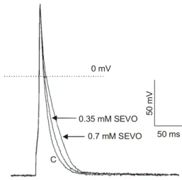
Fig. 1. Effect of sevoflurane (SEVO) on action potential duration in a rat ventricular myocyte. C represents control.
0 mV 0.35 mM SEVO 0.7 mM SEVO 50 ms 50 m V C
unpaired t-test를 사용하였다.
결 과
활동전위 0.35 (n = 6) 및 0.7 mM (n = 7) SEVO는 안정막 전위 및 활동전위의 크기에 영향을 미치지 않았으나, 활동전위 기간 의 50 및 90% (APD50, APD90)에서 의의있는 연장 효과를 보였다(Table 1, Fig. 1).Transient outward K+ current
0.35 mM SEVO는 +60 mV에서 측정한 Ito의 peak current 를 대조군에 비해 18 ± 2% 감소시켰으며(n = 7, P < 0.05) (Fig. 2D), 세척 후 대조치로 회복되었다. 0.35 mM SEVO는 +60 mV에서 Ito의 plateau current를 10 ± 3% 감소시켰으며 (P < 0.05), 세척 후 대조치로 회복되었다.
0.7 mM SEVO는 +60 mV에서 측정한 Ito의 peak current를 대조군에 비해 24 ± 2% 감소시켰으며(n = 11, P < 0.05)
(Fig. 2A-D), 세척 후 대조치로 회복되었다. 0.7 mM SEVO는 +60 mV에서 Ito의 plateau current를 10 ± 2% 감소시켰으며(P < 0.05) (Fig. 2C), 세척 후 대조치로 회복되었다. Ito의 peak와 plateau current에서 두 투여 농도 사이에 차이는 없었다. 0.35 및 0.7 mM SEVO 투여 군에서 Ito의 peak current의 대조치는 각각 3.82 ± 0.54 및 3.64 ± 0.30 nA였으며, plateau current의 대조치는 각각 1.51 ± 0.12 및 1.52 ± 0.10 nA였다
Sustained outward K+ current
Ito에서(Fig. 3A) 5 mM 4-aminopyridine을 투여 시 Isus만을 관찰할 수 있었으며(Fig. 3B, C), 0.7 mM SEVO 투여 시 변 화를 보이지 않았다(n = 6, NS) (Fig. 3C).
Steady-state inactivation of Ito (Itossi)
0.7 mM SEVO에서 Ito의 inactivation curve는 Boltzmann dis-tribution을 보였으며(Fig. 4A) 대조군의 half inactivation (V1/2) 은 -31.53 ± 0.68 mV, 0.7 mM SEVO 투여 시에는 -35.04 ± 0.84 mV로 두 군 간의 차이를 보이지 않았다(n = 4, NS) Fig. 2. Effects of sevoflurane (SEVO) on transient outward K+ currents (Ito) in rat ventricular myo-cytes. (A, B) Recordings of control (A) and 0.7 mM SEVO (B) in a rat ventricular myocyte. (C) Current- voltage relationships of Ito. Closed and open circles indicate the peak current of Ito at every potential in the control and in the presence of 0.7 mM SEVO, respectively. Triangles are the current levels at the end of the test pulses before (closed) and after (open) application of 0.7 mM SEVO. (D) Effects of 0.35 mM and 0.7 mM SEVO on the amplitude of Ito. *: P < 0.05 vs. control. Error bars indicate mean ± SEM. * * * * * *
A
B
D
C
1 A n 100 ms(Fig. 4B).
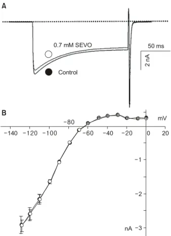
Inwardly rectifying K+ current
0.7 mM SEVO 투여 시 -130 mV에서 측정한 IkI는 대조군 과 차이를 보이지 않았다(n = 7, NS)(Fig. 5A, B). 두 투여 농 도간의 차이는 없었다. -130 mV에서의 대조치는 -2.92 ± 0.25 nA였다. L-type Ca2+ current 0.35 및 0.7 mM SEVO는 대조군에 비해 각각 28 ± 3% (n = 8, P < 0.05), 33 ± 1% (n = 7, P < 0.05)를 감소시켰으 며(Fig 6B), 세척 후 대조치로 회복되었다(Fig 6A). 두 투여 농도간의 차이는 없었다. 대조치는 0.35 및 0.7 mM 투여 군 에서 각각 -0.75 ± 0.09 및 -0.81 ± 0.20 nA였다.
고 찰
심근 세포의 활동전위 기간은 세포막을 통과하는 내향 및 외향전류의 상호간의 균형에 의해 결정되며, 이러한 균형의 변화에 따라 활동전위 기간이 연장 혹은 단축될 수 있다.14,15) 심실근 세포의 초기 재분극 시기(early repolarization phase)는 Ito의 활성에 의해 형성된다. 즉, 활동전위의 초기 재분극 시 기인 phase I 및 plateau phase의 초반은 Ito에 의해 결정되며, plateau phase는 주로 ICa, L에 의해 결정된다. Plateau phase가 Fig. 3. Effect of sevoflurane (SEVO) on sustained outward K+currents (Isus) in a rat ventricular myocyte. (A) A control recording of transient outward K+ currents (Ito). (B) Isus obtained following application of 5 mM 4-aminopyridine, a specific blocker of Ito. (C) After 0.7 mM SEVO exposure. SEVO had no effect on Isus.
l , Control to lSUS,Control l SEVOsus, 100 ms 1 nA
A
B
C
Fig. 4. Effect of sevoflurane (SEVO) on inwardly rectifying K+current (Ik1) in rat ventricular myocytes. (A) Closed and open circles indicate control and 0.7 mM SEVO in a rat ventricular myocyte, respectively, at a membrane potential of -130 mV. (B) Current-voltage rela-tionships for IkI before and after addition of 0.7 mM SEVO. Closed and open circles indicate control and 0.7 mM SEVO, respectively. Error bars indicate mean ± SEM.
-1 -2 -3 nA mV -140 -120 -100 -80 -60 -40 -20 0 20 0.7 mM SEVO Control 50 ms
A
B
2 n A끝날 무렵에 Ik는 심실근 세포의 재분극에 관여하고, IkI는 활 동전위의 마지막 재분극 시기의 조정을 담당하며14) 또한 휴 지막전위(resting membrane potential)를 유지시키는 역할을 한 다. 따라서, IkI가 억제되면 휴지 막전위가 탈분극 되는 현상 을 보이며 또한 활동전위 기간이 연장된다. 쥐의 심실근 세 포에서 재분극에 관여하는 전류로는 Ito, ICa, L 및 IkI 등이 있 다.
쥐 심실근 세포를 이용한 본 실험에서 SEVO는 활동전위 기간을 연장시키는 효과를 보였으며, 재분극에 관련된 전류 중 Ito와 ICa, L은 감소시켰으나, IkI와 Isus에는 별다른 영향을 보이지 않았다. Ito는 쥐,16) 개,17) 고양이,18) 토끼19) 및 사람의 심실근 세포 에20) 존재하는 반면 기니 픽 심실근 세포에는 거의 존재하 지 않으며, 쥐 심근 세포에서는 Ik가 매우 작거나 분포하지 않는 것으로 알려져 있다.21) Ito는 potassium에 의해 전도되 며, 빠른 활성(activation) 및 불활성(inactivation)을 보이고 전 체적으로 외향전류를 형성한다.22) 전압 의존적인 특성을 갖 고 있으며, 조직에서의 전류 밀도(current density)가 매우 높 고, 또한 4-aminopyridine에 선택적으로 차단되는 특성을 보 Fig. 5. Steady-state inactivation of Ito. (A) A representative control
recording in a rat ventricular myocyte. Cells were clamped to a range of conditioning potentials from -100 to 0 mV followed by a test pulse to +80 mV to evoke residual Ito. (B) Steady-state inactivation curves are shown under control conditions and in the presence of 0.7 mM sevoflurane (SEVO). Closed and open circles indicate control and 0.7 mM SEVO, respectively. Data are presented as mean ± SEM for four cells and were fitted with the Boltzmann function. The half-inactivations (V1/2) ofcontrol and0.7 mM SEVO were -31.53 ± 0.68 mV and -35.04 ± 0.84 mV, respectively, which showed no differences. -100 -80 -60 -40 -20 0 1.0 0.8 0.6 0.4 0 0.2 l/l m ax mV Control 0.7 mM SEVO Control 200 ms 1. 5 n A
A
B
Fig. 6. Effect of sevoflurane (SEVO) on L-type Ca2+ current (ICa, L). (A) A representative example of the effect of SEVO on ICa, L in a rat ventricular myocyte. The open circles represent the peak of an individual current record. The horizontal bar indicates the period when an anesthetic was applied. (Inset) an example of individual currents recorded in the presence of 0.7 mM SEVO. (B) Depression of ICa, L following application of 0.35 mM and 0.7 mM SEVO, respectively. *vs. control. Error bars indicate mean ± SEM.
인다. 현재 Ito는 두 가지의 종류가 있는 것으로 알려져 있 다. 한 종류는 calcium 의존성 Cl- 전류(Ca2+- dependent Cl- current)이며, 다른 한 종류는 calcium 비의존성 K+ 전류(Ca2+- independent K+ current)로서,23) calcium 비의존성 Ito는 활동전 위 기간의 결정에 중요한 역할을 하는 것으로 알려져 있 다.24) 쥐 심실근 세포에는 calcium 비의존성 Ito만 존재하는 것으로 보고되었다.25) 본 결과에서 Ito의 감소는 숫자상으로 볼 때 크지 않으나 쥐 심실근 세포에서 Ito의 전류 밀도가 매우 큰 것을(+60 mV에서 19.9 ± 2.8 pA/pF)26) 생각하여 볼 때 실제 재분극 에 미치는 영향은 클 것으로 생각된다. 또한, Ito의 활성화가 매우 빠르기 때문에 phase I 및 early plateau phase에 수백 pA의 전류의 변화는 plateau phase에 관여하는 다른 전류 (Ca2+ 내향 전류, Ik 등)에 영향을 미쳐 결과적으로 Ito의 변 화는 활동전위 기간에 영향을 미칠 수 있다. 따라서 본 실 험에서 SEVO에 의한 Ito의 의의있는 감소는 쥐 심실근 세포 의 활동전위 기간의 연장에 기여하였을 것으로 생각된다. 쥐 및 사람의 심실근 세포에서 Ito는 재분극에 관여하는 주 된 전류이며,16,26,27) Ito 및 Ik 차단제인 tedisamil은 쥐의 심실근 세포에서 활동전위 기간을 연장시키고,28) 이는 tedisamil을 투 여한 Langendorff preparation상의 쥐 심근에서도 QT 간격을 연장시키는 효과를 보이고 있다.29) 말기 심부전 환자에서 활 동전위의 연장은 Ito expression level의 감소와 밀접한 관련이 있다는 보고는30,31) Ito의 감소와 활동전위의 연장이 사람에서 도 또한 연관성이 있음을 입증하고 있다.32,33)
SEVO의 Ito에 대한 억제 효과가 steady state inactivation curve를 좌측으로 이동시키기 때문인지를 알아보기 위해 Itossi 를 측정해 보았다. SEVO 투여 시 half inactivation (V1/2)에 거 의 변화가 없는 것으로 보아 SEVO의 Ito에 대한 억제 효과 가 steady state inactivation curve를 좌측으로 이동시키기 때 문은 아닌 것으로 생각된다. 즉, SEVO는 voltage-dependency 와 inactivation kinetics를 변화시키지 않고 Ito의 크기만 변화시 켰는데 이것은 SEVO가 single channel conductance는 변화시키 지 않으면서 단지 single opening의 기간을 감소시키기 때문인 것으로 생각된다. Halothane을 사용한 실험에서 Davies 등은27) halothane이 Ito의 크기를 감소시키며, steady state inactivation curve도 좌측으로 이동시킴을 관찰하였는데, 이것은 SEVO 의 Ito에 대한 불활성화 기전이 halothane과는 다르다는 것을 의미한다고 하겠다.
Isus는 4-aminopyridine에 의해 Ito가 제거된 상태에서 남은 전류에 해당되며, potassium 통로 차단제인 TEA (tetraethy-lammonium chloride)에 의해 억제되는 것으로 보아 delayed outward K+ current로 생각되며,34) 빠르게 활성화되지만 불 활성화 되지 않는 전류로서 활동전위의 전체적인 재분극 과정에 영향을 미치는 것으로 생각되고 있다. 본 실험에서
SEVO가 Isus에 영향을 미치지 않은 점으로 보아 SEVO에 의 한 Ito의 감소는 순수하게 Ito에만 영향을 준 것으로 생각할 수 있다. IkI는 심실근 세포에서 휴지막전위의 유지에 우선적으로 작용하는 K+ 전류이다. IkI의 억제는 이완기 탈분극(diastolic depolarization)을 일으키고 이는 심실근 세포의 흥분도(car-diac excitability)를 증가시키며35) 이로 인하여 비정상적인 automaticity가 발생할 수 있다.36) IkI의 inward current의 이러 한 작용 외에 outward component는 활동전위의 재분극 끝부 분 형성에 영향을 미친다.37,38) 결과적으로 IkI의 억제는 early afterdepolarization을 일으킬 수 있으며 이로 인한 부정맥을 유발시킬 수 있다.36) 본 실험에서 SEVO는 IkI의 inward 및 outward component에 영향을 미치지 않았는데, IkI의 변화가 APD90에 해당하는 활동전위 기간과 휴지막 전위에 영향을 미칠 수 있는 점을 고려해 볼 때, SEVO 투여 시 휴지막전 위에 변화가 없었던 것은 IkI의 inward component에 별다른 영향이 없었기 때문인 것으로 생각된다. Agus 등은39) -40 mV를 고정 막전압으로 하였을 때 세포 외액에 CdCl2를 첨 가하지 않은 경우는 Ito가 활성화되지 않으나, CdCl2를 첨가 하면 Ito가 활성화됨을 관찰하였다. 이것은 CdCl2 (0.2 mM) 에 의한 steady state inactivation curve의 우측 이동으로 인하 여 -40 mV에서도 Ito channel의 ∼20%가 활성화되어 있기 때문이라고 하였다. 본 실험에서 Ik1 측정 시 세포외액에 0.2 mM의 CdCl2를 첨가하였으므로 -40 mV 이상의 전류에 Ito 가 포함되어 있을 가능성이 있으므로 본 실험으로는 SEVO 가 Ik1의 outward component에 미치는 영향은 평가하기 어렵 다. Park 등은6) 기니 픽 심실근 세포에서 0.35 mM(1.7%에 해당) SEVO가 Ik1에 거의 영향이 없다고 하였으며(inward component: ∼8% 감소, outward component: ∼7% 감소), 0.7 mM에서도 유사한 감소를 보임으로서 SEVO가 IkI에 별다른 영향이 없음을 보고하였다. Stadnicka 등은8) 기니 픽 심실근 세포에서 SEVO가 IkI의 inward component를 감소시키며(0.35 mM: 0%, 0.75 mM: ∼20%), -80∼0 mV 사이에서 관찰할 수 있는 outward component는 증가시키는(0.35 mM: ∼10%, 0.75 mM: ∼12%) 이중적인 효과를 보고하였으나 이 역시 억제 및 증가의 정도가 크지 않다. 기니 픽 심실근 세포에서 halo-thane 및 isoflurane은 IkI의 inward 및 outward component에 SEVO와 유사한 이중적인 효과를 보이기는 하나 이 역시 억 제 및 증가의 정도가 크지 않아 큰 영향이 없음을 알 수 있 다.40) Halogenated ether 계통의 약제인 SEVO와 isoflurane이 IkI에 유사한 효과를 보임으로써 기타 halogenated ether 계통 의 약제인 desflurane도 유사한 효과를 보일 것으로 추정되 기는 하나 이는 향후 실험을 통해 확인해 보아야 할 것이 다.
Ca2+ 통로를 통한 Ca2+ 내향전류로 중요한 역할을 하고 있 다. 활동전위의 plateau phase에 Ca2+은 세포내로 유입되며 유입된 Ca2+은 근소포체(sarcoplasmic reticulum)로부터 Ca2+ 배출을 유도하고, 또한 다음 배출을 위해 근소포체에 Ca2+을 공급하기도 한다.41) ICa, L이 억제되면 활동전위 기간은 감소 하게 된다. 본 실험에서 0.35 mM SEVO는 ICa, L을 ∼28%, 0.7 mM SEVO는 ∼33% 감소시켰으며, 또한 기니 픽6) 및 개의10) 심실근 세포에서도 ICa, L의 감소가 보고되어 있다. 심근세포의 활동전위 기간은 세포막을 통과하는 내향전 류와 외향전류 상호간의 균형에 의해 결정되며, 이러한 균 형의 변화에 따라 활동전위 기간이 단축 혹은 연장될 수 있다.14,15) 외향전류인 Ito의 감소는 활동전위 기간을 연장시 키며, 내향전류인 ICa, L의 감소는 활동전위 기간을 감소시키 는데, 본 연구에서 SEVO가 활동전위 기간을 연장시킨 것은 아마도 SEVO에 의한 ICa, L의 감소로 인한 활동전위 기간 단축 효과가 Ito의 감소로 인한 활동전위 기간 연장 효과에 의해 상쇄되었기 때문인 것으로 생각할 수 있을 것이다. Shibata 등은7) 기니 픽의 심실근 세포를 이용한 실험에서 0.65 mM의 SEVO가 휴지막 전위를 변화시키지 않으면서 활 동전위 기간을 연장시킴을 보고하였다. 이 연구에서 SEVO는 Ik를 구성하는 요소인 느리게 활성화되는 Iks 및 빠르게 활성 화되며 inward rectifier 경향을 보이는 Ikr 중 Iks를 더욱 현저 히 감소시키며, Ikr의 선택적 차단제인 E4031을 투여 후 활동 전위가 더욱 연장되는 점으로 보아 Iks의 감소가 활동전위 기 간 연장의 주된 원인으로 생각하였다. 동일종의 심실근 세 포에서 isoflurane 또한 유사한 결과를 보이고 있다.42) 사람의 심실근 세포에 분포하는 전류로는 Ito,43) Ikr44) 및 Ik1가 분포하고 있으며,45) 개나 토끼의 Iks에 비해 전류 의 크기가 작기는 하나 최근 Iks도 분포하고 있음이 보고 되었다.46) 사람의 심실근 세포에서 Ito의 전류 밀도는 쥐 에 비해 3∼4배 정도 낮으나(+60 mV에서 8.2 ± 0.7 pA/pF) 주된 K+ 전류이며, 또한 두 종간에 Ito의 전기생 리학적 특성(kinetics)이 매우 유사한 점과,26) IkI에 대한 약제의 효과가 동일한 결과를 보이는 점으로 보아,47) SEVO의 임상적인 QTc interval 연장 효과는 Iks의 억제 외에 Ito의 억제도 그 한 원인으로 생각해 볼 수 있을 것 이다. 결론적으로, 쥐 심실근세포에서 SEVO의 활동전위 기 간 연장 효과는 Ito의 감소 효과에 의한 것으로 생각된 다. SEVO에 의한 ICa, L의 감소에도 불구하고 활동전위 기 간이 연장된 것은 Ito의 감소가 이를 상쇄하였기 때문인 것으로 생각된다. 활동전위에서 SEVO투여시 휴지막전위 의 변화가 없었던 것은 SEVO가 IkI의 inward component에 영향이 없었기 때문인 것으로 생각된다.
참 고 문 헌
1. Kleinsasser A, Kuenszberg E, Loeckinger A, Keller C, Hoermann
C, Lindner KH, et al: Sevoflurane, but not propofol, significantly prolongs the Q-T interval. Anesth Analg 2000; 90: 25-7. 2. Kuenszberg E, Loeckinger A, Kleinsasser A, Lindner KH,
Puehrin-ger F, Hoermann C: Sevoflurane progressively prolongs the QT interval in unpremedicated female adults. Eur J Anaesthesiol 2000; 17: 662-4.
3. Loeckinger A, Kleinsasser A, Maier S, Furtner B, Keller C, Kueh-bacher G, et al: Sustained prolongation of the QTc interval after anesthesia with sevoflurane in infants during the first 6 months of life. Anesthesiology 2003; 98: 639-42.
4. Whyte SD, Booker PD, Buckley DG: The effects of propofol and sevoflurane on the QT interval and transmural dispersion of repo-larization in children. Anesth Analg 2005; 100: 71-7.
5. Gallagher JD, Weindling SN, Anderson G, Fillinger MP: Effects of sevoflurane on QT interval in a patient with congenital long QT syndrome. Anesthesiology 1998; 89: 1569-73.
6. Park WK, Pancrazio JJ, Suh CK, Lynch C III: Myocardial depressant effects of sevoflurane: mechanical and electrophysiologic actions in vitro. Anesthesiology 1996; 84: 1166-76.
7. Shibata S, Ono K, Iijima T: Sevoflurane inhibition of the slowly activating delayed rectifier K+ current in guinea pig ventricular cells. J Pharmacol Sci 2004; 95: 363-73.
8. Stadnicka A, Bosnjak ZJ, Kampine JP, Kwok WM: Effects of sevoflurane on inward rectifier K+ current in guinea pig ventric-ular cardiomyocytes. Am J Physiol 1997; 273: H324-32. 9. Azuma M, Matsumura C, Kemmotsu O: The effects of sevoflurane
on contractile and electrophysiologic properties in isolated guinea pig papillary muscles. Anesth Analg 1996; 82: 486-91. 10. Hatakeyama N, Momose Y, Ito Y: Effects of sevoflurane on
con-tractile responses and electrophysiologic properties in canine single cardiac myocytes. Anesthesiology 1995; 82: 559-65.
11. Rithalia A, Hopkins PM, Harrison SM: The effects of halothane, isoflurane, and sevoflurane on Ca2+ current and transient outward K+ current in subendocardial and subepicardial myocytes from the rat left ventricle. Anesth Analg 2004; 99: 1615-22. 12. Hamill OP, Marty A, Neher E, Sakmann B, Sigworth FJ: Improved
patch-clamp techniques for high-resolution current recordings from cells and cell-free membrane patches. Pflügers Arch 1981; 391: 85-100.
13. Hönemann CW, Washington J, Hönemann MC, Nietgen GW, Du-rieux ME: Partition coefficents of volatile anesthetics in aqueous electrolyte solutions at various temperatures. Anesthesiology 1998; 89: 1032-5.
14. Carmeliet E: Mechanisms and control of repolarization. Eur Heart J 1993; 14 Suppl: 3-13.
15. Coraboeuf E: Ionic basis of electrical activity in cardiac tissues. Am J Physiol 1978; 234: H101-16.
16. Josephson IR, Sanchez-Chapula J, Brown AM: Early outward current in rat single ventricular cells. Circ Res 1984; 54: 157-62.
17. Litovsky SH, Antzelevitch C: Transient outward current prominent in canine ventricular epicardium but not endocardium. Circ Res 1988; 62: 116-26.
18. Furukawa T, Myerburg RJ, Furukawa N, Bassett AL, Kimura S: Differences in transient outward currents of feline endocardial and epicardial myocytes. Circ Res 1990; 67: 1287-91.
19. Giles W, Shimoni Y: Comparison of sodium-calcium exchanger and transient inward currents in single cells from rabbit ventricle. J Physiol 1989; 417: 465-81.
20. Nabauer M, Beuckelmann DJ, Erdmann E: Characteristics of transient outward current in human ventricular myocytes from patients with terminal heart failure. Circ Res 1993; 73: 386-94. 21. Rees S, Curtis MJ: Which cardiac potassium channel subtype is
the preferable target for suppression of ventricular arrhythmias? Pharmacol Ther 1996; 69: 199-217.
22. Tristani-Firouzi M, Chen J, Mitcheson JS, Sanguinetti MC: Molec-ular biology of K+ channels and their role in cardiac arrhythmias. Am J Med 2001; 110: 50-9.
23. Nerbonne JM: Molecular basis of functional voltage-gated K+ channel diversity in the mammalian myocardium. J Physiol 2000; 525: 285-98.
24. Escande D, Coulomb A, Faivre JF, Deroubaix E, Coraboeuf E: Two types of transient outward currents in adult human atrial cells. Am J Physiol 1987; 252: H142-8.
25. Dukes ID, Morad M: The transient K+ current in rat ventricular myocytes: evaluation of its Ca2+ and Na+ dependence. J Physiol 1991; 435: 395-420.
26. Wettwer E, Amos G, Gath J, Zerkowski HR, Reidemeister JC, Ravens U: Transient outward current in human and rat ventricular myocytes. Cardiovasc Res 1993; 27: 1662-9.
27. Davies LA, Hopkins PM, Boyett MR, Harrison SM: Effects of halothane on the transient outward K+ current in rat ventricular myocytes. Br J Pharmacol 2000; 131: 223-30.
28. Dukes ID, Cleemann L, Morad M: Tedisamil blocks the transient and delayed rectifier K+ currents in mammalian cardiac and glial cells. J Pharmacol Exp Ther 1990; 254: 560-9.
29. Rees SA, Tsuchihashi K, Hearse DJ, Curtis MJ: Combined admin-istration of an Ik(ATP) activator and Ito blocker increases coronary flow independently of effects on heart rate, QT interval, and ischaemia-induced ventricular fibrillation in rats. J Cardiovasc Pharmacol 1993; 22: 343-9.
30. Kaprielian R, Wickenden AD, Kassiri Z, Parker TG, Liu PP, Backx PH: Relationship between K+ channel down-regulation and [Ca2+]i in rat ventricular myocytes following myocardial infarction. J Physiol 1999; 517: 229-45.
31. Gidh-Jain M, Huang B, Jain P, el-Sherif N: Differential expression of voltage-gated K+ channel genes in left ventricular remodeled myocardium after experimental myocardial infarction. Circ Res 1996; 79: 669-75.
32. Kaab S, Dixon J, Duc J, Ashen D, Nabauer M, Beuckelmann DJ,
et al: Molecular basis of transient outward potassium current downregulation in human heart failure: a decrease in Kv4.3 mRNA correlates with a reduction in current density. Circulation 1998; 98: 1383-93.
33. Li GR, Lau CP, Leung TK, Nattel S: Ionic current abnormalities associated with prolonged action potentials in cardiomyocytes from diseased human right ventricles. Heart Rhythm 2004; 1: 460-8. 34. Apkon M, Nerbonne JM: Characterization of two distinct
depolar-ization-activated K+ currents in isolated adult rat ventricular myocytes. J Gen Physiol 1991; 97: 973-1011.
35. Nichols CG, Makhina EN, Pearson WL, Sha Q, Lopatin AN: Inward rectification and implications for cardiac excitability. Circ Res 1996; 78: 1-7.
36. The Task Force of the Working Group on Arrythmias of the Euro-pean Society of Cardiology: The ‘Sicilian Gambit', a new approach to the classification of antiarrhythemic drugs based on their action on arrhythmogenic mechanisms. Eur Heart J 1991; 12: 1112-31. 37. Giles WR, Imazumi Y: Comparison of potassium currents in rabbit
atrial and ventricular cells. J Physiol 1988; 405: 123-45. 38. Koumi S, Wasserstrom JA, Ten Eick RE: Beta-adrenergic and
cholinergic modulation of the inwardly rectifying K+ current in guinea pig ventricular myocytes. J Physiol 1995; 486: 647-59. 39. Agus ZS, Dukes ID, Morad M: Divalent cations modulate the
transient outward current in rat ventricular myocytes. Am J Physiol 1991; 261: C310-8.
40. Stadnicka A, Bosnjak ZJ, Kampine JP, Kwok WM: Modulation of cardiac inward rectifier K+ current by halothane and isoflurane. Anesth Analg 2000; 90: 824-33.
41. Bers DM: Cardiac excitation-contraction coupling. Nature 2002; 415: 198-205.
42. Suzuki A, Aizawa K, Gassmayr S, Bosnjak ZJ, Kwok WM: Biphasic effects of isoflurane on the cardiac action potential: an ionic basis for anesthetic-induced changes in cardiac electrophysiology. Anesthe-siology 2002; 97: 1209-17.
43. Amos GJ, Wettwer E, Metzger F, Li Q, Himmel HM, Ravens U: Differences between outward currents of human atrial and subepicardial ventricular myocytes. J Physiol 1996; 491: 31-50. 44. Veldkamp MW, van Ginneken AC, Opthof T, Bouman LN: Delayed
rectifier channels in human ventricular myocytes. Circulation 1995; 92: 3497-504.
45. Beuckelmann DJ, Nabauer M, Erdmann E: Alterations of K+ currents in isolated human ventricular myocytes from patients with terminal heart failure. Circ Res 1993; 73: 379-85.
46. Virag L, Iost N, Opincariu M, Szolnoky J, Szecsi J, Bogats G, et al: The slow component of the delayed rectifier potassium current in undiseased human ventricular myocytes. Cardiovasc Res 2001; 49: 790-7.
47. Carnes CA, Muir WW 3rd, Van Wagoner DR: Effect of intrave-nous anesthetics on inward rectifier potassium current in rat and human ventricular myocytes. Anesthesiology 1997; 87: 327-34.