J Health Tech Assess 2015;3(1):26-34 ISSN 2288-5811 Copyright © 2015 The Korean Association for Health Technology Assessment
Introduction
Atrial fibrillation (AF) is triggered by ectopic beating of the pulmonary vein (PV), and catheter-based pulmonary vein iso-lation (PVI) using radiofrequency is accepted to be effective for treatment of patients in whom AF is refractory to anti-ar-rhythmic drugs.1,2) The preferred ablation strategy features
electrical isolation of the PV by creation of circumferential le-sions around the right and left PV ostia.3) Although
three-di-mensional (3D) mapping systems are routinely used for cathe-ter-based PVI, point-by-point ablation remains complex and time-consuming, requiring a high level of technical compe-tence. Such technical complexity and the need to visualize the
circular mapping and ablation catheters in real time, means that many centers perform only one or two procedures daily, and patients endure long-term procedural discomfort and are exposed to the risks associated with general anesthesia. More importantly, patients are subjected to significant levels of radia-tion (shared to some extent by the operators).4-7) The adverse
effects of radiation exposure during the catheter ablation are well documented in medical literature.8,9) To address such
tech-nical difficulties; and to render the procedure simpler, shorter, and safer; multielectrode catheters enabling simultaneous map-ping and ablation have been developed. Such catheters allow an operator to create lesions over much of the circumference of each PV via a single radiofrequency (RF) application, without
Comparative Efficacy and Procedural Durability
of Radiofrequency Ablation for Atrial Fibrillation:
A Systematic Review and a Meta-Analysis
Hyunsook Choi, MPH1, Eun-Cheol Park, MD, PhD2,3, Sun Ha Jee, PhD4, and Sohee Park, PhD3,5
1Medtronic Korea Ltd., Seoul, Korea
2Department of Preventive Medicine and 3Institute of Health Services Research, Yonsei University College of Medicine, Seoul, Korea
4Department of Epidemiology and Health Promotion, Institute for Health Promotion, Graduate School of Public Health, Yonsei University, Seoul, Korea 5Department of Biostatistics, Graduate School of Public Health, Yonsei University, Seoul, Korea
Received March 19, 2015 Revised April 7, 2015 Accepted April 23, 2015 Address for Correspondence: Sohee Park, PhD
Department of Biostatistics, Graduate School of Public Health, Yonsei University, 50 Yonsei-ro, Seodaemun-gu, Seoul 120-752, Korea
Tel: +82-2-2228-1532 Fax: +82-2-392-7734 E-mail: soheepark@yuhs.ac
Objectives: In patients with atrial fibrillation (AF), the success rate of pulmonary vein isolation procedure has improved with advances in three-dimensional mapping systems but procedure re-mains complex and time-consuming. AF ablation using a multielectrode catheters enabling both mapping and ablation have been developed to address the technical difficulties. Our objective was to systematically review current knowledge on the efficacy and procedural durability of AF ablation using a multielectrode catheter (MEA), compared to conventional pulmonary vein isolation (CPVI).
Methods: We systematically searched PubMed, EMBASE, Cochrane, and Korean domestic databas-es for studidatabas-es on MEA and CPVI. Results: Our meta-analysis showed that procedural time [stan-dardized mean difference (SMD)=-1.17, 95% confidence interval (CI): -1.67, -0.67] and fluoroscopic time (SMD=-0.64, 95% CI: -1.04, -0.24) were significantly shorter in MEA. The risk of AF recurrence [relative risk (RR)=0.85, 95% CI: 0.76, 0.94] was significantly lower and repeat procedures (RR=0.73, 95% CI: 0.53, 1.00) tended to be lower in MEA without statistical significance. No significant be-tween-treatment difference in complication rates was evident with a trend toward higher complica-tion rate in MEA (RR=1.04, 95% CI: 0.55, 1.93). Conclusion: In patients undergoing catheter abla-tion to treat AF, the efficacies of MEA and CPVI were comparable in terms of acute procedural success and repeat procedures. However, MEA afforded the benefit of reduced procedure-related time, including procedural time, fluoroscopic time, and radiofrequency application time and lower AF recurrence. MEA was associated with a slightly higher risk of thromboembolism, but nonethe-less afforded patient benefits, when skilled physicians carefully performed all procedures.
Key Words Atrial fibrillation · Catheter ablation · Multielectrode ablation ·
Conventional pulmonary vein ablation · Radiofrequency ablation · Meta-analysis.
Original Article
JoHTA
having to change position. Thus, procedural time is reduced; the need for fluoroscopy minimized; procedural efficacy and safety maintained or improved; and AF ablation rendered more accessible, associated with shorter surgical learning curves.10)
To date, only three systematic reviews on the use of catheter-based ablation to treat AF have appeared, of which two com-pared catheter ablation with medical treatment.11,12) One
re-view assessed the safety and efficacy of AF ablation using phased RF energy and multielectrode catheters, but presented only quantitative acute procedural and 6/12 month success rates.13)
No previous study has systematically reviewed the efficacy and safety of multielectrode catheter ablation (MEA) com-pared to conventional PV isolation (CPVI). Therefore, the ob-jective of our present study was to systematically review the ef-ficacy and procedural durability of AF ablation using multielectrode catheters (the MEA procedure), and CPVI.
Methods
We systematically reviewed available data using a predeter-mined protocol established by reference to the Preferred Report-ing Items for Systematic Reviews and Meta-Analysis statement (http://www.prisma-statement.org/).
Search strategy
To identify and retrieve all relevant literature describing the outcomes of AF patients treated via MEA, we searched PubMed, EMBASE, Cochrane, and four Korean domestic databases in April 2015. Search terms included both keywords and corre-sponding Medical Subject Headings; thus “atrial fibrillation” AND “multielectrode radiofrequency ablation” AND “conven-tional point-by-point ablation”. Inclusion criteria were: 1) the work was an original article on AF patients; 2) the research de-sign was controlled; 3) MEA was compared with conventional ablation using 3D mapping methods; and, 4) the language of publication was English or Korean. Exclusion criteria were: 1) research on conditions other than paroxysmal or persistent AF; 2) treatment with other than MEA; 3) comparison of MEA with conditions other than CPVI; 4) non-reporting of MEA outcomes reported; 5) non-reporting of MEA efficacy; 6) a case series or a case report; 7) an animal study or an ab-stract-only publication; and, 8) an evidence level lower than two upon analysis of bias risk. Secondary texts were identified via manual review of the reference lists.
Study selection and assessment of bias risk
Two investigators independently assessed publications con-sidered to be eligible for inclusion at the title and/or abstract level. Full-text reviews were conducted if it was difficult to
as-certain from the abstract whether the article met our inclu-sion/exclusion criteria. The risk of bias in each study was eval-uated by two independent investigators using the Scottish Intercollegiate Guideline Network checklist. The questions posed explored concealment of patient allocation and the presence of other potential sources of bias.
Data collection and analysis
A standardized data extraction form was used to extract outcomes of interest and two investigators independently ex-tracted data from selected full-text articles using this form (which reinforced the inclusion and exclusion criteria). Out-come variables included procedural, fluoroscopic time, and RF application times, acute success rates upon treatment of pa-tients, AF recurrence, need for repeat procedures and compli-cations, in an effort to evaluate the procedural durability and efficacy of MEA compared to CPVI. If the two reviewers dis-agreed on any topic, the disagreement was resolved by consul-tation. We performed a meta-analysis to estimate pooled esti-mates of standardized mean differences (SMDs) or relative risks (RRs) among outcome variables. Pooled estimates were obtained using fixed-effect or random-effects models, depend-ing on the extent of heterogeneity evident among studies. Het-erogeneity was assessed by derivation of Q statistics and quan-tified using Higgin’s I2 statistic. Also, all of sensitivity analysis,
subgroup analysis, and meta-regression were used to assess Fig. 1. Flowchart showing search and selection of studies for
re-view.
Records identified through database search (n=337, Pubmed 78, EMBASE 233, Cochrane 16, Domestic 10)
Additional records identified through hand-searching (n=4)
Records duplicated (n=56)
Failed to meet inclusion criteria (n=270)
Excluded due to quality assessment (lever of evidence
2-below) (n=0) Records after duplicates
removed (n=285)
Records selected for detailed full-text review (n=15)
Studies included in final analysis (n=15)
heterogeneity. Forest plots and pooled estimates were pro-duced using Review Manager version 5.2 (the Cochrane Col-laboration). Funnel plot and meta-regression analysis were performed with the aid of Stata version 12.0 (Stata Corpora-tion, College StaCorpora-tion, TX, USA).
Results
Literature search and study characteristics
Of the 337 articles reviewed, 15 including 2732 patients sat-isfied our predetermined inclusion criteria;10,14-27) these
includ-ed seven randomizinclud-ed controllinclud-ed trials15,17,18,21,24-26) and eight
non-randomized comparative studies10,14,16,19,20,22,23,27) (Fig. 1).
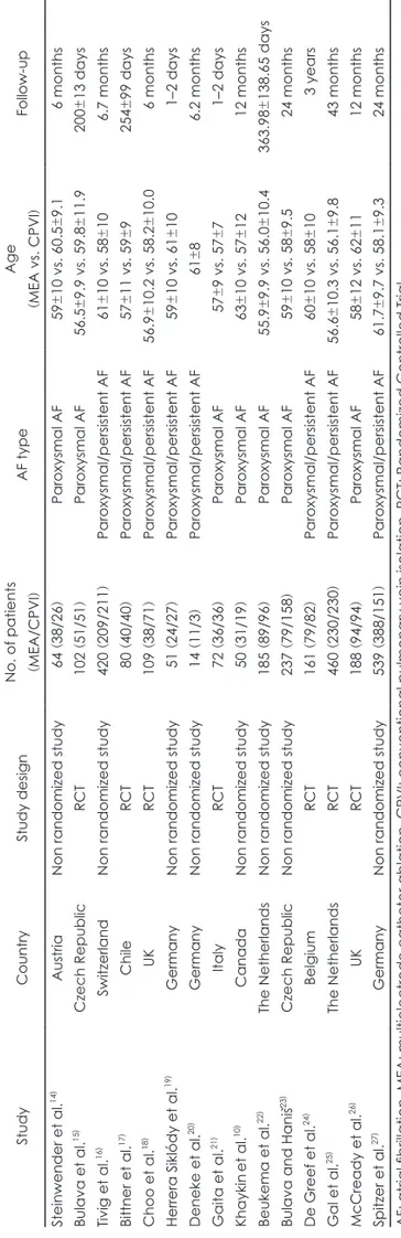
Study characteristics are listed in Table 1. Overall, 1437 pa-tients underwent MEA procedures, and 1295 papa-tients CPVI, to treat paroxysmal or persistent AF. Of the 15 selected studies, 7 dealt with paroxysmal AF patients and 8 included those with paroxysmal or persistent AF.
Procedural outcomes
Procedural and fluoroscopic times
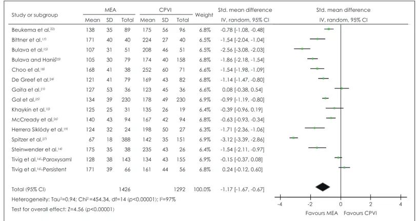
The total MEA procedural time was 45 min less than that of CPVI [SMD=-1.17, 95% confidence interval (CI): -1.67, -0.67, p<0.00001] (Fig. 2). The total fluoroscopic time was also sig-nificantly less (by 7.16 min) when MEA rather than CPVI was performed (SMD=-0.64, 95% CI: -1.04, -0.24, p=0.002). The MEA RF application time was 11.96 min shorter than that as-sociated with CPVI, and this difference was significant (SMD=-0.70, 95% CI: -1.26, -0.14, p=0.01).
Table 2 shows our subgroup analysis of the pooled estimates of SMDs in procedural time between MEA and CPVI strati-fied by study design, AF type, and catheter type. In terms of study design, the MEA procedural time was significantly less than that of CPVI in both randomized (SMD=-1.17, 95% CI: -1.63, -0.71, p<0.00001) and non-randomized controlled trials (SMD=-1.16, 95% CI: -2.07, -0.25, p=0.01), and the pooled SMD was larger for Randomized Controlled Trials. In terms of AF type, MEA was associated with a procedural time signifi-cantly shorter than required for CPVI (SMD=-0.97, 95% CI: -1.54, -0.40, p=0.0009) in patients with paroxysmal AF, but not in those with persistent AF (SMD=0.24, 95% CI: -0.12, 0.60, p=0.19). The MEA procedural time was significantly shorter than that of CPVI when either catheter type was used for mul-tielectrode ablation. The catheters used were a duty-cycled multipolar ablation catheter (SMD=-1.15, 95% CI: -1.67, -0.62, p<0.0001) and a high-density mesh ablator catheter (SMD= -1.54, 95% CI: -2.11, -0.97, p<0.00001).
Table 1.
Characteristics of studies included in the meta-analysis
Study
Country
Study design
No. of patients (MEA/CPVI
) AF type Age (MEA vs. CPVI ) Follow-up Steinwender et al. 14 ) Austria
Non randomized study
64 (38/26 ) Paroxysmal AF 59 ±10 vs. 60.5 ±9.1 6 months Bulava et al. 15 ) Czech Republic RCT 102 (51/51 ) Paroxysmal AF 56.5 ±9.9 vs. 59.8 ±11.9 200 ±13 days Tivig et al. 16 ) Switzerland
Non randomized study
420 (209/211 ) Paroxysmal/persistent AF 61 ± 10 vs. 58 ± 10 6.7 months Bittner et al. 17 ) Chile RCT 80 (40/40 ) Paroxysmal/persistent AF 57 ± 11 vs. 59 ± 9 254 ± 99 days Choo et al. 18 ) UK RCT 109 (38/71 ) Paroxysmal/persistent AF 56.9 ±10.2 vs. 58.2 ±10.0 6 months
Herrera Siklódy et al.
19
)
Germany
Non randomized study
51 (24/27 ) Paroxysmal/persistent AF 59 ±10 vs. 61 ±10 1–2 days Deneke et al. 20 ) Germany
Non randomized study
14 (11/3 ) Paroxysmal/persistent AF 61 ± 8 6.2 months Gaita et al. 21 ) Italy RCT 72 (36/36 ) Paroxysmal AF 57 ±9 vs. 57 ±7 1–2 days Khaykin et al. 10 ) Canada
Non randomized study
50 (31/19 ) Paroxysmal AF 63 ±10 vs. 57 ±12 12 months Beukema et al. 22 ) The Netherlands
Non randomized study
185 (89/96 ) Paroxysmal AF 55.9 ± 9.9 vs. 56.0 ± 10.4 363.98 ± 138.65 days
Bulava and Haniš
23
)
Czech Republic
Non randomized study
237 (79/158 ) Paroxysmal AF 59 ± 10 vs. 58 ± 9.5 24 months De Greef et al. 24 ) Belgium RCT 161 (79/82 ) Paroxysmal/persistent AF 60 ±10 vs. 58 ±10 3 years Gal et al. 25 ) The Netherlands RCT 460 (230/230 ) Paroxysmal/persistent AF 56.6 ±10.3 vs. 56.1 ±9.8 43 months McCready et al. 26 ) UK RCT 188 (94/94 ) Paroxysmal AF 58 ± 12 vs. 62 ± 11 12 months Spitzer et al. 27 ) Germany
Non randomized study
539 (388/151 ) Paroxysmal/persistent AF 61.7 ± 9.7 vs. 58.1 ± 9.3 24 months
Clinical outcomes
Ten studies10,14-19,21-23) reported the success rates of acute
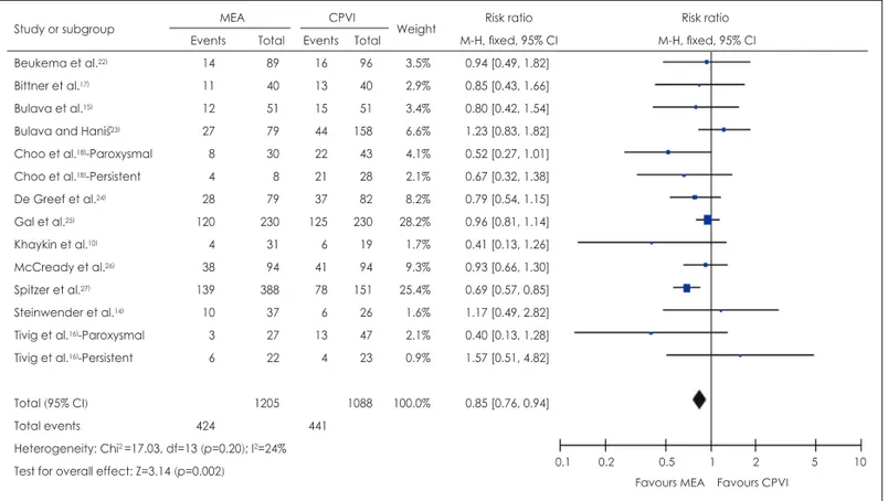
pro-cedures in terms of complete PV isolation immediately after ablation. No significant between-treatment difference in acute procedural success rate was evident (RR=0.99, 95% CI: 0.97, 1.01, p=0.35). After ablation, the risk of AF recurrence was lower if MEA had been employed (RR=0.85, 95% CI: 0.76, 0.94, p=0.002) (Fig. 3) with statistical significance from twelve studies10,14-18,22-27) reporting AF recurrence. Also, the number of
repeat procedures performed after initial ablation that did not completely isolate the PV was somewhat less after MEA than CPVI (RR=0.73, 95% CI: 0.53, 1.00, p=0.05), but the differ-ence was not significant.
Table 3 presents our subgroup analysis results for AF
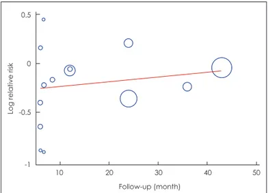
recur-rence stratified by follow-up period, study design, AF type, and catheter type used during MEA. AF recurred less often af-ter MEA in studies with shoraf-ter follow-up durations (less than 6 months; RR=0.69, 95% CI: 0.45, 1.06, p=0.09, and 6–12 months; RR=0.80, 95% CI: 0.53, 1.19, p=0.27). Also, MEA was associated with a reduced AF recurrence more than 1 year af-ter the procedure (RR=0.87, 95% CI: 0.78, 0.97, p=0.01) and the difference was statistically significant. Meta-regression of the RRs for AF recurrence after MEA vs. CPVI in terms of fol-low-up duration showed a tendency toward establishment of a slightly positive linear relationship between the RR and follow-up duration without significance (β=0.005, 95% CI: -0.007, 0.017, p=0.395) (Fig. 4).
Table 2. Subgroup analysis on the mean difference of procedure time between MEA and CPVI
Sub-group No.
of study
No. of patients Std. mean difference (95% CI)
MEA CPVI
Total 14 1426 1292 -1.17 (-1.68, -0.67)*
Sensitivity analysis† 12 1181 1045 -1.48 (-1.98, -0.98)*
Study design RCT 7 568 604 -1.17 (-1.63, -0.71)*
Non randomized study 7 858 688 -1.16 (-2.07, -0.25)*
AF type Paroxysmal AF 8 561 635 -0.97 (-1.54, -0.40)*
Persistent AF 1 66 56 0.24 (-0.12, 0.60)
Catheter type Duty-cycled multipolar ablation catheter 14 1388 1266 -1.15 (-1.67, -0.62)*
High-density mesh ablator catheter 1 38 26 -1.54 (-2.11, -0.97)*
*Pooled estimates are from random-effects model, †Sensitivity analysis was performed by excluding extreme studies. AF: atrial
fi-brillation, MEA: multielectrode catheter ablation, CPVI: conventional pulmonary vein isolation, CI: confidence interval, RCT: ran-domized controlled trial
Fig. 2. Forest plot for standardized mean difference (SMD) for procedure time between multielectrode catheter ablation (MEA) and
conventional pulmonary vein isolation (CPVI). CI: confidence interval, SD: standard deviation.
Study or subgroup MEA CPVI Weight Std. mean difference Std. mean difference
Mean SD Total Mean SD Total IV, random, 95% CI IV, random, 95% CI
Beukema et al.22) 138 35 89 175 56 96 6.8% -0.78 [-1.08, -0.48]
Bittner et al.17) 171 40 40 224 27 40 6.5% -1.54 [-2.04, -1.04]
Bulava et al.15) 107 31 51 208 46 51 6.5% -2.56 [-3.08, -2.03]
Bulava and Haniš23) 105 30 79 174 40 158 6.8% -1.86 [-2.18, -1.54]
Choo et al.18) 168 41 38 252 60 71 6.6% -1.54 [-1.98, -1.09] De Greef et al.24) 121 41 79 169 43 82 6.8% -1.14 [-1.47, -0.80] Gaita et al.21) 127 53 36 123 45 36 6.6% 0.08 [-0.38, 0.54] Gal et al.25) 134 39 230 178 49 230 6.9% -0.99 [-1.19, -0.80] Khaykin et al.10) 125 25 31 135 26 19 6.4% -0.39 [-0.96, 0.19] McCready et al.26) 140 43 94 167 42 94 6.8% -0.63 [-0.93, -0.34]
Herrera Siklódy et al.19) 124 32 24 198 50 27 6.3% -1.71 [-2.36, -1.06]
Spitzer et al.27) 67 18 388 142 35 151 6.9% -3.12 [-3.39, -2.86]
Steinwender et al.14) 175 35 38 235 43 26 6.4% -1.54 [-2.11, -0.97]
Tivig et al.16)-Paroxysaml 128 38 143 134 43 155 6.9% -0.15 [-0.37, 0.08]
Tivig et al.16)-Persistent 171 39 66 161 44 56 6.8% 0.24 [-0.12, 0.60]
Total (95% CI) 1426 1292 100.0% -1.17 [-1.67, -0.67]
Heterogeneity: Tau2=0.94; Chi2 =454.34, df=14 (p<0.00001); I2=97%
Test for overall effect: Z=4.56 (p<0.00001) -4 -2 0 2 4
Complications
Reported complications associated with PV isolation via catheter ablation included embolisms, pericardial effusion, stroke, tamponade, pulmonary vein puncture, transient isch-emic attack, femoral hematoma, and pseudoaneurysms. No significant between-treatment difference in the frequency of occurrence of any complication was evident, but a trend to-ward a higher complication rate after MEA was apparent (RR=1.04, 95% CI: 0.55, 1.93, p=0.91) (Fig. 5). Eight studies
17,19-21,24-27) reported thromboembolic complications, the most
frequent and potentially devastating. Of eight relevant studies, three19,21,26) reported thromboembolic events in the first 1–2
days after the procedure and the other five17,20,24,25,27) gave
fol-low-up results at more than 6 months up to 3 years. The risk of thromboembolic events tended to be slightly lower after MEA (RR=0.99, 95% CI: 0.23, 4.19, p=0.99) at follow-up times of over 6 months, in contrast to a higher incidence (RR=4.84, 95% CI: 2.04, 11.47, p=0.0003) observed during the acute peri-od (1–2 days after the procedure) (Supplementary Fig. 1).
Discussion
Our present systematic review and meta-analysis found that RF ablation using multielectrode catheters reduced procedural
Table 3. Subgroup analysis on the RRs of AF recurrence between MEA and CPVI
Sub-group No. of study No. of patients RR (95% CI)† MEA CPVI Total 14 1205 1088 0.85 (0.76, 0.94)
Follow-up period ≤6 month 3 75 97 0.69 (0.45, 1.06)
>6 month, <1 year 4 140 161 0.80 (0.53, 1.19)
≥1 year 7 990 830 0.87 (0.78, 0.97)*
Study design RCT 7 532 568 0.87 (0.77, 1.00)
Non randomized study 7 673 520 0.81 (0.68, 0.96)*
AF type Paroxysmal AF 8 438 534 0.88 (0.72, 1.07)
Persistent AF 2 30 51 0.93 (0.51, 1.72)
Catheter type Duty-cycled multipolar ablation catheter 13 1168 1062 0.84 (0.76, 0.93)*
High-density mesh ablator catheter 1 37 26 1.17 (0.49, 2.82)
*Statistically significant, †Mantel-Haenszel pooled estimate. AF: atrial fibrillation, MEA: multielectrode catheter ablation, CPVI:
con-ventional pulmonary vein isolation, RR: relative risk, CI: confidence interval, RCT: Randomized Controlled Trial
Fig. 3. Forest plot for relative risk (RR) of AF recurrence between multielectrode catheter ablation (MEA) and conventional pulmonary
vein isolation (CPVI). CI: confidence interval.
Study or subgroup MEA CPVI Weight Risk ratio Risk ratio
Events Total Events Total M-H, fixed, 95% CI M-H, fixed, 95% CI
Beukema et al.22) 14 89 16 96 3.5% 0.94 [0.49, 1.82]
Bittner et al.17) 11 40 13 40 2.9% 0.85 [0.43, 1.66]
Bulava et al.15) 12 51 15 51 3.4% 0.80 [0.42, 1.54]
Bulava and Haniš23) 27 79 44 158 6.6% 1.23 [0.83, 1.82]
Choo et al.18)-Paroxysmal 8 30 22 43 4.1% 0.52 [0.27, 1.01]
Choo et al.18)-Persistent 4 8 21 28 2.1% 0.67 [0.32, 1.38]
De Greef et al.24) 28 79 37 82 8.2% 0.79 [0.54, 1.15] Gal et al.25) 120 230 125 230 28.2% 0.96 [0.81, 1.14] Khaykin et al.10) 4 31 6 19 1.7% 0.41 [0.13, 1.26] McCready et al.26) 38 94 41 94 9.3% 0.93 [0.66, 1.30] Spitzer et al.27) 139 388 78 151 25.4% 0.69 [0.57, 0.85] Steinwender et al.14) 10 37 6 26 1.6% 1.17 [0.49, 2.82]
Tivig et al.16)-Paroxysmal 3 27 13 47 2.1% 0.40 [0.13, 1.28]
Tivig et al.16)-Persistent 6 22 4 23 0.9% 1.57 [0.51, 4.82]
Total (95% CI) 1205 1088 100.0% 0.85 [0.76, 0.94]
Total events 424 441
Heterogeneity: Chi2 =17.03, df=13 (p=0.20); I2=24%
Test for overall effect: Z=3.14 (p=0.002) 0.1 0.2 0.5 1 2 5 10
times and AF recurrence compared to those of CPVI using 3D mapping, but the overall efficacies were in terms of acute pro-cedural success and the need for repeat procedures were com-parable. The procedural time was significantly reduced in the MEA group, but differed among the studies reviewed. Two studies16,21) reported that MEA procedural time was longer
than that of CPVI, contrary to other studies. Perhaps the sur-geons performing MEA were at early stages of their learning curves, whereas the surgeons performing CPVI were not. The result has also shown a similarity in fluoroscopy time and RF application time representing longer time in MEA. Some
ab-stracts28-31) reported that the learning curve for use of
multi-electrode catheters was relatively short. Over the first 20 cases, procedural, fluoroscopic, and application times decreased by 33%, 45%, and 21%, respectively; and acute success rates in-creased with procedure experience. Subgroup analysis by AF type showed that all of procedural, fluoroscopic, and RF appli-cation times during treatment of paroxysmal AF patients were shorter when MEA was applied, but, in contrast, procedural times for persistent AF patients were longer. Such differences between AF type may be attributable to the need to perform additional ablation dictated by complex fractionated atrial electrograms obtained from the left atrial septum and body, performed at the discretion of the operator in patients with persistent AF.
The acute success rates were comparably high between treatments, being 82–100% in most studies when either MEA or CPVI was employed. However, the acute success rate of 79– 93% for persistent AF patients was slightly lower than that for paroxysmal AF patients, attributable to the need for additional ablation to treat persistent AF, as mentioned above.
The RRs of AF recurrence after MEA and CPVI varied by follow-up period, and tended to be lower short-term (≤1 year) after MEA, and the difference was significant between MEA and CPVI upon longer-term follow-up (>1 year); this tenden-cy was also captured via meta-regression analysis. Likewise, the risk of a need for a repeat procedure was lower after MEA than CPVI. Such a finding was expected because the need for Fig. 4. Meta-regression for relative risk of AF recurrence on
fol-low-up duration in the study. AF: atrial fibrillation.
10 20 30 40 50 Follow-up (month) 0.5 0 -0.5 -1
Log relative risk
Fig. 5. Forest plot for the pooled RR of complications between multielectrode catheter ablation (MEA) and conventional pulmonary vein
isolation (CPVI). RR: relative risk, CI: confidence interval.
Study or subgroup MEA CPVI Weight Risk ratio Risk ratio
Events Total Events Total M-H, random, 95% CI M-H, random, 95% CI
Beukema et al.22) 3 89 2 96 7.3% 1.62 [0.28, 9.46]
Bittner et al.17) 1 40 2 40 5.0% 0.50 [0.05, 5.30]
Bulava et al.15) 0 51 0 51 Not estimable
Bulava and Haniš23) 2 79 11 158 8.9% 0.36 [0.08, 1.60]
Choo et al.18)-Combined 2 38 2 71 6.6% 1.87 [0.27, 12.74]
De Greef et al.24) 4 79 5 82 10.2% 0.83 [0.23, 2.98]
Deneke et al.20) 2 11 1 3 6.2% 0.55 [0.07, 4.16]
Gaita et al.21) 14 36 3 36 11.0% 4.67 [1.47, 14.86]
Gal et al.25) 3 230 11 230 10.3% 0.27 [0.08, 0.96]
McCready et al.26) 2 94 0 94 3.4% 5.00 [0.24, 102.77]
Herrera Siklódy et al.19) 9 24 2 27 9.2% 5.06 [1.21, 21.16]
Spitzer et al.27) 6 388 5 151 10.9% 0.47 [0.14, 1.51]
Tivig et al.16) 5 209 6 211 10.9% 0.84 [0.26, 2.71]
Total (95% CI) 1368 1250 100.0% 1.04 [0.55, 1.93]
Total events 53 50
Heterogeneity: Tau2=0.57; Chi2=21.86, df=11 (p<0.03); I2=50%
Test for overall effect: Z=0.11 (p=0.91) 0.01 0.1 1 10 100
a repeat procedure is triggered by AF recurrence, which is more common after CPVI. Tivig et al.16) reported that
low-power MEA ablation reduced the need for repeat procedures compared to use of standard RF ablation; low-power ablation to a defined lesional depth (using appropriate generator set-tings) may prevent extensive tissue injury. The success of MEA is explained by the high-level lesional integrity afforded by use of circumferential ablation catheters compared to the point-by-point ablation of 3D systems. Improved lesional integrity reduces the opportunities for reconnection of trigger foci. Al-though 3D systems allow precise visualization of the positions of mapping and ablation catheters, construction of such com-plex maps is associated with a steep learning curve, compro-mising success rates in newer centers.
Of the various complications associated with the MEA and CPVI procedures, thromboembolic events were the most common and the risk of such events was somewhat higher af-ter MEA, significantly different from the risk afaf-ter CPVI. However, sub-group analysis by follow-up period revealed that the risk of such events tended to be slightly lower upon longer-term follow-up after MEA, in contrast to the higher incidence observed during the acute period (1–2 days) after the proce-dure. Deneke et al.20) evaluated the clinical consequences and
longer-term characteristics of lesions in patients with acutely detected ischemic embolic lesions after catheter ablation of AF. Only acute lesions of maximum diameters >10 mm were de-tected on longer-term follow-up MRI; 94% of lesions resolved without identifiable scar formation. The mechanisms by which silent brain injuries are caused remain unclear; blood clots, air, tissue, or fat, may be in play. Alternatively, such injuries may be produced upon sheath manipulation prior to left atrial cathe-terization, or by ablation per se. Asymptomatic cerebral MRI lesions may trigger subclinical deterioration, but no patient had any detectable neurological deficit immediately after abla-tion or on follow-up evaluaabla-tion performed by an experienced neurologist.20) Also, no AF ablation study revealed any
connec-tion between development of silent ischemic MRI lesions and adverse neuropsychological outcomes.32,33) Although the
de-velopment of thromboembolic events after AF ablation is of concern, no relationship between such events and an adverse outcome or deterioration in neuropsychological performance was substantiated in any trial. Further long-term studies are needed to evaluate patient outcomes. The catheter type used during MEA was associated with development of thrombo-embolic events. Recently, Verma et al.34) described several ways
by which the development of silent intracranial embolisms may be minimized via catheter manipulation and appropriate electrode use with maintenance of good success rates.
Conclusions
Upon meta-analysis, multielectrode ablation used to treat AF afforded beneficial reductions in all of procedural, fluoro-scopic, and RF application times, but the outcomes in terms of acute procedural success, AF recurrence and the need for re-peat procedure were comparable to those of traditional treat-ment, CPVI. Although the thromboembolism risk at 1–2 days post-procedure was slightly elevated, the long-term safety pro-file of MEA was comparable to that of CPVI, indicating that MEA benefits patients with AF when skilled physicians per-form the procedure carefully. Further, larger, well-organized randomized trials combined with analysis of cost-effectiveness are needed.
REFERENCES
1) Haïssaguerre M, Jaïs P, Shah DC, Takahashi A, Hocini M, Quiniou G, et al. Spontaneous initiation of atrial fibrillation by ectopic beats originating in the pulmonary veins. N Engl J Med 1998;339:659-666.
2) Fuster V, Rydén LE, Cannom DS, Crijns HJ, Curtis AB, Ellenbogen KA, et al. ACC/AHA/ESC 2006 Guidelines for the Management of Patients with Atrial Fibrillation: a report of the American College of Cardiology/American Heart Association Task Force on Practice Guidelines and the European Society of Cardiology Committee for Practice Guidelines (Writing Committee to Revise the 2001 Guilines for the Management of Patients With Atrial Fibrillation): de-veloped in collaboration with the European Heart Rhythm Associa-tion and the Heart Rhythm Society. CirculaAssocia-tion 2006;114:e257-e354.
3) Calkins H, Brugada J, Packer DL, Cappato R, Chen SA, Crijns HJ, et al. HRS/EHRA/ECAS expert consensus statement on catheter and surgical ablation of atrial fibrillation: recommendations for per-sonnel, policy, procedures and follow-up. A report of the Heart Rhythm Society (HRS) Task Force on Catheter and Surgical Abla-tion of Atrial FibrillaAbla-tion developed in partnership with the Europe-an Heart Rhythm Association (EHRA) Europe-and the EuropeEurope-an Cardiac Arrhythmia Society (ECAS); in collaboration with the American College of Cardiology (ACC), American Heart Association (AHA), and the Society of Thoracic Surgeons (STS). Endorsed and ap-proved by the governing bodies of the American College of Cardiol-ogy, the American Heart Association, the European Cardiac Ar-rhythmia Society, the European Heart Rhythm Association, the Society of Thoracic Surgeons, and the Heart Rhythm Society. Euro-pace 2007;9:335-379.
4) Birnie D, Healey JS, Krahn AD, Ahmad K, Crystal E, Khaykin Y, et al. Prevalence and risk factors for cervical and lumbar spondylosis in interventional electrophysiologists. J Cardiovasc Electrophysiol 2011;22:957-960.
5) Estner HL, Deisenhofer I, Luik A, Ndrepepa G, von Bary C, Zrenner B, et al. Electrical isolation of pulmonary veins in patients with atri-al fibrillation: reduction of fluoroscopy exposure and procedure du-ration by the use of a non-fluoroscopic navigation system (NavX). Europace 2006;8:583-587.
6) Lakkireddy D, Nadzam G, Verma A, Prasad S, Ryschon K, Di Biase L, et al. Impact of a comprehensive safety program on radiation ex-posure during catheter ablation of atrial fibrillation: a prospective study. J Interv Card Electrophysiol 2009;24:105-112.
7) Rotter M, Takahashi Y, Sanders P, Haïssaguerre M, Jaïs P, Hsu LF, et al. Reduction of fluoroscopy exposure and procedure duration
during ablation of atrial fibrillation using a novel anatomical naviga-tion system. Eur Heart J 2005;26:1415-1421.
8) Nahass GT. Fluoroscopy and the skin: implications for radiofre-quency catheter ablation. Am J Cardiol 1995;76:174-176.
9) Kovoor P, Ricciardello M, Collins L, Uther JB, Ross DL. Risk to patients from radiation associated with radiofrequency ablation for supraventricular tachycardia. Circulation 1998;98:1534-1540. 10) Khaykin Y, Zarnett L, Friedlander D, Wulffhart ZA, Whaley B,
Giewercer D, et al. Point-by-point pulmonary vein antrum isolation guided by intracardiac echocardiography and 3D mapping and duty-cycled multipolar AF ablation: effect of multipolar ablation on pro-cedure duration and fluoroscopy time. J Interv Card Electrophysiol 2012;34:303-310.
11) Terasawa T, Balk EM, Chung M, Garlitski AC, Alsheikh-Ali AA, Lau J, et al. Systematic review: comparative effectiveness of radio-frequency catheter ablation for atrial fibrillation. Ann Intern Med 2009;151:191-202.
12) Chen HS, Wen JM, Wu SN, Liu JP. Catheter ablation for paroxys-mal and persistent atrial fibrillation. Cochrane Database Syst Rev 2012;4:CD007101.
13) Andrade JG, Dubuc M, Rivard L, Guerra PG, Mondesert B, Macle L, et al. Efficacy and safety of atrial fibrillation ablation with phased radiofrequency energy and multielectrode catheters. Heart Rhythm 2012;9:289-296.
14) Steinwender C, Hönig S, Leisch F, Hofmann R. Clinical experience with routine use of a single combined mapping and ablation catheter for isolation of pulmonary veins in patients with paroxysmal atrial fibrillation. Wien Klin Wochenschr 2010;122:146-151.
15) Bulava A, Haniš J, Sitek D, Ošmera O, Karpianus D, Snorek M, et al. Catheter ablation for paroxysmal atrial fibrillation: a randomized comparison between multielectrode catheter and point-by-point ab-lation. Pacing Clin Electrophysiol 2010;33:1039-1046.
16) Tivig C, Dang L, Brunner-La Rocca HP, Özcan S, Duru F, Scharf C. Duty-cycled unipolar/bipolar versus conventional radiofrequency ablation in paroxysmal and persistent atrial fibrillation. Int J Cardiol 2012;157:185-191.
17) Bittner A, Mönnig G, Zellerhoff S, Pott C, Köbe J, Dechering D, et al. Randomized study comparing duty-cycled bipolar and unipolar radiofrequency with point-by-point ablation in pulmonary vein iso-lation. Heart Rhythm 2011;8:1383-1390.
18) Choo WK, Farwell D, Harris S. Experience of atrial fibrillation ab-lation in a new cardiac centre using three-dimensional mapping and multielectrode duty-cycled radiofrequency ablation. Arch Cardio-vasc Dis 2011;104:396-402.
19) Herrera Siklódy C, Deneke T, Hocini M, Lehrmann H, Shin DI, Mi-yazaki S, et al. Incidence of asymptomatic intracranial embolic events after pulmonary vein isolation: comparison of different atrial fibrillation ablation technologies in a multicenter study. J Am Coll Cardiol 2011;58:681-688.
20) Deneke T, Shin DI, Balta O, Bünz K, Fassbender F, Mügge A, et al. Postablation asymptomatic cerebral lesions: long-term follow-up using magnetic resonance imaging. Heart Rhythm 2011;8:1705-1711.
21) Gaita F, Leclercq JF, Schumacher B, Scaglione M, Toso E, Halimi F,
et al. Incidence of silent cerebral thromboembolic lesions after atrial fibrillation ablation may change according to technology used: com-parison of irrigated radiofrequency, multipolar nonirrigated catheter and cryoballoon. J Cardiovasc Electrophysiol 2011;22:961-968. 22) Beukema RJ, Elvan A, Smit JJ, Delnoy PP, Misier AR, Reddy V.
Pulmonary vein isolation to treat paroxysmal atrial fibrillation: con-ventional versus multi-electrode radiofrequency ablation. J Interv Card Electrophysiol 2012;34:143-152.
23) Bulava A, Haniš J. The influence of the technology on the success of the treatment of paroxysmal atrial fibrillation-single center expe-rience. Cor et vasa 2012;54:e393-e400.
24) De Greef Y, Buysschaert I, Schwagten B, Stockman D, Tavernier R, Duytschaever M. Duty-cycled multi-electrode radiofrequency vs. conventional irrigated point-by-point radiofrequency ablation for re-current atrial fibrillation: comparative 3-year data. Europace 2014; 16:820-825.
25) Gal P, Aarntzen AE, Smit JJ, Adiyaman A, Misier AR, Delnoy PP, et al. Conventional radiofrequency catheter ablation compared to multi-electrode ablation for atrial fibrillation. Int J Cardiol 2014;176:891-895.
26) McCready J, Chow AW, Lowe MD, Segal OR, Ahsan S, de Bono J, et al. Safety and efficacy of multipolar pulmonary vein ablation cath-eter vs. irrigated radiofrequency ablation for paroxysmal atrial fibril-lation: a randomized multicentre trial. Europace 2014;16:1145-1153. 27) Spitzer SG, Karolyi L, Weinmann T, Scharfe F, Rämmler C, Otto T,
et al. Multielectrode phased radiofrequency ablation compared with point-by-point ablation for pulmonary vein isolation-outcomes in 539 patients. Res Rep Clin Cardiol 2014;5:11-20.
28) Dang L, Boersma L, Oral H, Morady F. Multi-electrode catheters using low energy phased radiofrequency for ablation of chronic atri-al fibrillation. (abstract) Europace 2008;10:i84.
29) Mulder A, Wijffels M, Wever E, Boersma L. Long term results of multi-electrode pulmonary vein isolation with bipolar/unipolar RF energy for paroxysmal AF. (abstract) Heart Rhythm 2009;6:S277. 30) Brunelli M, Raffa S, Grosse A, Regoli F, Geller JC. Efficacy of a
novel circular multielectrode ablation catheter for pulmonary vein isolation: an anatomic anlaysis using 3D CT reconstruction of the left atrium. (abstract) Heart Rhythm 2009;6:405.
31) Brunelli M, Raffa S, Grosse A, Wauters K, Geller JC. Acute efficacy and safety of a novel circular multielectrode radiofrequency ablation catheter for pulmonary vein isolation in paroxysmal atrial fibrilla-tion. (abstract) Eur Heart J 2009;30:814.
32) Schwarz N, Kuniss M, Nedelmann M, Kaps M, Bachmann G, Neu-mann T, et al. Neuropsychological decline after catheter ablation of atrial fibrillation. Heart Rhythm 2010;7:1761-7167.
33) Vermeer SE, Prins ND, den Heijer T, Hofman A, Koudstaal PJ, Bre-teler MM. Silent brain infarcts and the risk of dementia and cogni-tive decline. N Engl J Med 2003;348:1215-1222.
34) Verma A, Debruyne P, Nardi S, Deneke T, DeGreef Y, Spitzer S, et al. Evaluation and reduction of asymptomatic cerebral embolism in ablation of atrial fibrillation, but high prevalence of chronic silent in-farction: results of the evaluation of reduction of asymptomatic cere-bral embolism trial. Circ Arrhythm Electrophysiol 2013;6:835-842.
Supplementary Fig. 1. Forest plot for stratified relative risks (RR) of thromboembolic events between multielectrode catheter ablation
(MEA) and conventional pulmonary vein isolation (CPVI) by follow-up duration. CI: confidence interval.
Study or subgroup MEA CPVI Weight Risk ratio Risk ratio
Events Total Events Total M-H, fixed, 95% CI M-H, fixed, 95% CI
7.2.1 1–2 days
Gaita et al.21) 14 36 3 36 34.6% 4.67 [1.47, 14.86]
McCready et al.26) 2 94 0 94 5.8% 5.00 [0.24, 102.77]
Herrera Siklódy et al.19) 9 24 2 27 21.7% 5.06 [1.21, 21.16]
Subtotal (95% CI) 154 157 62.1% 4.84 [2.04, 11.47]
Total events 25 5
Heterogeneity: Chi2=0.01, df=2 (p=1.00); I2=0%
Test for overall effect: Z=3.58 (p=0.0003) 7.2.2 ≥6 months
Bittner et al.17) 1 40 1 40 11.5% 1.00 [0.06, 15.44]
De Greef et al.24) 0 79 0 82 Not estimable
Deneke et al.20) 2 11 1 3 18.1% 0.55 [0.07, 4.16]
Gal et al.25) 0 230 0 230 Not estimable
Spitzer et al.27) 2 388 0 151 8.3% 1.95 [0.09, 40.46]
Subtotal (95% CI) 748 506 37.9% 0.99 [0.23, 4.19]
Total events 5 2
Heterogeneity: Chi2=0.52, df=2 (p=0.77); I2=0%
Test for overall effect: Z=0.01 (p=0.99)
Total (95% CI) 902 663 100.0% 3.38 [1.67, 6.85]
Total events 30 7
Heterogeneity: Chi2=4.65, df=5 (p=0.46); I2=0%
Test for overall effect: Z=3.38 (p=0.0007)
Test for subgroup differences: Chi2=3.41, df=1 (p=0.06); I2=70.7%
0.01 0.1 1 10 100