A THESIS
FOR THE DEGREE OF MASTER OF SCIENCE
MOLECULAR CLONING AND CHARACTERIZATION
OF THIOL DEPENDENT ANTIOXIDANT ENZYMES
FROM DISK ABALONE (Haliotis discus discus)
Wickrama arachchilage Anoja Pushpamali
Department of Marine Biotechnology GRADUATE SCHOOL
Molecular Cloning and Characterization of Thiol
Dependent Antioxidant Enzymes from Disk
Abalone (Haliotis discus discus)
Wickrama arachchilage Anoja Pushpamali
(Supervised by Professor Jehee Lee)
A thesis submitted in partial fulfillment of the requirement of the degree of Masters of Science
2006.07.10
This thesis has been examined and approved by
……… Thesis director,
Choon-Bok Song, Professor of Marine Biotechnology
……… You-Jin Jeon, Professor of Marine Biotechnology
……… Jehee Lee, Professor of Marine Biotechnology
………. Date
Department of Marine Biotechnology GRADUATE SCHOOL
CONTENT
요 약 문 ……….. IV
List of Figure……….. VI
Part I
Characterization of two thioredoxin peroxidase clones from abalone 6
1. ABSTRACT………... 6
2. MATERIALS AND METHODS………. 8
2.1. Cloning and sequencing of abalone thioredoxin peroxidase (HdTPx1 and HdTPx2)………. 8
2.2. Amplification of the coding sequences………. 9
2.3. Ligation into pMAL-c2X expression vector……… 9
2.4. Purification of HdTPx1 and HdTPx2……….. 9
2.5. In vitro enzyme activity assay………..……… 10
2.6. Peroxidase assay………... 11
2.7. In vivo H2O2 tolerance assay……….….. 12
2.8. Sequence analysis and comparison………. 12
4.2. Analysis of deduced amino acid sequence………. 29
4.3. Alignment and secondary structure of HdTPx1 and HdTPx2……… 30
4.4. In vitro enzyme activity……… 30
4.5. Peroxidase acivity………. 31
4.6. In vivo H2O2 tolerance activity……….. 32
4.7. Optimum pH………. 33
4.8. Optimum temperature……….… 33
4.9. Phylogenetic analysis………...… 33
Part II Cloning and Characterization of Abalone Thioredoxin 2……… 34
1. ABSTRACT……… 34
2. MATERIALS AND METHODS……….. 36
2.1. Cloning and sequencing of abalone thioredoxin 2 (HdTxn2)……….. 36
2.2. Amplification of HdTxn2……….……… 36
2.3. Ligation of the clone in to into pMAL-c2X expression vector……… 37
2.4. Protein expression and purification………...… 37
2.6. In vitro enzyme activity ……….. 39
2.7. Sequence analysis and comparison………. 39
3. RESULTS………... 40
4. DISCUSSION……….. 49
4.1. Cloning, sequence analysis and comparison of abalone thioredoxin 2 (HdTxn2)……….. 49
4.2. Analysis of deduced amino acid sequence………. 49
4.3. Enzymatic activity assay……….. 51
4.4. Optimum temperature………. 51
4.5. Optimum pH………. 52
4.6. Metal catalyzed oxidation (MCO) DNA cleavage protection assay……….. 52
4.7. Phylogenetic analysis………... 53
SUMMERY……….. 54
REFERENCES……… 57
요 약 문
Thioredoxin peroxidase 와 thioredoxin 는 peroxiredoxin family 에 속하는 thiol 의존성 항산화 효소이다. 그들은 다양한 활성 산소종에 의해 발생하는 산화적 스트레스로부터 지질, 단백질 그리고 DNA 들을 보호하기 위해 살아있는 세포에서 중요한 역할을 한다. 연 체 동 물 에 서 catalases 와 superoxide dismutases 그 리 고 glutathione peroxidas 는 산화적 스트레스에 대항하여 세포를 보호 하는 중요한 항산화 효소이다. 하지만 thioredoxin 효소들은 동물세 포 이외에 무척추동물에서도 항산화 방어 시스템처럼 작용하는 것으 로 보고되어져 있다. 까막전복으로부터 두 개의 thioredoxin peroxidase 효소들이 확인되어고 각각 756 bp 와 600 bp 의 코딩영역을 포함하는 1318 bp (HdTPx1) 와 900 bp (HdTPx2)의 유전자 서열이 확인되었다. HdTPx1 는 분자량이 28 kDa 인 252 개의 아미노산 서열을 가지고 있었고 HdTPx2 는 분자량이 22 kDa 인 199 개의 아미노산 서열을 가지고 있었다. 두 효소 모두 N-terminal Cystein (HdTPx1 - Cys98; HdTPx2 – Cys52) 과 C-terminal Cystein (HdTpx1 – Cys219; HdTPx2 – Cys173)을 가지는 2Cys-peroxiredoxins 로 분류 할 수 있다. 두 효소의 주된 차이점으로 HdTPx1 은 target 단백질을 세포외로 분비시키기 위해 N-말단에 신호서열로 작용하는 30 개의 아미노산 서열을 가지고 있지만 HdTPx2 는 신호서열을 가지고 있지 않다.
HdTPx1 은 Biomphalaria glabrata와 Xenopus tropicalis 의 TPx 에 각각 85%와 80%의 유사성을 나타냈고, HdTPx2 는 Haliotis
discus hannai and Branchiostoma belcheri tsingtaunese의 TPx 에 각각 98%와 78% 의 유사성을 나타냈다. HdTPx1 와 HdTPx2 는 인간의 TPx 와도 각각 79%와 72%의 유사성을 보였다.
HdTPx1 와 HdTPx2 는 둘 다 효소농도 25 μg/ml 이상에서 supercoiled plasmid DNA 의 nick 형성을 50%이상 억제하였고 이는 항산화 효과를 통해 DNA 를 보호한 것으로 보여졌다. 두 효소의 hydrogen peroxide 제거 효과는 효소 농도 증가에 따라 증가하였고 DTT 의 존 재 는 peroxidase 활 성 을 촉 진 시 켰 다 . HdTPx1 와 HdTPx2 의 효소 활성은 pH 8 에서 최적에 도달했고 알카리성 pH 에 서는 낮은 활성을 보였다. 최적 온도는 두 효소 모두 37 oC 로 나타났 고 90 oC 에서 조차도 활성을 보였다. 전 복 의 HdTPx1 와 HdTPx2 는 기 능 적 으 로 2-Cys peroxiredoxins 그룹에 포함되지만 계통유전학적으로는 두 유전자가 서로 다르게 나타났다. 그 두 TPx 유전자들은 서로 계통유전학적 거 리가 멀다. 하지만 이 두 유전자들은 복족류과에 속하는 다른 생물 (HdTPx1: Biomphalaria glabrata; HdTPx2: Haliotis discus
hannai) 들과 유사하였다. 미토콘드리아에서는 다른 세포기관들보다 활성 산소종의 생산 이 많이 이루어진다. 그 때문에 thioredoxin 2 는 미토콘드리아에서 중요한 항산화 효소 중 하나로 작용한다. 전복의 thioredoxin 2 (HdTxn2) 유전자는 483 bp 의 코딩 서열을 포함하는 전체 1,171 bp 의 염기서열이 밝혀졌고 이는 162 개의 아미노산서열을 인식하며 19
가 4 개의 α helical 영역에 의해 둘러싸여 있 형태의 이차원 구조를 가 진 다 . 전 복 의 Txn2 는 활 성 영 역 (motif WCGPC) 이 다 른 thioredoxin 들과 일치하며 아미노산 서열은 Xenopus tropicalis,
Mus musculus 그리고 Homo sapiens의 thioredoxin 2 에 각각 61%,
56% 그리고 43%의 유사성을 나타내었다.
HdTxn2 는 25 oC 에서 DTT 에 의해 인슐린의 이황화 결합을 촉진하였다. 그 효소의 specific activity 는 1.825 Umg-1 이었고, 1 Unit 은 650nm 에서 1 분동안 흡광도값이 1 증가하는 것을 나타낸다. HdTxn2 는 염기성 조건에서 높은 oxidoreductase 활성을 보였다. 그 효소는 30 oC 에서 최고 활성을 보였고 90 oC 같은 높은 온도 조건 에서도 빠른 활성의 감소를 보이지 않았다. 항산화 역할을 하는 그 효 소의 능력은 MCO 시스템으로부터 supercoiled plasmid DNA 를 50% 이상 유지시킬 수 있음을 보여 주었다. 계통유전학적으로 HdTxn2 는 Schistosoma mansoni thioredoxin 2 에 가까웠지만 포 유동물의 thioredoxin 과는 거리가 멀었다. 이 연구에서는 처음으로 까막전복으로부터 thiol 의존성 항산 화 물 질 을 cloning 하 고 발 현 하 였 다 . 까 막 전 복 의 Thioredoxin peroxidase 와 미토콘드리아 thioredoxin 은 처음으로 이 연구에서 기능적으로 특징되어졌고, 이 연구는 전복으로부터 다른 중요한 유전 자들의 분리하는 시발점이 될 수 있을 것이다.
List of Figures
Fig.1.1 Nucleotide sequence and the deduced amino acid sequence of HdTPx1.
Fig.1.2 Multiple sequence alignments of HdTPx1 amino acid sequence with known thioredoxin peroxidase sequences.
Fig.1.3 Nucleotide sequence and the deduced amino acid sequence of HdTPx2.
Fig.1.4 Multiple sequence alignments of HdTPx2 amino acid sequence with known Thioredoxin peroxidase sequences. Fig.1.5 Alignment and secondary structure prediction of HdTPx1 and
HdTPx2.
Fig.1.6 SDS-polyacrylamide gel electrophoresis of HdTPx1 and HdTPx2.
Fig.1.7 Protection of metal-catalysed oxidation (MCO) dependant DNA cleavage by HdTPx1 and HdTPx2.
Fig.1.8 Catalysis of H2O2 removal by HdTpx2 in the presence of DTT
in concentration-dependant manner
Fig.1.9 Catalysis of H2O2 removal by HdTpx2 in the presence of DTT
in concentration-dependant manner
Fig.1.10 Removal of butyl hydroperoxide (BHP) by HdTPx1 and HdTPx2 in concentration dependant manner
Fig.1.11 Optimal temperature of HdTPx1 and HdTPx2 enzyme activity. Fig.1.12 Optimal pH of HdTPx1 and HdTPx2 enzyme activity.
Fig.1.13 H2O2 tolerance of BL21 (DE3) E. coli containing HdTPx1
Fig. 2.1 Nucleotide sequence and the deduced amino acid sequence of HdTxn2.
Fig. 2.2 Secondary structure prediction of HdTxn2.
Fig. 2.3 Multiple sequence alignment of HdTxn2 with known thioredoxin 2 sequences.
Fig. 2.4 Analysis of HdTxn2 in a 12% denaturing SDS-polyacrylamide gel.
Fig. 2.5 Oxidoreductase activity of HdTxn2 by catalyzing the reduction of insulin disulfide bonds by DTT.
Fig. 2.6 Optimal temperature of HdTxn2. Fig. 2.7 Optimal pH of HdTxn2.
Fig. 2.8 Protection of metal-catalysed oxidation (MCO) dependant DNA cleavage by HdTxn2.
Fig. 2.9 Neighbor-joining phylogram showing the phylogenetic relationships between HdTxn2 and thioredoxin 2 sequences of 19 organisms.
INTRODUCTION
Oxygen is an essential element of living organisms for their aerobic cell metabolism. Though it is not toxic in ground state, it has the capability of excitation or partially breakdown into number of reactive species during the physiological processes as well as exposure to UV radiation (Trotter and Grant, 2005). Commonly these are called reactive oxygen species (ROS) including superoxide anion (O2–), singlet oxygen (1O2) and hydroxyl radicals
(·OH), which generate oxidative stress when their production reaches above the threshold level. Not only ROS, but also reactive nitrogen intermediates including NO , ·NO2, NO2 , N2O3, N2O4, S-nitrosothiols, peroxynitrite
(OONO ), dinitrosyl-iron complexes (Nathan and Shiloh, 2000) and sulfur radicals can damage membrane lipids, unfold or inactivate proteins, degrade nucleotides and ultimately kill the whole cell (Rouhier et al., 2001). They take part in the production of hydrogen peroxides or other alkyl hydroperoxides, which leads to the above-mentioned damages.
In biological systems Ni, Cr, Co, Cu, and Fe are known as toxic metals as they promote the oxidation of biomolecules (Kasprzah, 2002). In the presence of electron donors (thiol groups), reduced metal ions generate superoxide anions by reducing oxygen. These superoxide anions initiated the production of hydrogen peroxide (H2O2), ultimately the production of
hydroxyl radicals (.OH) through Fenton reaction (Salazar-Calderón et al., 2000). In addition to the reactive species and metal ions, exposure to water pollutants (Lam and Gray, 2003; Walsh and O'Halloran, 1997) and the seasonality changes can initiate oxidation stress in marine organisms
stress, aerobic organisms possess an antioxidant defense system utilizing non-enzymatic detoxication involving glutathione, ascorbate, λ-tocopherol and transitional metal chelators as well as the enzymatic mechanism depend on catalases, glutathione peroxidases and enzymatic scavengers like superoxide dismutases (Demasi et al., 2001; Rouhier et al., 2001; Hatao et
al., 2006). To detoxify the ROS and other oxidant molecules, oxidation
defense mechanisms with low molecular scavengers and antioxidant enzymes have been evolved in marine mollusks (Regoli et al., 1997).
Thiol specific enzymes belong to the glutathione system and thioredoxin systems are the leading antioxidant enzymes exist in all organisms. Thioredoxin peroxidase, thioredoxin reductase and thioredoxin are key compounds of the thioredoxin system. These enzymes possess thiol/dithiol groups on conserved cysteine residues as their active domains (Powis et al., 2000; Koo et al., 2002; Eddy et al., 2004).
Thioredoxin peroxidase (TPx) is ubiquitous low molecular weight protein, which belongs to a new family of antioxidant enzymes peroxiredoxin (Jos et al., 2005). This enzyme is first reported from
Saccharomyces cerevisiae (Li et al., 2004) and later it was isolated from
cDNA libraries of various mammalians (Lim et al., 1994; Ichimia et al., 1997; Leyens et al., 2003; Eddy et al., 2005), insects (Lee et al., 2005), Teleostei (Fujiki et al., 1999), plants (Bernier-Villamor et al., 2004) and prokaryotes (Do et al., 2003; Hughes et al., 2003), showing the availability in wide range of organisms. TPx is distinguished by other conventional peroxidases as they use thiol groups on conserved cysteine residues instead of metal or other redox cofactors to detoxify either hydrogen peroxides or other various alkyl hydroperoxides into water and corresponding alcohol (Hansen et al., 2006). In some cases they decompose highly toxic reactive nitrogen species (RNS) like peroxinitrite (Dubuisson et al., 2004; Monteiro
et al., 2004; Banmeyer et al., 2005) in the presence of thioredoxin as the
physiological electron donor (Chae et al., 1999).
Also TPx is known as a cellular redox enzyme, which can inhibit the apoptosis (Kim et al., 2000; Berggren et al., 2001). It is reported that the members of this family are directly involved with the activation of nuclear factor κB (NF-κB), cell differentiation (Li et al., 2004) and cell proliferation (Jin et al., 1997). TPx is highly conserved in eukaryotes and prokaryotes suggesting the biological importance of this type of antioxidant enzyme (Jin
et al., 1997).
In general peroxiredoxin family members are divided into two groups: 1-Cys peroxiredoxins and 2-Cys peroxiredoxins, according to the number of active cystein residues present. 1-Cys peroxiredoxins bear only one Cys residue in N-terminus whilst 2-Cys peroxiredoxins contain the additional second Cys residue in the C-terminus (Chae et al., 1994). In 2-Cys peroxiredoxins, N-terminal 2-Cys residue forms an intermolecular disulfide bond with the additional C-terminal Cys residue of another subunit. The disulfide bond is reduced by the thioredoxin and oxidized thioredoxin followed by the further reduction by thioredoxin reductase in the thioredoxin system (Kawakami et al., 2004).
The mitochondria are critical physiological source for the production of ROS within the cell as it produces energy by consuming oxygen. Production of cellular energy in the form of adinosin triphosphate (ATP) via oxidative phosphorylation results ROS from 0.4-4% of the consumed oxygen (Nonn et al., 2003). The aberrant increase in production of ROS within the mitochondria induces apoptosis by releasing various apoptotic-inducing
first from Escherichia coli (Laurent et al., 1964) it has been identified from wide varieties of prokaryotes (Chae et al., 1993; Andersson et al., 1998; Tanaka et al., 2004) and eukaryotes including fungi (Hall et al., 1971), plants (Gelhayae et al., 2002; Maeda et al., 2003), mammals (Damidomopoulos et
al., 2002; Samoilov et al., 2002) as an antioxidant enzyme.
Thioredoxins (Txn) are small ubiquitous protein that participates in various intracellular redox reactions (Miranda-Vizuete et al., 1997; Stroev et
al., 2004; Smeets et al., 2005). It is composed of ~200 amino acid residues in
a single peptide chain having the configuration of two cystein residues in characteristic -Cys-Gly-Pro-Cys- form (Lee et al., 2001). The redox mechanism allows Txn a wide scope of biological functions. Txn is induced by viral infections and oxidative stress, and it acts as a regulatory factor of transcription factors. Recently it was discovered that the Txn has the function of an electron donor for the antioxidant enzymes belongs to the super family peroxiredoxin, which involve in reducing hydrogen peroxide and other organic peroxides (Lee et al., 2001). In eukaryotic cells Txns function as a hydrogen donor for methionine sulfoxide reductase (Arner and Holmgren, 2000) and ribonucleotide reductase (RNR), which is an essential enzyme supplying deoxyribonucleotides for DNA replication (Holmgren, 1989; Aslund et al., 1994; Miranda-Vizuete et al., 1997). In addition Txn itself performs antioxidant activity by reducing hydrogen peroxide and scavenging free radicals (Nakamura et al., 1994; Spector et al., 1988).
There are two isoforms of Txns known as Txn 1 and Txn 2 where the former is primarily present in cytoplasm and nucleous while the latter is localized in mitochondria (Damdimopoulos et al., 2002).
Abalones are attractive aquaculture species that are univalve (single-shelled) marine gastropods from the genus Haliotis and the family Haliotidae (Ragg and Taylor, 2006). In their natural habitat, they are slow-feeding
nocturnal herbivores, with the adults grazing predominantly on seaweeds and the juveniles on microalgae and diatoms found on the surfaces on which they settle (Elliott, 2000). The disk abalone (Haliotis discus discus) also known as Japanese abalone are well distributed deeper areas up to depths of 50 m in the Japanese costs alone the Pacific Ocean and Japan sea south of Hokaido (Sawabe et al., 2004; Ahmed et al., 2005) and southern Korea. Abalones become one of most commercially important species as a food and a source of pearl (Elliott, 2000).
Carefoot et al. (2000) has reported that the digestive gland of abalone has a protective role by its production of antioxidants apart from the sequestration environmentally derived heavy metals and pollutants. It is a common phenomenon occurring additional oxidative stress in aquatic organisms due to the chronic exposure to environmental pollution (Orbea et
al., 2000). Winston (1991) reported that the enzymatic antioxidant defense
mechanism of aquatic organisms mainly depends on SOD, catalase and glutathione peroxidase.
We isolated cDNA fragments of thiol dependant antioxidant genes from the disk abalone (Haliotis discus discus) digestive gland cDNA library. During this study we cloned two thioredoxin peroxidases and a thioredoxin 2, expressed in E. coli expression system and performed the functional characterization of each enzyme. Here we present data of antioxidant activity of purified abalone thioredoxin peroxidase and thioredoxin2 genes.
Part I
Characterization of two clones of thioredoxin peroxidase from
disk abalone
1.
ABSTRACT
Thioredoxin peroxidase (TPx) is low molecular weight antioxidant enzyme, which is first isolated from Saccharomyces cerevisiae and at present it has been identified from wide variety of prokaryotic and eukaryotic organisms. TPx also termed as peroxiredoxin 4, natural killer enhancing factor or thiol-specific antioxidant enzyme is belongs to peroxiredoxin, an antioxidant family. These enzymes own one (Cys52) or two conserved cystein residues (Cys52 and Cys173) in their amino acid sequence. Cys52 is responsible for reduction of hydrogen peroxide and oxidized cystein binds with Cys173 to form the intermolecular disulfide bond. The typical 2-Cys TPxs have two conserved redox active cysteins i.e., peroxidatic cysteine and the resolving cysteine. The tertiary structure of TPx can be homo or hetero-dimer under normal cellular conditions.
Two thioredoxin peroxidases (HdTPx1 and HdTPx2) isolated from disk abalone (Haliotis discus discus) showed highest sequence similarity to TPxs isolated from mollusks rather than the mammalian TPxs. Especially, HdTPx2 shared high sequence similarity (98%) with the TPx of Haliotis
discus hannai, which is a sub species of H. discus discus. Similarly,
HdTPx1 sequence showed 85% identity with the TPx of Biomphalaria
glabrata. HdTPx1 (756 bp) is a 252 amino acid residue protein with 28kDa
of estimated molecular mass whilst HdTPx2 (600 bp) is 199-amino acid-residue protein with 22 kDa of molecular weight. In vitro antioxidant activity of purified recombinant proteins were analyzed by peroxidase assay and
metal catalyzed oxidation (MCO) DNA protection assay. Peroxidase activity of both enzymes was conducted using hydrogen peroxide and butyl hydroperoxide (BHP). Both enzymes showed higher hydrogen peroxide reduction rather than BHP reduction. HdTPx2 showed higher H2O2 removal
ability (2.65 mM/min/mg protein) than the HdTPx1 (2.55 mM/min/mg protein). However, both enzymes at the concentration ≥25 µg/ml recovered ≥50% of supercoiled DNA concentration from MCO system. Maximum peroxidase activity of both enzymes were obtained at pH 8-10 and showed thermal-sensitivity at higher temperature conditions (70 – 90 oC). Although HdTPx1 and HdTPx2 are functionally realated, they shared different phylogenetic relationships, but can be clustered in to a group of organisms sharing the same ancestor.
2. MATERIALS AND METHODS
2.1. Cloning and sequencing of abalone thioredoxin peroxidase (HdTPx1 and HdTPx2)
Two putative thioredoxin peroxidase clones (HdTPx1 and HdTPx2) were identified from the abalone cDNA library and analyzed the homology with the full length coding sequence using the BLAST program available at National center for Biotechnology Informations (NCBI) Gene bank corresponding to the function (http://www.ncbi.nlm.nih.gov/BLAST/). The cDNA clone, transformed in to Escherichia coli DH10b and plasmid DNA was isolated by AccuprepTM plasmid extraction kit (Bioneer Co., Korea). After the restriction digestion with KpnI and BamHI insert sizes were determined by agarose gel electrophoresis. The full length of the HdTPx1 was determined by sequencing with the inner primer 5’-CCGATGAGCTATAAACAATCG-3’, while the HdTPx2 is by 5’-
AGGACGACATGCTTAGTCACGACA – 3’. The derived full-length
sequences were compared with the known sequences and the open reading frames were verified having the lengths of expected size. The primers of the coding sequences of HdTPx1 and HdTPx2 were designed with the
appropriate restriction enzyme sites. 5’- GAGAGAGAATTCATGGCGGGAGCAACAAC - 3’ (forward) and 5’-
GAGAGAAAGCTTTTATAGCTCATCGGAAGTTTTCTG - 3’ (reverse) primers including EcoRI and HindIII restriction sites were designed from HdTPx1 coding sequence. Forward (5’-GAGAGAGGATCCATGGCCCAAGTCGGAAAC - 3’) and reverse (5’ - GAGAGAAAGCTTTCAGTTGACCTTGGAGAAGTAGTTC - 3’) primers were designed including BamHI and HindIII restriction sites at the N terminus and C terminus of the HdTPx2 coding sequence respectively.
2.2. Amplification of the coding sequences
The clones (HdTPx1 and HdTPx2) were amplified by polymerase chain reaction (PCR) in a total volume of 50 µl of reaction mixture contained 5 units of Ex Taq polymerase (Takara Korea Biomedical Inc., Korea), 5 µl of 10 x Ex Taq buffer, 4 µl of 2.5 mM dNTP, 50 ng of each templates, 50 pmol of each primer. After initial denaturation at 94 oC for 2 min, the reaction was subjected to 25 cycles of denaturation at 94 oC for 30 sec, 30 sec of annealing at 55 oC, and 90 sec elongation at 72 oC. The final extension was carried out at 72 oC for 5 min and the PCR product was analyzed using 1% agarose gel.
2.3. Ligation into pMAL-c2X expression vector
The PCR products were purified by AccuprepTM gel purification kit (Bioner Co., Korea) and undergone phosphorelation. The purified products and pMAL-c2X vector (New England Biolabs, USA.) were digested by corresponding restriction enzymes and the inserts were ligated in to the pMAL-c2X vector at 16 oC, overnight in a reaction mixture containing 100 ng of pMAL-c2X vector, 70 ng of each PCR product, 1 µl of 10X ligation buffer and 0.5 µl 1X T4DNA ligase (Takara Korea Biochemical Inc., Korea). The ligated products were transformed into XL1-Blue cells and the transformants with the recombinant clones were verified by colony cracking and sequencing reaction. The recombinant plasmids with inserts were transformed into the competent cells, E. coli BL21(DE3) for protein expression.
(2%) medium with 0.01% amphicillin at 37 oC until the OD600nm approached
0.5. Synthesis of the fusion protein was induced with 0.5 mM (final concentration) isopropylthio-β-galactoside (IPTG) for 3 hrs at 30 oC. The cells were collected by centrifugation at 3500 rpm, at 4 oC for 30 min and resuspended in 1 ml Tris-HCl, pH 7.4, 200 mM NaCl, 0.5 M EDTA (column buffer) and stored in -20 oC. Then cells were sonicated (Bandalin Sonopulse, Bandalin Electronics, Germany.) in short pulses of 10 sec under the maximum power of 30 W for 5 times. The supernatant was cleared after centrifugation at 9500 rpm, 4 oC for 30 min. The soluble proteins (HdTPx1 and HdTPx2) were purified by affinity chromatography on amylose resin column. The proteins having maltose binding protein (MBP) tag were eluted (elution buffer: column buffer with 10mM maltose) and the purity and the molecular masses were determined by SDS-polyacrylamide gel electrophoresis (Laemmli, 1970). Concentrations of the purified proteins were determined by the method of Bradford, (1976) using bovine serum albumin (BSA) as the standard.
2.5. In vitro enzyme activity assay
Metal-catalyzed oxidation (MCO) DNA cleavage protection assay was performed as described by Lim et al., (1993) and Sauri et al., (1995) with modifications described by Li et al., (2004) and Jian et al., (2005). Briefly 50 µl of reaction mixture containing 33 µM FeCl3, 3.3 mM
dithriothreitol (DTT) and concentrations of the purified HdTPx1 and HdTPx2 ranging 6.25 - 100 µg/ml were incubated at 37 oC. After 2 hours 300 ng of pUC19 supercoiled plasmid DNA was added to each reaction mixtures and incubated for 2.5 h at 37 oC. 10 µl of each sample was run on 0.8% (w/v) agarose gel with ethidium bromide.
2.6. Peroxidase assay
In vitro hydrogen peroxide removing ability of the recombinant
HdTPx1 and HdTPx2 was determined according to the method described by Thurman et al. (1971); Stadtman et al. (1990); Lim et al. (1993). Briefly, the reaction mixture (100 µl) containing, various concentrations (0 - 50 µg/ml) of HdTPx1 / HdTPx2 and the reaction buffer with 50 mM Tris-HCl (pH 8.0) and 5 mM DTT were incubated at 37 oC for 30 min. After incubation, H2O2
was added at a final concentration of 50 µM, and the mixture was incubated for another 30 min. 8% (w/v) trichloroacetic acid 0.9 ml was added to stop the reaction and protein was removed by centrifugation. 0.2 ml 10 mM ferrous ammonium sulfate and 0.1 ml 2.5 M potassium thiocyanate were added and the absorbance was measured at 480 nm and results were expressed using known amounts of H2O2 as standard.
The removal of organic hydroperoxide substrates by the abalone TPxs was analyzed spectrophotometrically by the method described by Kawakami et al. (2004) and Chauhan and Mande (2001) with slight modifications. The reaction mixture (0.5 ml) containing 50 mM Tris/HCl buffer (pH 8.0), 1 mM EDTA, 10 mM DTT and different concentrations of TPxs were incubated 30 min at 37 oC. The reaction was started by adding 2 mM tert-Butyl hydroperoxide and incubated another 30 min at 37 oC further. After adding 10% TCA the enzymatic reaction was stopped and proteins were removed by centrifugation. 50 µl of reaction mixture was mixed with 0.65 ml H2O, 0.2 ml 10 mM ferrous ammonium sulfate and 0.1 ml 2.5 M
potassium thiocyanate and the remaining peroxide content was measured spectrophotometrically at 480 nm.
2.7. In vivo H2O2 tolerance assay
An antioxidant activity of recombinant HdTPx1 and HdTPx2 in vivo against hydrogen peroxide was monitored by testing the sensitivity of E. coli cells to H2O2 according to the method described by Li et al., (2004). BL21
(DE3) E. coli cells transformed with pMAL-HdTPx1 and pMAL-HdTPx2 were grown and induced by IPTG at a final concentration of 0.5 mM in LB glucose medium. The cell concentration was diluted up to the OD600nm = 0.2.
One-third serial dilutions of the cell suspensions were prepared and 5 µl droplet of each was plated on LB agar medium containing different concentrations of H2O2 (0, 0.4 and 0.8 mM). Plates were incubated at 37oC
overnight and the diameters of the cell clusters of TPx induced E. coli cells were compared with cells with pMAL-c2X expressing maltose binding protein (MBP) plasmids and cells without plasmids.
2.8. Sequence analysis and comparison
The neucleotide sequence was analyzed using DNAssist program (version 2.2) and the deduced amino acid sequence analysis was performed using CLUSTAL W multiple sequence alignment program - version 1.83 (Thompson, 1994). The similarity of the nucleotide and amino acid sequence was searched using National Center for Biotechnology Information (NCBI) Basic Local Alignment Search Tool (BLAST) program
(http://www.ncbi.nlm.nih.gov/blast/). The phylogenetic analysis was
conducted using the Neighbor Joining method by MEGA 3.0 program (Kumar et al., 2004).
Fig.1.1: The composite cDNA sequence and deduced amino acid sequence of HdTPx1. Both nucleotide sequence and amino acid sequences are in bold-face. The coding sequence is numbered starting from ATG, the initial codon (left) and the deduced amino acid sequence from Met (left in parenthesis). N-terminal and C-terminal active motifs are boxed. Polyadenylation signals are in underlined bold letters and poly (A) tail is in bolded italic.
Fig.1.2: The multiple sequence analysis of HdTPx1 with known thioredoxin peroxidases. RnTPx: Rattus norvegicus (AAH59122); IsTPx:
Ixodus scapularis (AAY66580); HsTPx: Homo sapiens (AAH16770);
BtTPx: Bos taurus (AAG53660). The identical residues are shaded by dark gray and semi-conserved sites are in light gray. Gaps (-) are introduced to maximize the similarity of sequences.
Fig.1.3: The composite cDNA sequence and deduced amino acid sequence of HdTPx2. Both nucleotide sequence and amino acid sequences are in bold-face. The coding sequence is numbered starting from ATG, the initial codon (left) and the deduced amino acid sequence from Met (left in parenthesis). The 5’ UTR was numbered in minus from the initial codon. The polyadenylation signal was bolded and underlined and the poly (A) tail is in bold italic. N-terminal and C-terminal active motifs are boxed. The asterisk (*) indicates the stop codon that cannot be translated into any amino acid.
Fig.1.4: The multiple sequence analysis of HdTPx2 with known thioredoxin peroxidases. BbTPx: Branchiostoma belcheri (AY737279); TnTPx: Tetraodon nigroviridis (AAY21814); CcTPx: Cyprinus carpio (BAA32086); SmTPx: Scophthalmus maximus (ABF01135). The identical residues are shaded in dark gray and semi-conserved sites are in light gray. Gaps (-) are introduced automatically to maximize the similarity of sequences.
Fig.1.5: Alignment and Secondary structural elements of HdTPx1 and HdTPx2. The identical residues are marked in asterisk (*) where as the conserved sites depend on the functionality is indicated in colon and
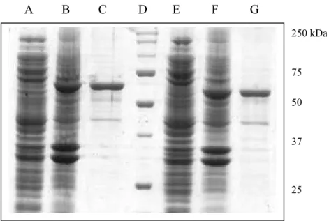
A B C D E F G 250 kDa 75 50 37 25
Fig. 1.6: The SDS-polyacrylamide gel electrophoresis of HdTPx1 and HdTPx2. Lane A: E. coli cells with pMAL-HdTPx1 plasmids without induction; lane B: induced E. coli cells with pMAL-HdTPx1 plasmids; lane C: purified MBP-HdTPx1 fusion protein (70.5 kDa); lane D: molecular markers (BIO-RAD) with the molecular weights in kDa; lane E: E. coli cells with pMAL-HdTPx2 plasmids without induction; lane F: induced E. coli cells with pMAL-HdTPx2 plasmids; lane G: purified MBP-HdTPx2 fusion protein (67.5 kDa). Molecular weight of maltose binding protein (MBP) is 42.5 kDa.
A B C D E F G H I J K II A B C D E F G H I NF SF NF SF I
Fig.1.7: Protection of MCO dependant DNA cleavage by HdTPx1 (I) and HdTPx2 (II). Lane A: pUC19 DNA alone without incubation; lane B: incubated pUC19 DNA in water; lane C: pUC19 DNA in FeCl3 alone; lane
D: pUC19 in DTT alone; lane E: pUC19 with MCO system; lane F: pUC19 with BSA (0.6mg/ml) as control protein; lane G-K: pUC19 with varying concentrations (6.25, 12.5, 25, 50 and 100 µg/ml) of HdTPx1 or HdTPx2
0 20 40 60 80 100 0 10 20 30 40 50 HdTPx1 concentration (ug/ml) % H 2 O2 + DTT - DTT
Fig.1.8: Catalysis of H2O2 removal by HdTpx1 in the presence of DTT in
concentration-dependant manner. The values are the means of three replicates. Same experiment was conducted with (+ DTT) and without (-DTT) thiols.
0 20 40 60 80 100 0 10 20 30 40 50 HdTPx2 concentration (ug/ml) % H 2 O2 + DTT - DTT
Fig.1.9: Catalysis of H2O2 removal by HdTpx2 in the presence of DTT in
concentration-dependant manner. The values are the means of three replicates. Same experiment was conducted with (+ DTT) and without (-DTT) thiols. 0 20 40 60 80 100 120 0 10 20 30 40 50 TPx Concentration (ug/ml) % B H P HdTPx2 HdTPx1
0 20 40 60 80 100 4 6 8 10 pH % r em ov al of H 2O 2 HdTPx1 + DTT HdTPX2 + DTT
Fig.1.11: The optimum pH of HdTPx1 and HdTPx2 enzyme activity. The ferrithiocyanate assay was conducted with the presence and absence of DTT. HdTPx1 / HdTPx2 (50 µg/ml) at different pH (4, 6, 6.8, 8 and 10) were incubated at 37 oC for 30 min prior to add the substrate.
0 20 40 60 80 100 20 30 37 50 60 70 80 90 Temperature (oC) % removal of H 2 O2 HdTPx1 + DTT HdTPX2 + DTT
Fig.1.12: The optimum temperature condition of HdTPx1 and HdTPx2 enzyme activity. The ferrithiocyanate assay was conducted with the presence and absence of DTT. HdTPx1 / HdTPx2 (50 µg/ml) was incubated at different temperature conditions (20, 30, 37, 50, 60, 80 and 90) for 30 min prior to start the reaction.
II I (a) 1 2 1 2 3 1 2 3 II II I (c) I (b)
Fig.1.13: H2O2 tolerance of BL21 (DE3) E. coli containing HdTPx1 (I)
and HdTPx2 (II) fusion proteins. (a) Control plate showing growth of cells with no H2O2 present. (B) Plate containing 0.4 mM H2O2. (C) Plate
Ictalurus punctatus (AAU29515) Cyprinus carpio (BAB39202) Myotis lucifugus (AAT79401)
Cricetulus griseus (AAF32369) Tetraodon nigroviridis (ABC59169) Ostertagia ostertagi (CAD20737)
Onchocerca volvulus (AAC48312) Acanthocheilonema viteae (AAL91102) Dirofilaria immitis (AAC38831) Branchiostoma belcheri tsingtaunese (AAU84951) HdTPx2
Haliotis discus hannai (AAZ22925) Aedes aegypti (AAL37254) Bombyx mori (AAR15420)
Drosophila melanogaster (AAK06771)
Apis mellifera ligustica (AAP93584) Drosophila melanogaster (AAK06769) Ixodes scapularis (AAY66580)
HdTPx1
Biomphalaria glabrata (AAK26236) Xenopus tropicalis (AAH76692)
Bos taurus (AAG53660) Homo sapiens (NP_006397) Rattus norvegicus (AAH59122) Mus musculus (AAH19578)
93 100 100 100 98 99 55 89 99 99 82 98 64 90 59 86 81 81 43 34 35 37 0.05
Fig.1.14: Phylogenetic analysis of HdTPx1 and HdTPx2 with known thioredoxin peroxidase sequences from 23 species in the NCBI data base. Amino acid sequences were aligned by CLUSTAL W program in MEGA 3.0 server and used for phylogenetic inference using Neighbor-Joining method. The NCBI accession numbers of each sequence was indicated within parenthesis. Bootstrap values are indicated on each branch (500 replicates).
4. DISCUSSION
4.1. Sequence analysis of abalone thioredoxin peroxidases (HdTPx1 and HdTPx2)
The partial cDNA fragment of HdTPx1 (839 bp) and HdTPx2 (859 bp) were sequenced to obtain each full-length cDNA sequences. The complete sequence (1,318 bp) of HdTPx1 contains an open reading frame of 756 bp preceeded by 25 bp corresponding to the 5’ untranslated region (UTR) and followed by 537 bp of 3’ - UTR (Fig.1.1). The coding sequence encodes a protein of 252 deduced amino acid residues with the predicted molecular mass and theoretical isoelectric point of 28 kDa (Fig.1.6) and 5.97 respectively. The 3’- UTR sequence contains polyadenylation (AATAAA) signal at 1,222 bp upstream from a stretch of 13 adinosins corresponding to the poly (A) tail.
The complete nucleotide sequence, determined the HdTPx2 was found to be 1,045 bp long. The open reading frame of 600 codons was started by ATG (86-686 bp) preceeded by 85 bp 5’ - UTR. The translational stop codon TGA locates in 598 bp followed by 360 bp of 3’ - UTR (Fig.1.3). The 3’ UTR contain polyadenylation signal at 1002 position of the nucleotide sequence with 32 bp of poly (A) tail. The resulting coding sequence has the coding capacity for a 199 amino acid residue polypeptide with the predicted molecular mass of 22 kDa (Fig.1.6) and theoretical isoelectric point of 5.7 respectively.
Peroxiredoxin family is subdivided based on the number of conserved, redox active cystein residue present in the amino acid sequence.
HdTPx1 and HdTPx2 contain both N-terminal and C-terminal cystein residues in their amino acid sequences. In HdTPx2, two active cysteins are at 52 and 173 of the amino acid sequence (Fig.1.3) where as in HdTPx1 they are at 98 and 219 places (Fig.1.1). Since the members bearing only N-terminal motif are termed 1Cys-peroxiredoxin and those having both are 2Cys-peroxiredoxins (Alphy et al., 2000), two TPxs (HdTPx1 and HdTPx2) isolated from abalone can be termed as 2Cys-peroxiredoxins. Amino acid sequence of the HdTPx1 slightly deviated from the HdTPx2, as it contains 30 amino acid residues long signal peptide (Fig.1.1).
4.2. Analysis of deduced amino acid sequence
To identify the homology of the HdTPx1, the deduced amino acid sequence was aligned with those other thioredoxin peroxidases from other organisms reported in then protein data bank of NCBI. HdTPx1 showed 85% identity with the Bloodfluke Planorb snail (Biomphalaria glabrata), 80% with the pipid frog (Xenopus tropicalis) and 79% of human (Homo
sapiens) thioredoxin peroxidase. Overall it shows high similarity with the
reported organisms. The multiple sequence analysis was performed using the CLUSTAL W program (version 1.83), comparing thioredoxin peroxidases from R. norvegicus (78%), H. sapiens (74%), I. scapularis (79%) and B.
taurus (78%) (Fig.1.2). The amino acid sequences of HdTpx1 and
thioredoxin peroxidase of other organism are well conserved downstream from the second methionine (Me29).
Analysis of the amino acid sequence revealed high sequence homology of HdTPx2 (98%) with the Haliotis discus hannai and 72% with
H. sapiens TPx. Apart from that HdTPx2 showed 78% identity to TPx of Branchiostoma belcheri tsingtaunese, which is another sea invertebrate. As
Ictalurus punctatus and 75% sequence similarity to thiol peroxidase of Bombyx mori. The multiple sequence alignment of HdTPx2 and TPx from, T. nigroviridis, C. carpio and S. maximus is illustrated in Fig.1.4. The amino
acid sequence homogeneity was critical in the region surrounding Cys52 and Cys173 the corresponding active site of TPx.
4.3. Alignment and secondary structure of HdTPx1 and HdTPx2
The amino acid sequence of HdTPx1 has extra 47 amino acids upstream compared to HdTPx2 and it is well conserved with HdTPx2 downstream from the second methionine (Me29). 2-Cys TPx has compact, spherical structure with seven-stranded β sheets surrounded by five α helices (Choi et al., 2005). The structure of HdTPx1 and HdTPx2 contain the typical peroxiredoxin fold, where the Cys52 active residue locates between β3 and α2 (Fig.1.5). The most notable difference of the HdTPx1 and HdTPx2 with the typical TPx structure is the presences of an extra α helix (α7) in the C-terminal end (Choi et al., 2005).
4.4. In vitro enzyme activity
The antioxidant activity of HdTPx1 and HdTPx2 was determined by metal-catalyzed oxidation (MCO) assay using pUC19 plasmid DNA. Metal ions such as iron, zinc, manganese, copper and cobalt generate ROS through fenton reaction causing deleterious effects on proteins, DNA and lipids (Salazar-Calderón et al., 2000). The radicals produced by the MCO system caused nicking of the DNA, with an evidence of shifting in the gel mobility of the supercoiled plasmid (Fig.1.7). Thiol containing electron donors
nicking of supercoiled pUC19 DNA (Fig.1.7). In the presence of DTT, the thiol containing electron donor, HdTPx1 (25 µg/ml or above concentrations) and HdTPx2 (50 µg/ml or above concentrations) maintained approximately half or more of supercoiled DNA concentration after 2.5 hours of incubation period at 37 oC.
4.5. Peroxidase acivity
Cys52 cystein residue directly responsible for the peroxidase activity of TPxs (Montemartini et al., 1999) and it accepts the hydrogen bond from Arg128 and donates to the carboxylate of Glu55 (Alphy et al., 2000). Alphy et
al. (2000) predicted that Arg128 is to stabilize the ionized state of Cys52 and
increase the activity. Cys52 residue in one molecule of the dimer forms a di-sulfide bond with Cys173 in the other molecule (Hirotsu et al., 1999) and it is an intermediate of the peroxidation reaction catalysed by Cys52 (Chae et al., 1994). TPx catalyses the reduction of hydrogen peroxide or alkyl peroxides at the present of DTT. Cys173 recycles the catalytic activity of Cys52 in active TPxs (Chae et al., 1994). The peroxide removal of HdTPx1 and HdTPx2 was evaluated by ferrithiocyanate system. The recombinant proteins have the ability of removing H2O2 and the presence of DTT (thiol group)
promoted the activity more efficiently (Fig.1.8; Fig.1.9). The increasing concentrations of HdTPx1 and HdTpx2 enhanced the reduction of substrate concentration in the reaction mixture. However, HdTPx1 showed lower reduction rate (Fig.1.8) compared to the HdTpx2 (Fig.1.9).
The peroxidase activity of HdTPx1 and HdTPx2 on alkyl peroxide substrates was measured by the ferrithiocyanate assay using tert-Butyl hydroperoxide (BHP). Abalone TPxs catalyzed decrease in substrate concentration in concentration dependant manner (Fig.1.10) using DTT as an electron-transfer partner. The specific activity of the purified HdTPx1 and
HdTPx2 was estimated to be 0.249 mM/min/mg and 0.182 mM/min/mg respectively when BHP was used as the substrate. The specific activities for the hydrogen peroxide reducing ability of HdTPx1 and HdTPx2 are 2.55 mM/min/mg and 2.65 mM/min/mg respectively. Kawakami et al. (2004) have demonstrated the specific activity of peroxiredoxin from Pyrococcus
horikoshii is about 0.0098 µmol/min/mg for hydrogen peroxide reduction.
2-Cys peroxiredoxin of Arabidopsis have showed the specific activity of 6.5 mol H2O2 mol Prx min-1 towards hydrogen peroxide reduction (Horling et al.,
2003).
4.6. In vivo H2O2 tolerability
The antioxidant activity of HdTPx1 / HdTPx2 in vivo was evaluated by determining the sensitivity of E. coli cells to hydrogen peroxide.The viability of the E. coli cells with no plasmids was drastically reduced, when increases the H2O2 concentration of the growth medium from 0.4 - 0.8 mM
(Fig.1.13). The growth medium with 0.8 mM H2O2 showed no colonies of
cells with no plasmids (Fig.1.13) while E. coli cells containing pMAL vector expressing HdTPx1 / HdTPx2 showed survival. However, at 1.2 mM H2O2
concentration no colonies were detected (data not shown). These results collectively suggested that the abalone TPxs (HdTPx1 and HdTPx2) act as antioxidants in vivo.
4.7. Optimum pH
The optimal pH of peroxiredoxin isolated from rat lungs and kidney was around pH 8 and gives high activity in alkaline pH range (Fujii et al.,
8 (Banmeyer et al., 2005). The optimum pH of HdTPx1 and HdTPx2 enzymatic activity was evaluated by peroxidase assay in different pH conditions (pH 4-10). Both enzymes showed their optimum activity at pH 8 and reduction of the enzymatic activity at lower pH levels (Fig.1.11) explained the highest activity at alkaline conditions.
4.8. Optimum temperature
The peroxiredoxin isolated from P. horikoshii reported having extreme thermostability as it remained full activity at 90 oC and 75% activity at 100 oC (Kawakami et al., 2004). The mammalian TPx has shown highest activity at 40 oC and was thermostable even at 90 oC (Banmeyer et al., 2005). The enzyme activity of HdTPx1 and HdTPx2 showed 23% and 19% peroxidase reducing activity even at 90 oC suggested these are thermostable enzymes. The optimum temperature of both enzymes activity is 37 oC (Fig.1.12).
4.9. Phylogenetic analysis
The phylogenetic analysis of both HdTPx1 and HdTPx2 recovered the eukaryotic clades, i.e. mammalian, teloest, insect and nematode TPx sequences. Although functionally related as members of 2-Cys peroxiredoxins, phylogenetic analysis of HdTPx1 and HdTPx2 suggests that these two enzymes may have different evolutionary origin (Fig.1.14). HdTPx2 shows 100% similarity to TPx of H. discus hannai (Japanese abalone) (AAZ22925). Phylogenetically HdTPx1 is distant from the H.
discus hannai TPx, but shares a greater similarity to the TPx of B. glabrata
(AAK26236), a fresh water mollusk. However, both HdTPx1 and HdTPx2 can be clustered in to a same group of organisms that share a common ancestor.
Part II
Cloning and Characterization of Abalone Thioredoxin 2
1. ABSTRACT
Thioredoxin is a small ubiquitous protein with disulfide/di-thiol motifs, composed of ~200 amino acids in a single peptide chain having the configuration of two cystein residues in characteristic -Cys-Gly-Pro-Cys- form. It acts as an electron donor for the antioxidant enzymes belongs to the super family peroxiredoxin, which involve in reducing hydrogen peroxide and other organic peroxides. Apart from that, thioredoxin itself acts as an antioxidant compound. Two isoforms of thioredoxin have been identified present in cytoplasm (thioredoxin 1) and mitochondria (thioredoxin 2).
This study was focused on cloning, expression and functional characterization of Thioredoxin 2 from the disk abalone (Haliotis discus
discus) cDNA library (HdTxn2). 1171 bp of full length nucleotide sequence
was obtained by sequencing 729 bp of cDNA fragment homologous (61%) to Xenopus tropicalis thioredoxin 2. The verified open reading frame (522 bp) was amplified by PCR with designed primers 5’- GAGAGAGAATTCATGTCTAGTGTGTGCATGCAAG -3’ (forward) and 5’- GAGAGAAAGCTTTCAGTTGATTAGTTTCTCAACAAAAG -3’ (reverse) possessing EcoRI and HindIII sites at N-terminus and C-terminus respectively.
19 kDa of purified HdTxn2 showed high oxidoreductase activity by catalyzing disulfide reduction of insulin by DTT. Similar to the previously
catalysed oxidation system. ≥25 µg/ml of HdTxn2 recover ≥50% supercoiled plasmid DNA without converting in to nicked form. The phylogenetic analysis of the HdTxn2 with nineteen thioredoxins from phylogenetically close and/or distance organisms reveals the 35% relationship with Schistosoma mansoni thioredoxin. Most of the thioredoxins from mammalians and fish species are phylogenetically distance from the HdTxn2.
2. MATERIALS AND METHODS
2.1. Cloning and sequencing of abalone thioredoxin 2 (HdTxn2)
The cDNA fragment of putative Thioredoxin 2 (HdTxn2) clone was obtained from the disk abalone digestive gland cDNA library. The similarity of the sequence was analyzed by comparing with the known sequences using the BLAST X program available at National center for Biotechnology Information (NCBI) (http://www.ncbi.nlm.nih.gov/BLAST/). This putative clone was transformed in to Escherichia coli DH10b and the plasmid DNA was isolated by AccuprepTM plasmid extraction kit (Bioneer Co., Korea). Plasmid was digested with Kpn1 and BamHI (New England Biolabs, USA) restriction enzymes and the insert size was determined by agarose gel electrophoresis. The clone was sequenced using internal primer 5’ -
GAACTGGTGTGTGGACATGTTGGT – 3’ and poly (T) primer. The
derived full-length sequence was compared with the known sequences in the protein database and the open reading frame with the expected size was
verified. The forward primer (5’- GAGAGAGAATTCATGTCTAGTGTGTGCATGCAAG -3’) and reverse
primer (5’- GAGAGAAAGCTTTCAGTTGATTAGTTTCTCAACAAAAG -3’) were designed with EcoRI and HindIII sites at N terminus and C terminus respectively.
2.2. Amplification of HdTxn2.
The coding sequence of the clone was amplified by polymerase chain reaction (PCR) in 50 µl of reaction mixture contained 5 units of Ex Taq
cycles of denaturation at 94 oC for 30 sec, 30 sec of annealing at 55 oC, and 90 sec elongation at 72 oC. The final extension was carried out at 72 oC for 5 min and the PCR product was analyzed using 1% agarose gel with 1% ethidium bromide. The purified PCR product (39 µl) using AccuprepTM gel purification kit (Bioner Co., Korea) was phosphorelated at 37 oC for 1 hr in a 50 µl of a total mixture containing 5 µl 10x kinase buffer, 5 µl of 10 mM ATP, 1 µl Takara kinase (Takara Korea Biomedical Inc., Korea). The phosphorelated product was purified from a 1% agarose gel using QiaexII gel purification kit (QIAGEN Inc., USA). The purified product and the pMAL-c2X vector (New England Biolabs, USA) were digested using the same restriction enzymes and the vector was dephosphorylated with calf intestine phosphate (New England Biolabs, USA) according to the manufacture’s protocol.
2.3. Ligation of the clone in to into pMAL-c2X expression vector
The digested insert was ligated in to the pMAL-c2X vector at 16 oC, overnight in a reaction mixture containing 100 ng of pMAL-c2X vector, 70 ng of PCR product, 1 µl of 10X ligation buffer and 0.5 µl 1X T4DNA ligase (Takara Korea Biochemical Inc., Korea). The ligated product was transformed in to XL1-Blue cells and the transformants with the correct recombinant clone were ensured by colony cracking and sequencing reaction.
2.4. Protein expression and purification
E. coli BL21 (DE3) was transformed with the pMAL-HdTxn2
construct and a single positive colony was inoculated in 10 ml of LB medium with 0.01% amphicillin and 2% glucose at 37 oC until the OD600nm
approached 0.5. The fusion protein was induced with 0.5 mM (final
cells were harvested by centrifugation at 3500 rpm, at 4 oC for 30 min, and resuspended in 1 ml Tris-HCl, pH 7.4, 200 mM NaCl, 0.5 M EDTA (column buffer) and stored in -20 oC. The protein was recovered by sonication in short pulses of 10 sec under the maximum power of 30 W for 5 times. The supernatant, cleared by centrifugation at 9500 rpm, 4 oC for 30 min was loaded on to amylose resin column equilibrated with the column buffer. The soluble protein was eluted (elution buffer: column buffer containing 10mM maltose) and the size and the purity of the HdTxn2 were determined by SDS - polyacrylamide gel electrophoresis. The concentration was determined by the method of Bradford (1976) using bovine serum albumin (BSA) as the standard.
2.5. Enzymatic activity assay
Thiol-disulfide oxidoreductase activity assay was conducted using insulin (sigma I5500) as an electron acceptor according to the method described by Holmgren, 1979; Collet et al., 2003; Mossner et al., 1999. Briefly, the reaction mixture was prepared containing 100 mM potassium phosphate, 2 mM EDTA, 130 µM Insulin. Various concentrations of HdTxn2 and 0.6 mM dithriothreitol (DTT) were incubated at 25 oC for 10 min prior to start the reaction by adding insulin. The increase in turbidity of the mixture due to the precipitation of reduced insulin was measured by optical density at 650 nm (SmartSpecTM Plus Spectrophotometer, BIO-RAD,
2.6. In vitro enzyme activity
Metal-catalyzed oxidation (MCO) DNA cleavage protection assay was performed as described by Lim et al., (1993) and Sauri et al., (1995) with modifications described by Li et al., (2004) and Jian et al., (2005). Briefly the total volume of reaction mixture containing 33 µM FeCl3, 3.3
mM DTT and concentrations of the purified HdTxn2 ranging 6.25 - 200 µg/ml were incubated at 37 oC. After 2 hours 300 ng of pUC19 supercoiled plasmid DNA was added to each reaction mixtures and incubated for another 2.5 h at 37 oC. 10 µl of each sample was run on 0.8% (w/v) agarose gel.
2.7. Sequence analysis and comparison
The neucleotide sequence was analyzed using DNAssist program (version 2.2) and the deduced amino acid sequence analysis was performed using CLUSTAL W multiple sequence alignment program - version 1.83 (Thompson, 1994). The similarity of the nucleotide and amino acid sequence was searched using National Center for Biotechnology Information (NCBI) Basic Local Alignment Search Tool (BLAST) program
(http://www.ncbi.nlm.nih.gov/blast/). The phylogenetic analysis was
conducted using the Neighbor Joining method by MEGA 3.0 program (Kumar et al., 2004).
Fig.2.1: Nucleotide and deduced amino acid sequence of HdTxn2. The coding sequence is in bold-face and numbered from the first base of the initiator codon, ATG. The deduced amino acid residues are numbered (in parentheses) beginning with methionine the initiator. The mitochondrial targeting peptide is underlined. The characteristic active site WCGPC is in bold-faced and boxed. The polyadinylation signals (AATAAA) are bolded and underlined. The poly (A) tail is in bold and italic.
Fig.2.2: Predicted secondary structure of HdTxn2 protein. Typical five β strand core surrounded by four α helixes in β1 α1 β2 α2 β3 α3 β4 β5 α4
pattern is indicated. Areas of thioredoxin loops in the structure are marked in L. Amphipathic α helix region is indicated (α helix) and the
arrow indicates the probable mitochondrial signal peptide protease
cleavage site as determined by the consensus motif forthe cleavage by the two-protease model (Hendrick et al., 1989). In bold are; arginine -10; hydrophobic residues at position -8; residues present in position-5 from
******
Fig.2.3: Multiple sequence alignment of HdTxn2 amino acid sequence with the known Txn2 sequences in NCBI database. MmTxn2: Mus musculus (AAH68182); HsTxn2: Homo sapiens (AAN05576); BtTxn2: Bos taurus (AAI12877); XtTxn2: Xenopus tropicalis (AAH82341). Identical residues are shaded in dark gray and conserved substitutions are in gray. The gaps are introduced automatically to maximize the homogeneity. The characteristic conserved site WCGPC is indicated by asterisk (*).
250 kDa 150 100 75 50 37 25 20 A B C D
Fig.2.4: SDS-PAGE analysis of the HdTxn2 protein. Lane A: E. coli pellets with thioredoxin 2 gene before induction; lane B: after induction; lane C: purified fusion protein (19 kDa); lane D: molecular weight markers (BIO-RAD) in kDa. The fusion protein of HdTxn2 (19 kDa) and Maltose binding protein (42.5 kDa) showed a single band corresponding to 61.5 kDa of molecular mass.
0 0.004 0.008 0.012 0.016 0.02 0 8 16 24 HdTxn2 (µg) A cti vi ty (n m/ mi n )
Fig.2.5: Oxidoreductase activity of HdTxn2 by catalyzing the reduction of insulin disulfide bonds by DTT. Insulin (130 µM) was incubated at 25
oC with 0.6 mM DTT only (no enzyme) or with various concentrations of
HdTxn2 (8 µg, 16 µg and 24 µg). 0 20 40 60 80 100 120 0 20 40 60 80 100 Temperature (oC) R el ati ve a cti vi ty (% )
0 20 40 60 80 100 4 5 6 7 8 9 10 11 12 1 pH Re la ti ve Ac ti vi ty (% 3 )
Fig.2.7: Optimum pH of HdTxn2 oxidoreductase activity.
1 2 3 4 5 6 7 8 9 10 11 12
NF SF
Fig.2.8: Protection of MCO dependant DNA cleavage by HdTxn2. Lane 1: pUC19 DNA alone without incubation; lane 2: incubated pUC19 DNA in water; lane 3: pUC19 DNA in FeCl3 alone; lane 4: pUC19 in DTT
Bos taurus (AAI12877) Canis familiaris (XM_531748) Homo sapiens (NP_036605) Rattus norvegicus (NP_445783) Mus musculus (AAH68182)
Melopsittacus undulatus (AAO72715) Gallus gallus (XM_424587) Xenopus tropicalis (AAH82341) Xenopus laevis (AAH43794)
Danio rerio (AAH65316)
Oreochromis mossambicus (AAX61130) Haliotis discus discus
Schistosoma mansoni (AAX51223) Apis mellifera (XP_393603)
Bombyx mori (ABD36368) Aedes albopictus (AAV90706) Strongylocentrotus purpuratus (XP_790171)
Pseudomonas fluorescens (ABA76760) Rhodobacter sphaeroides (ZP_00916888) 100 59 96 95 35 61 22 99 89 94 89 82 90 64 49 99 0.2
Fig.2.9: Phylogenetic analysis of HdTxn2 with known thioredoxin sequences from 18 species in the NCBI database. Amino acid sequences were aligned by CLUSTAL W program in MEGA 3.0 server and used for phylogenetic inference using Neighbor-Joining method. The NCBI accession numbers of each sequence was indicated within parenthesis. Bootstrap values are indicated on each clade (500 replicates).
4. DISCUSSION
4.1. Cloning, sequence analysis and comparison of abalone thioredoxin 2 (HdTxn2)
The cDNA fragment (729 bp) encoding HdTxn2 was isolated from the digestive gland cDNA library of disk abalone H. discus discus. The putative HdTxn2 clone displayed high sequence similarity (61%) to pipid frog (Xenopus tropicalis) thioredoxin 2.
The full-length sequence and the deduced amino acid sequence of HdTxn2 are given in figure 2.1. The complete sequence (1,231 bp) consists of 59 bp of 5’-untraslated region (UTR) followed by an open reading frame consists of 483 bp. The 3’-UTR is a 689 bp fragment, including three polyadenylation signals (AATAAA) at 588, 872 and 1,179 positions followed by the 26 bp long poly (A) tail (Figure 2.1). The open reading frame extends from methionine codon, ATG to the termination codon TGA, which codes for a protein of 162 amino acids carrying the conserved domain WCGPC identical to thioredoxins. The N terminal sorting by PSORT program predicted that the initial 30 amino acid residues of the HdTxn2 amino acid sequence are belonging to the mitochondrial signal peptide (http://hc.ims.u-tokyo.ac.jp/iPSORT/#predict) (Fig.2.1). This analysis proves the mitochondrial intracellular localization of HdTxn2.
4.2. Analysis of deduced amino acid sequence
which is identical to all thioredoxins (Mirenda-Visuate et al., 1997; Smeets
et al., 2005; Wahl et al., 2005). The N-terminal part of the protein contained
high amount of positive charged residues and the secondary structure predicted a potential α helix following a β strand, which is reported as a common feature for mitochondrial targeting signal peptides (Spyrou et al., 1997).
The N-terminal part of the HdTxn2 is rich in positively charged amino acid residues and the predicted secondary structure comprised of a potential α helix followed by a β sheet (Fig.2.2). Hendrick et al. (1989) has demonstrated few distinct features of the amino termini of mitochondrial proteins: absence or lack of acidic amino acid residues, presence of great number of arginine, serine and leucine residues compared to cytosolic peptides and a predicted large fragment of helix, suggesting the presence of positively charged amphiphilic α helices.
HdTxn2 possesses 61% identity to western-clawed frog (X.
tropicalis). As well as it shows 56%, 43% and 42% sequence similarity to
mammalian thioredoxin 2 i.e., Mus musculus, Homo sapiens and Bos taurus. Multiple alignment of HdTxn2 with thioredoxin 2 from X. tropicalis, H.
sapiens, B. taurus and M. musculus showed highest C-terminal homology
with many conserved amino acid residues (Fig.2.3). The amino acid residues surrounded by the active WCGPC site is identical to all species.
The estimated molecular weight of the HdTxn2 is 19 kDa (Fig.2.4) and the theoretical isoelectric point is 8.19 (Expasy proteomic tools). The molecular weight of the human mitochondrial thioredoxin 2 (Jin et al., 2002) and the mitochondrial thioredoxin isolated from rat heart (Spyrou et al., 1997) were reported as 18 kDa and 18.2 kDa respectively. In contrast cytoplasmic thioredoxin, possesses lower molecular mass of around 12 kDa (Gleason and Holmgren, 1981; Spector et al., 1988; Maeda et al., 2003; Sadek et al., 2003).
4.3. Enzymatic activity assay
An in vitro model reaction of thioredoxin 2 redox potential was established with respect to its ability to catalyze the reduction of insulin by DTT at 25 oC and pH 7.0. Reduction of the disulfide bonds generates free α and β chains of insulin (Reckenfelderbäumer et al., 2000). The rate of increasing the turbidity due to the precipitation of insoluble β chain of insulin was measured at 650 nm (Jeon and Ishikawa, 2002). The cystein residues of active domain are known to be mainly responsible for the dithio reducing enzymatic activity (Jin et al., 2002). In contrast to the blank (without protein), in the presence of varying concentrations (8, 16 and 24 µg/ml) of the recombinant Trx2 gave rise to the rate of insulin reduction proving the catalytic activity is dose dependant (Figure 2.5). According to the catalysis activity on insulin disulfide reduction by DTT, the specific activity of the HdTxn2 is 1.825 Umg-1 (One unit (insulin reduction unit) is defined as the amount of enzyme that will cause an absorbance change of 1.0 at 650 nm in 500 µl of reaction buffer containing 100 mM potassium phosphate buffer, 2 mM EDTA, 330 µM DTT, 130 µM bovine insulin, pH 7 per minute at 25°C). The specific activity of commercial human thioredoxin is two fold greater than the abalone thioredoxin. Abalone and other marine organisms primarily depend on catalases and superoxide dismutases like antioxidant enzymes. Therefore the activity of HdTxn2 can be low relative to other organisms.
4.4. Optimum temperature
The optimum temperature of the enzyme activity was monitored by incubating the protein at different temperature conditions for 10 minutes
lead the thermal degradation of thioredoxin 95-97 oC, which rarely occurs at temperatures lower than 84.2 oC (Pedone et al., 2003). The highest residual catalytic activity of the HdTxn2 is reported at 30-37 oC of temperature range and it catalyzes the reduction of insulin even at 90 oC (Fig.2.6). It proves the thermal stability of abalone thioredoxin 2, which is similar to the previously reported thioredoxins. This thermal stability can be due to the existence of parallel 4 α sheets surrounding the 5 β strands in the secondary structure.
4.5. Optimum pH
Since the thiol groups are alkylated in their anion form (Kallis and Holmgren, 1980) increasing activity can be expected with the increasing pH. The residual activity of HdTxn2 was assayed in the pH range 4-12 (Fig.2.7). The close to constant catalytic activity of insulin reduction was resulted in the pH range 7-12. Kallis and Holmgran (1980) further reported, the increase in E. coli thioredoxin activity with the increase in pH (5.7 – 9) and they elaborated this increment in activity at pH 9 is due to the independent additional reaction of the second thiol in thioredoxin active site.
4.6. Metal catalyzed oxidation (MCO) DNA cleavage protection assay
Exposure to certain metal derivatives (iron, zinc, manganese, copper and cobalt) results variety of lethal effects to live cell constituents like proteins and DNA. Certain metal ions may act as catalysts of redox reactions in the presence of oxygen radicles (Kasprazak, 2002). Metal-catalyzed oxidation (MCO) system generates reactive radicals, which damages the plasmid DNA by nicking the supercoil form into nicked form. The ability of HdTxn2 to elminate the lethal effect of MCO on DNA was determined by using pUC19 plasmid DNA. Figure 2.8 interpreted that the absence (lane 5 and 6) or low concentration (lane 7) of the fusion protein in
the MCO system causes nicking of supercoiled pUC19 DNA. The addition of HdTxn2 at a concentration between 6.25 and 200 µg/ml of the reaction mixture prevented nicking of the DNA caused by ROS, generated by the MCO system. Presence of 25 µg/ml or above the concentration of HdTxn2 maintained approximately half or more of supercoiled DNA after 2.5 hours of incubation period at 37 oC.
4.7. Phylogenetic analysis
The phylogenetic analysis illustrated that the HdTxn2 protein grouped within a clade that contained thioredoxin 2 from both phylogenetically closed and distant organisms (Figure 2.9). Nineteen sequences including HdTxn2 are grouped into their respective taxa i.e., thioredoxin 2 from mammals, fishes, arthropods and nematodes using the Neighbor-joining method. HdTxn2 has very distant relationship with prokaryotic thioredoxin2 as well as the mammalian thioredoxin 2. But it showed close relationship to thioredoxin2 isolated from Schistosoma
SUMMERY
Thioredoxin peroxidase and thioredoxin 2 are thiol dependant antioxidant enzymes belong to the family peroxiredoxin. They play an important role in living cells to protect lipids, proteins and DNAs from oxidative stress caused by various ROS. In mollusks catalases, superoxide dismutases and glutathione peroxidases are the prominent antioxidant enzymes that protects cells against oxidative stress. However, enzymes of thioredoxin system have been reported as taking part in the antioxidant defense system of invertebrates other than mammals.
Two TPx, identified from disk abalone (H. discus discus) carried 1318 bp (HdTPx1) and 900 bp (HdTPx2) of composite sequences with 756 bp and 600 bp of coding sequences respectively. HdTPx1 is a 252-amino acid residue protein with 28 kDa of molecular weight while HdTPx2 is a 199-amino acid residue protein having the molecular weight of 22 kDa. Both enzymes can be categorized into 2Cys-peroxiredoxins, as they carry both N-terminal Cystein (HdTPx1 - Cys98; HdTPx2 – Cys52) and C-terminal Cystein (HdTpx1 – Cys219; HdTPx2 – Cys173). The main difference of the HdTPx1 from HdTPx2 is the presence of 30-amino acids long signal peptide at the N terminal domain.
HdTPx1 showed 85% and 80% of sequence similarity to the TPx from B. glabrata and X. tropicalis respectively, whilst HdTPx2 showed 98% and 78% identity to TPx from H. discus hannai and B. belcheri tsingtaunese respectively. HdTPx1 and HdTPx2 showed 79% and 72% homogeneity to human TPx respectively.
Both HdTPx1 and HdTPx2 showed the antioxidant activity by protecting DNA from MCO system. ≥25 µg/ml of the enzyme concentration maintained ≥50% of the supercoiled plasmid DNA without converting in to