저작자표시-비영리-변경금지 2.0 대한민국 이용자는 아래의 조건을 따르는 경우에 한하여 자유롭게 l 이 저작물을 복제, 배포, 전송, 전시, 공연 및 방송할 수 있습니다. 다음과 같은 조건을 따라야 합니다: l 귀하는, 이 저작물의 재이용이나 배포의 경우, 이 저작물에 적용된 이용허락조건 을 명확하게 나타내어야 합니다. l 저작권자로부터 별도의 허가를 받으면 이러한 조건들은 적용되지 않습니다. 저작권법에 따른 이용자의 권리는 위의 내용에 의하여 영향을 받지 않습니다. 이것은 이용허락규약(Legal Code)을 이해하기 쉽게 요약한 것입니다. Disclaimer 저작자표시. 귀하는 원저작자를 표시하여야 합니다. 비영리. 귀하는 이 저작물을 영리 목적으로 이용할 수 없습니다. 변경금지. 귀하는 이 저작물을 개작, 변형 또는 가공할 수 없습니다.
I
심장 섬유아세포에서 기계적 신장에 따른 효과
지도교수 김 기 석
황 새 벽
이 논문을 의학 석사학위 논문으로 제출함
2014년 12월
황새벽의 의학 석사학위 논문을 인준함
심사위원장 ○
인위 원 ○
인위 원 ○
인제주대학교 대학원
2014년 12월
II
The effect of mechanical stretch on cardiac fibroblast
Sae-Byeok Hwang
Supervised by professor Ki-Seok Kim
A thesis submitted in partial fulfillment of the requirement for the degree of Master in Medicine
December, 2014
This thesis has been examined and approved.
Date Approved:
Department of Medicine
GRADUATE SCHOOL
CHEJU NATIONAL UNIVERSITY
III
ABSTRACT
Hypertrophy results in cardiac infarction that makes higher fatality. There are many causes inducing hypertrophy. One of reasons is high blood pressure that lets mechanical stretch apply to a heart. Mechanical stretch could lead to hypertrophy. The markers of hypertrophy are characterized by differentiation of fibroblast to myofibroblast and extracellular matrix (ECM) formation. Angiotensin Ⅱ (Ang Ⅱ) is a vasoactive hormone and a strong agent that initiates hypertrophic response and myofibroblast formation. Losartan is an angiotensin Ⅱ type 1 receptor (AT1R) blocker that makes AT1R not work. Transforming Growth Factor-β1 (TGF-β1) is another activator that triggers differentiation inhibited by SB431542. So the present study investigated that mechanical stretch causing hypertension could be blocked by Losartan and SB431542.
AT1R existed in cardiac fibroblasts and it was activated by Ang Ⅱ in proportion to Ang Ⅱ concentration. pERK, one of crucial signaling cascades for Ang Ⅱ activation, could be attenuated by Losartan. However, the phosphorylation of ERK under mechanical stretch was not diminished by Losartan. Interestingly, pSmad2, one of downstream of TGF-β1 pathway is always activated and mechanical stretch downregulates the expression of pSmad2. Also SB431542, blocker of TGF-β1 receptor, could not block pERK activation under mechanical stretch.
Taken together, pERK activation induced by mechanical stretch could not be reduced by Losartan and SB431542, and mechanical stretch doesn’t activate TGF-β1 receptor on cardiac fibroblasts. From these results, even if AT1R is blocked by Losartan, the pERK doesn’t attenuate in cardiac fibroblasts, and the stretch doesn't prompt differentiation of cardiac fibroblasts through TGF-β1 receptors.
IV
CONTENTS
ABSTRACT……….….………Ⅲ CONTENTS……….….………Ⅳ LIST OF FIGURES……….….………Ⅴ 1. Introduction……….………12. Materials and Methods 2-1. Experimental animals……….….………3
2-2.Isolation of rat cardiac fibroblasts..…….………….…………..….………3
2-3. Mechanical strain……….………4
2-4. Western blot……….………4
2-5. Statistical analysis……….……….……….………5
3. Results 3-1. Expression of Vimentin……….………..………6
3-2. Effects of Ang Ⅱ on cardiac fibroblasts………….………6
3-3. Effects of the mechanical stretch on AT1R activation in cardiac fibroblasts..…6
3-4. Effect of TGF-β1 on Smad2 phosphorylation in cardiac fibroblasts……..….…6
3-5. Effect of starvation on Smad2 phosphorylation in cardiac fibroblasts…..……13
3-6. Effect of mechanical stretch on Smad2 phosphorylation in cardiac fibroblasts..…13
3-7. Effect of mechanical stretch time periods on Smad2 phosphorylation in cardiac fibroblasts……….…………13
4. Discussion……….……….……….………20
5. References……….………..……….………23
V
LIST OF FIGURES
Figure 1. Expression of Vimentin………7 Figure 2. Effect of Ang Ⅱ on cardiac fibroblasts………8 Figure 3. Effect of Ang Ⅱ on cardiac fibroblasts………9 Figure 4. Effect of mechanical stretch on AT1R activation in cardiac fibroblasts………10 Figure 5. Effect of mechanical stretch on AT1R activation in cardiac fibroblasts………11 Figure 6. Effect of TGF-β1 on Smad2 phosphorylation in cardiac fibroblasts………12 Figure 7. Effect of starvation on Smad2 phosphorylation in cardiac fibroblasts…………14 Figure 8. Effect of mechanical stretch on Smad2 phosphorylation in cardiac fibroblasts…15 Figure 9. Effect of mechanical stretch on Smad2 phosphorylation in cardiac fibroblasts…16 Figure 10. Effect of mechanical stretch on Smad2 phosphorylation in cardiac fibroblasts…17 Figure 11. Effect of mechanical stretch on Smad2 phosphorylation in cardiac fibroblasts…18 Figure 12. Effect of mechanical stretch time periods on Smad2 phosphorylation in cardiac fibroblasts………19
1
1. Introduction
Ventricular hypertrophy is the increase in the volume of the ventricular walls and associated with heart failure due to stress or disease such as hypertension and myocardial infarction (Maulik et al, 2012). In these pathological conditions, the normal heart environment gets changed to make heart fibrosis and differentiation from a variety of causes. Angiotensin Ⅱ (Ang Ⅱ), transforming growth factor-β (TGF-β), mechanical stretch and
several factors are known to activator of fibrosis and making transformation to myofibroblast. It has been reported that Ang Ⅱ/AT1 receptor contributes to fibrosis and hypertrophy (Janet
et al, 1999). It has been indicated that TGF-β1 derived from platelet attributes to cardiac fibrosis, and systolic dysfunction (Alexander et al, 2011). However there are few studies in respect of mechanical stretch and heart diseases on cardiac fibroblasts.
High blood pressure, one of reasons for hypertrophy, makes heart stressed. Mechanical tension initiates several pathways, TGF-β, Ang Ⅱ, Endothelin 1 and transient receptor
potential channels signaling (Jennifer et al, 2013). Angiotensin Ⅱ is neuroendocrine factor
that dominantly affects the cardiac fibrotic response. It has been documented that Ang II induces fibroblast proliferation and collagen deposition (Olson et al, 2005). Ang Ⅱ activates
extracellular signal-regulated kinases (ERK), a subgroup of the mitogen-activated protein kinase (MAPK) family initiating angiotensin Ⅱ type Ⅰ receptor (AT1R) signaling (Schorb
et al, 1995). And some studies show that angiotensin Ⅱ stimulates NAD(P)H oxidase
2
Other signaling comes from mechanical tension is TGF-β signaling. TGF-β receptor is heterodimeric complex consisting of TGF-β receptor type Ⅰ βRⅠ) and Ⅱ
(TGF-βRⅡ). TGF-β1 starts canonical and non-canonical signaling (Derynck et al, 2003). In
canonical TGF-β signaling, through TGF-βRⅠ, Smad2/3 gets phosphorylated by TGF-β
receptor followed by Smad4 interaction. Phosphorylated Smad2/3 and Smad4 go into the nucleus. In non-canonical signaling, through TGF-βRⅡ, TGF-β activates kinase (TAK1) and
TAK1 binding protein (TAB) are recruited. These proteins activate the MAPK signaling such as c-Jun N-terminal kinase (JNK) and p38 kinase. These canonical and non-canonical signaling of TGF-β receptor potently regulate myofibroblast transformation and fibrosis. Using mechanical stretch machine, Flexcell Tension System (FX-5000T) as a stress from disease such as high blood pressure, the present study was designed for cardiac fibroblasts to be stretched with inhibitors to block the pathology signaling making hypertrophy. We treated Losartan to block AT1R because hypertension is main cause of hypertrophy and heart failure. Also, we treated SB431542 to block TGF-β receptor whether these inhibitors could block the process of hypertrophy.
3
2. Materials and Methods
2-1. Experimental animals.
The present study was approved by the Animal Care and Use Committee at Jeju National University. Sprague-Dawley rats were obtained from physiology lab of Jeju National University. These rats were housed in the animal facility of Jeju National University medicine school Ⅱ. The animal facility maintained 23±4 ℃, 50±10% humidity. And the
light (lights on from 07:00 to 19:00, for 12 hours) was turned on. These animals were moved in at least 1 week ago before experiment.
2-2. Isolation of rat cardiac fibroblasts.
The rats (male, 8 weeks old) were anesthetized by ethyl ether, and the spine of the rats was dislocated. The hearts were removed out and placed in PBS. For decomposition, hearts were gently rinsed, chopped. These finely digested with 0.1% collagenase, 0.1% trypsine in PBS (Maryse et al, 1994). The supernatant from the first step of the digestion procedure (20 min) was thrown away. Cells from the eight subsequent digestions (30 min each) were gathered in 15 ml sterile polypropylene tube with Dulbecco's modified essential medium (DMEM) (Lfe Tchnologies, Carlsbad, CA, US). 4 ml with 20% FBS is for each 2 steps. And then centrifuge it at 200 g for 4 min. The cell pellets were resuspended in 10 ml of DMEM with 20% FBS and placed in a culture dish. The dishes were incubated for 2 h in a 5% CO2,
37 ℃ incubator. And changed the media with 10% FBS (Hiroaki et al, 1994). After five days, the dishes were ready to seed.
4
2-3. Mechanical strain
BioFlex culture plates are 6-well culture plates consisting of Flexible rubber membrane precoated with collagen Ⅰ (BF-3001C, Flexcell International Corp., US). After plated cells
are to be at a confluence of 70 to 80%, before do mechanical strain, transfer to serum free media for overnight. The computer-regulated vaccum strain equipment (Flxcell FX-5000 tension system, Flexcell International Corp., US) was used for mechanical strain. The sine shape applied with 0.4 frequency at 1% to 20% elongation for 5 min to 4 h that we want to exert.
2-4. Western Blot
After incubated in serum free media overnight, the cells were treated blocker at least for 30 minutes. Then the cells were exerted stretch. Cells were washed with PBS (Biosesang, KR), scrapped with a lysis buffer (RIPA, Biosesang, KR) contained protease inhibitor (Protease inhibitor cocktail, Sigma, US) and collected in micro tube (1.5 ml). Collected cells were did into vortex during 10 seconds for 6 times every 10 minutes. Lysate was centrifuged at 14,000 rpm at 4℃ for 15 min and transfered only the supernatant to new micro tube. Protein can be measured using BCA protein assay kit (Thermo Scientific, Rockford, US). 25 - 30 μg of total protein was separated by 8-10% SDS-polyacrylamide gel and transferred onto nitrocellulose transfer (NC) membrane (WhatmanTM, a part of GE Healthcare Life Science, Ltd., UK) in transfer buffer. After transfer, the membrane are cut properly and blocked with 5% non-fat dry milk or 5% albumin traction V (Biobasic Canada Inc, CA) in TBS-T buffer for 2 hours or more. The membrane incubated for 2 hours with primary antibody. Primary antibody; phospho-p44/42 MAPK (Erk1/2) (Thr202/Tyr204) (pERK)
5
(Cell Signaling, US), p44/42 MAPK (Erk1/2) antibody (tERK) (Cell Signaling, US), Anti-NADPH oxidase 4 antibody (Abcam, UK), GAPDH (Santa Cruz Biotechnology, US), pSmad2 (Cell Signaling, US). Then the membrane are fully washed with TBS-T buffer, and incubated with secondary antibodies. Secondary antibody; goat anti-rabbit IgG (H+L) (Invitrogen, Carlsland, US). After washed with TBS-T, Western lightning Plus-ECL reagents (NEN Life Science Products Inc, Boston, US) were used to detection immunoreactive protein bands of membrane and exposed on to a X-ray film blue.
2-5. Statistical analysis
Image J software were used to transform images into numerical values. Student's t-test was used to determine the statistical significance of differences compare to control. Data represent the mean ± standard deviation.
6
3. Results
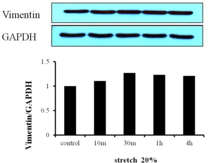
3-1. Expression of Vimentin
To confirm whether the primary cultured cells were fibroblasts, we did experiment, western blot for vimentin expression on the cells. Vimentin was expressed. So, we assumed that the cell was fibroblasts (fig. 1).
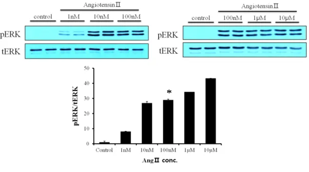
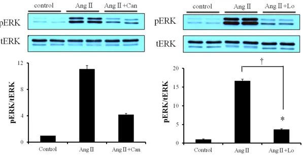
3-2. Effect of Ang Ⅱ on cardiac fibroblasts
To examine whether Ang Ⅱ activates ERK phosphorylation, we treated Ang Ⅱ in proportion to its concentration. The more concentration of Ang Ⅱ (1 nM~ 10 µM) increased, the more ERK was phosphorylated (Fig. 2). We knew that AT1R exists in cardiac fibroblasts. To clarify, we treated Ang Ⅱ with Losartan (100 µM) and Candesartan (1 µM) that are AT1R blocker. These blockers attenuated the phosphorylation of ERK (Fig. 3).
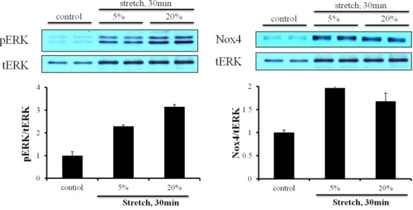
3-3. Effect of mechanical stretch on AT1R activation in cardiac fibroblasts
When the cells were stretched, phosphorylation of ERK and Nox4 expression were activated by the mechanical stretch (Fig. 4). Nox4 expression is related with AT1R signaling. To determine whether Losartan could block increased pERK by the mechanical stretch, we exerted the mechanical stretch with pretreated Losartan. But there was no significant difference between only stretch and stretch with Losartan on ERK phosphorylation (Fig. 5).
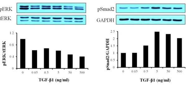
3-4. Effect of TGF-β1 on Smad2 phosphorylation in cardiac fibroblasts
To know whether TGF-β1 triggers its signaling, pSmad2 and pERK, TGF-β1 was treated on the cells with five different concentration (0.05 ~ 500 ng/ml). Smad2 was phosphorylated according to TGF-β1 concentration. So we knew TGF-β receptor existed on the primary cultured cardiac fibroblasts, but ERK was shown decreased phosphorylation (Fig. 6).
7
Figure 1. Expression of Vimentin. The primary cultured cells were taken by as described in ‘Materials and Methods’. The cells were serum-starved over night. Cells were lysed,
separated on SDS-polyacrylamide gels, and immunoblotted as described in ‘Materials and Methods’.
8
Figure 2. Effect of Ang Ⅱ on cardiac fibroblasts. Cardiac fibroblasts were serum-starved overnight and Ang Ⅱ was treated with five different concentrations (1 nM ~ 10 µM) for 5 min. Cells were lysed, separated on SDS-polyacrylamide gels, and immunoblotted as described in ‘Materials and Methods’. *P < 0.05, compared to control.
9
Figure 3. Effect of Ang Ⅱ on cardiac fibroblasts. Cardiac fibroblasts were serum-starved over-night and Ang Ⅱ was treated with Candesartan (3 x 1 µM) and Losartan (3 x 100 µM) respectively. Cells were lysed by RIPA buffer, separated on SDS-polyacrylamide gels, and immunoblotted as described in ‘Materials and Methods’ after 5 min (* P > 0.05, compared to control. † P < 0.05, Ang Ⅱ+ Losartan compared to Ang Ⅱ).
10
Figure 4. Effect of mechanical stretch on AT1R activation in cardiac fibroblasts. Cardiac fibroblasts were plated on bioflex plate coated with collagen Ⅰ, serum-starved overnight, and stretched with different elongation, 5% and 20% for 30 min. Cells were lysed by RIPA buffer and separated on SDS-polyacrylamide gels as described in ‘Materials and Methods’.
11
Figure 5. Effect of mechanical stretch on AT1R activation in cardiac fibroblasts. Cardiac fibroblasts were serum-starved overnight. Angiotensin Ⅱ (100 nM) were treated for 3 min 30 sec. Losartan (1 mM) was pretreated for 3 min. Others were stretched 20% elongation for 5 min.
12
Figure 6. Effect of TGF-β1 on Smad2 phosphorylation in cardiac fibroblasts. Cardiac fibroblasts were serum-starved for overnight. TGF-β1 was treated with five different concentrations (0.05 ~ 500 ng/ml) for 5 min. Cells were lysed by RIPA buffer and separated on SDS-polyacrylamide gels as described in ‘Materials and Methods’.
13
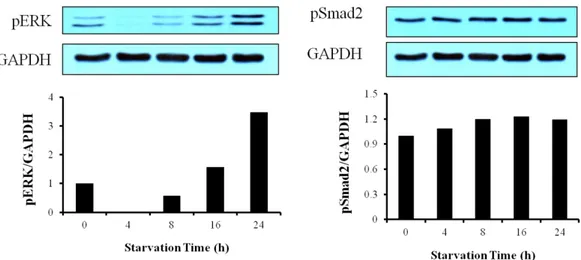
3-5. Effect of starvation on Smad2 phosphorylation in cardiac fibroblasts
To know the base line of Smad2 phosphorylation, we investigated starvation-dependent phosphorylation of Smad2 and ERK. Although pERK was decreased at 4h starvation and then increased, psmad2 kept expressed until 24 hour starvation (Fig. 7).
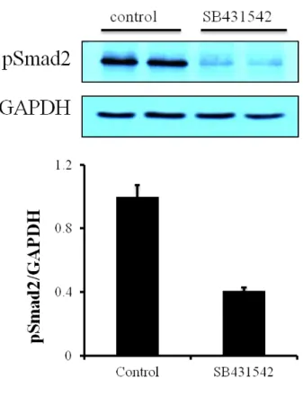
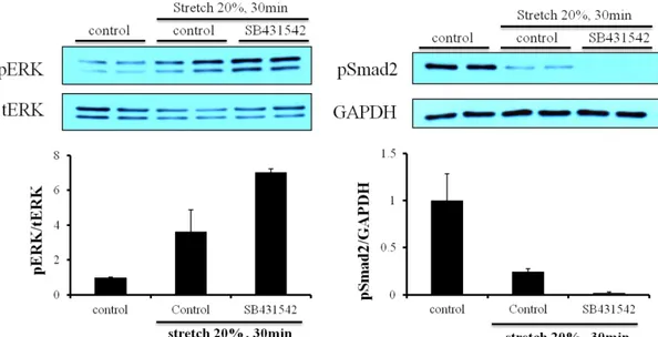
3-6. Effect of mechanical stretch on Smad2 phosphorylation in cardiac fibroblasts SB431542 (TGF-β1 receptor inhibitor) (10 µM) can block pSmad2, one of TGF-β1 receptor-induced protein in cardiac fibroblasts (Fig. 8). And we stretched the cells to examine whether SB431542 could block the phosphorylation of ERK or not. SB431542 could not block the raise of pERK and the level of pERK between stretch-control and SB431542 had no big difference (Fig. 9, 10). As SB431542 could block the pSmad2, SB431542 blocked TGF-β receptor. Interestingly, when cardiac fibroblasts were stretched,
the pSmad2 was diminished. In 20% elongation stretch, pSmad2 was more decreased than in 5% (Fig. 11).
3-7. Effect of mechanical stretch time periods on Smad2 phosphorylation in cardiac fibroblasts
To know the expression of ERK and Smad2 phosphorylation, cardiac fibroblasts were stretched depend on stretch time (10 min ~ 4 h). Phosphorylation of ERK was increased than control at 10 min and decreased until 1 hour and again increased at 4 h. Smad2 was also phosphorylated in a similar fashion. But Smad2 kept decreased than control until 1 h (Fig. 12).
14
Figure 7. Effect of starvation on Smad2 phosphorylation in cardiac fibroblasts. Cardiac fibroblasts were serum-starved for 0h, 4h, 8h, 16h, and 24h respectively. Cells were lysed by RIPA buffer and separated on SDS-polyacrylamide gels as described in ‘Materials and Methods’.
15
Figure 8. Effect of mechanical stretch on Smad2 phosphorylation in cardiac fibroblasts. Cardiac fibroblasts were serum-starved for 16 h. SB431542 (TGF-β1 receptor inhibitor) (10 µM) were treated for 30min before cells were lysed.
16
Figure 9. Effect of mechanical stretch on Smad2 phosphorylation in cardiac fibroblasts. Cardiac fibroblasts were serum-free and stretched for 10 min with 20% elongation.
SB431542 (TGF-β1 receptor inhibitor) (10 µM) were treated for 30 min before the cells were stretched. †P < 0.02, compared to stretch control.
17
Figure 10. Effect of mechanical stretch on Smad2 phosphorylation in cardiac fibroblasts. Cardiac fibroblasts were serum-free and stretched for 30 min with 20% elongation. SB431542 (TGF-β1 receptor inhibitor) (10 µM) were treated for 30 min before the cells were stretched.
18
Figure 11. Effect of mechanical stretch on Smad2 phosphorylation in cardiac fibroblasts. Cardiac fibroblasts were seeded on bioflex plate coated with collagen Ⅰ, serum-starved overnight, and stretched with different elongation, 5% and 10% for 30 min. Cells were lysed by RIPA buffer and separated on SDS-polyacrylamide gels as described in ‘Materials and Methods’.
19
Figure 12. Effect of mechanical stretch time periods on Smad2 phosphorylation in cardiac fibroblasts. Cardiac fibroblasts were seeded on bioflex plate coated with collagen Ⅰ, serum-starved overnight, and stretched with different elongation, 5% and 10% for 30 min. Cells were lysed by RIPA buffer and separated on SDS-polyacrylamide gels as described in ‘Materials and Methods’.
20
4. Discussion
The objective of this study was to investigate the effect of mechanical stretch on the fibroblasts. It is controversial whether several inhibitors could block the effect of stretch to start pathologic signal. There are few studies about mechanical stretch and cardiac fibroblasts in contrast to cardiac myocytes.
Angiotensin Ⅱ is a peptide hormone that induces vasoconstriction, a subsequent increase in blood pressure and hypertrophic and fibrotic response. Losartan is antagonist of AT1R. There are some studies that Losartan could reduce fibrotic and hypertrophic remodeling. It has been proposed that Losartan administration reduce blood pressure and myocardial mass in spontaneously hypertensive rats (Koprdová et al, 2009) and that Losartan suppresses expression of basal and enhanced cardiac angiotensin receptor (Sim et al, 2006). Also, there are results that Losartan can prevent long-term intensive exercise induced heart fibrosis in an animal model (Gay-Jordi et al, 2013). However, although some Losartan prevents stretch-induced electrical remodeling in cultured atrial neonatal myocytes (Erol et al, 2007) in cultured cardiac fibroblasts, when cells were stretched, Losartan had no effect on the pERK, one of AT1R down pathway (fig. 5). Or else other undiscovered elements might offset the effect of Losartan.
TGF-β signaling is very relevant to proliferation, cellular differentiation, and other functions. In cardiac fibroblasts, TGF-β can activate differentiation from fibroblast to myofibroblast. In the heart, there are some ways to generate myofibroblasts. Mechanical and neurohumoral stimuli could be. Several cytokines, chemokines, and growth factors can generate the myofibroblast. Injured hearts also induce several growth factors like TGF-β. Elevated wall tension and mechanical stretch are other causes for myofibroblast differentiation (Jennifer et al, 2013). Myofibroblasts are characterized by secreting
21
extracellular matrix (ECM) components such as collagen Ⅰ, collagen Ⅲ, fibronectin (Joel et al, 2004). In part, contractile activity of the myofibroblasts is observed. The expression of α-smooth muscle actin (α-SMA) is a marker of myofibroblast transformation. Properties of myofibroblast relate to heart disease.
After a myocardial infarction, myofibroblasts play a protective role by generating and remodeling of a fibrotic scar that prevents wall rupture of heart. There is chance that after protective differentiation of fibroblasts, some of fibroblasts undergo senescence or apoptosis. However, being in a long-term differentiation state due to high wall tension and dysfunction of neuroendocrine leads to chronic cardiac fibrosis and hypertrophic heart (Jennifer et al, 2013).
Most studies indicate that mechanical stretch or tension on cells activate TGF-β receptor and initiate differentiation to myofibroblast. It has been suggested that TGF-β1 and mechanical stretch are known to major mediators of differentiation into myofibroblast (Vincent et al, 2014) and that human cardiac fibroblast contraction activate integrins αvβ5 and αvβ3 that promote latent TGF-β1 activation. However, mechanical stretch diminishes Smad2 phosphorylation of TGF-β receptor down pathway (Fig. 10). In this present study, only few studies are similar to those found. It is studied that while fluid-induced shear stress activate TGF-β receptor through producing TGF-β1 and activate Smad2 expressing α-SMA and myofibroblast transition, cyclic strain activates strain sensitive receptor and block Smad2 (Galie et al, 2012).
In some point of view, 5% and more than 20% elongation affect differently cells. The vascular gene expression show differently. 5% elongation is physiological and 20% elongation make pathological response. For instance, in vascular smooth muscle cells, FGF-2, a growth factor involved in cellular reparation after injury is induced at 14% and 33% elongation without at 5% elongation (Konstantin et al, 2009). But In this study, there are no differences between 5% and 20% elongation in mechanical stretched cardiac fibroblasts.
22
ERK phosphorylation became higher at 20% elongation than 5% elongation. pSmad2 diminished more at 20% elongation (Fig. 11).
The main limitation of this study was that when the hearts are primary cultured, the cells were impacted so some cells would felt the heart scared. So, the control of experiment is quite variable. Another one is the method of stretch. There are many methods of stretching to cells. Therefore there are difficult in setting up some principle among much thesis. Also, because the flexcell plates couldn't be changed, various experiments are limited.
Taken together, TGF-β1, angiotensin Ⅱ, mechanical stretch induce heart dysfunction such as fibrosis and hypertrophy. Mechanical stretch activates ERK phosphorylation. However, mechanical stretch induced ERK phosphorylation couldn’t be attenuated by angiotensin receptor blocker (ARB), Losartan. So, Losartan couldn’t decrease cell proliferation in cardiac fibroblasts. Mechanical stretch also decreases pSmad2. SB431542 also couldn’t diminish pERK but pSmad2. Smad2 phosphorylation was diminished at 5% elongation and 20% elongation in cardiac fibroblasts.
23
5. References
(Maulik et al, 2012).
Subir Kumar Maulik, and Santosh Kumar. Oxidative stress and cardiac hypertrophy: a review. toxicology mechanisms and methods Vol. 22, No. 5, 359-366. (2012)
(Janet et al, 1999).
Janet M. Schnee, Willa A. Hsueh. Angiotensin II, adhesion, and cardiac fibrosis. Cardiovascular Research 46 (2000) 264–268. (1999)
(Alexander et al, 2011).
Alexander Meyer, Wei Wang, Jiaxiang Qu, Lori Croft, Jay L. Degen, Barry S. Coller, and Jasimuddin Ahamed. Platelet TGF-β1 contributions to plasma TGF-β1, cardiac fibrosis, and systolic dysfunction in a mouse model of pressure overload. Blood: 119 (4). (2011)
(Jennifer et al, 2013)
Jennifer Davis, Jeffery D. Molkentin. Myofibroblasts: Trust your heart and let fate decide. Journal of Molecular and Cellular Cardiology, Volume 70, May 2014, Pages 9–18. (2013)
(Olson et al, 2005)
Olson ER, Naugle JE, Zhang X, Bomser JA, Meszaros JG. Inhibition of cardiac fibroblast proliferation and myofibroblast differentiation by resveratrol. Am. J. Physiol. Heart Circ. Physiol. 288, H1131–8. (2005)
(Schorb et al, 1995)
24
stimulator of MAP-kinase activity in neonatal rat cardiac fibroblasts. J. Mol. Cell. Cardiol., 27, 1151–60. (1995)
(Wei et al, 2012)
Wei Zhang, Xiang-Fan Chen, Yan-Juan Huang, Qing-Qing Chen, Yuan-Jian Bao and Weizhong Zhu. 2,3,4′,5-Tetrahydroxystilbene-2-O-β-D-glucoside inhibits angiotensin II-induced cardiac fibroblast proliferation via suppression of the reactive oxygen species– extracellular signal-regulated kinase 1/2 pathway. Clinical and Experimental Pharmacology and Physiology 39, 429–437. (2012)
(Derynck et al, 2003)
Derynck R, Zhang YE. SMAD-dependent and SMAD-independent pathways in TGFbeta family signalling. Nature, 425, 577–84. (2003)
(Maryse et al, 1994)
Maryse Crabos, Michael Roth, Alfred W. A. Hahn, and Paul Eme. Characterization of Angiotensin Ⅱ Receptors in Cultured Adult Rat Cardiac Fibroblasts. The American Society for Clinical Investigation, Inc. Volume 93, 2372-2378. (1994)
(Hiroaki et al, 1994)
Hiroaki Matsubara, Mikihiko Kanasaki, Satoshi Murasawa, Yasuyuki Tsukaguchi, Yutaka Nio, and Mitsuo Inada. Differential Gene Expression and Regulation of Angiotensin Ⅱ Receptor Subtypes in Rat Cardiac Fibroblasts and Cardiomyocytes in Culture. The American Society for Clinical Investigation, Inc. Volume 93, 1592-1601 (1994)
25
R. KOPRDOVÁ , M. CEBOVÁ , F. KRISTEK, Long-Term Effect of Losartan Administration on Blood Pressure, Heart and Structure of Coronary Artery of Young Spontaneously Hypertensive Rats, Physiol. Res. 58: 327-335 (2009)
(Sim et al, 2006)
Meng-Kwoon Sim, Woei-Shin Chen, Effects of losartan on angiotensin receptors in the hypertrophic rat heart, Regulatory Peptides 137, 140–146 (2006)
(Gay-Jordi et al, 2013)
Gemma Gay-Jordi, Eduard Guash, Begon˜ a Benito, Josep Brugada, Stanley Nattel, Lluı´s Mont, Anna Serrano-Mollar, Losartan Prevents Heart Fibrosis Induced by Long-Term Intensive Exercise in an Animal Model, PLoS ONE 8(2): e55427. doi:10.1371/journal.pone.0055427 (2013)
(Erol et al, 2007)
Erol Saygili, Obaida R. Rana, Esra Saygili, Hannes Reuter, Konrad Frank, Robert H. G. Schwinger, Jochen Mu¨ller-Ehmsen, and Carsten Zobel, Losartan prevents stretch-induced electrical remodeling in cultured atrial neonatal myocytes, Am J Physiol Heart Circ Physiol 292: H2898–H2905 (2007)
(Joel et al, 2004)
Joel Atance, Michael J. Yost, and Wayne Carver, Influence of the Extracellular Matrix on the Regulation of Cardiac Fibroblast Behavior by Mechanical Stretch, J. Cell. Physiol. 200: 377–386 (2004)
26
Vincent Sarrazy, Anne Koehler, Melissa L. Chow, Elena Zimina, Chen X. Li, Hideyuki Kato, Christopher A. Caldarone, and Boris Hinz, Integrins αvβ5 and αvβ3 promote latent TGF-β1 activation by human cardiac fibroblast contraction, Cardiovascular Research 102, 407–417 (2014)
(Galie et al, 2012)
PA Galie, MW Russell, MV Westfall, and JP Stegemann, Interstitial fluid flow and cyclic strain differentially regulate cardiac fibroblast activation via AT1R and TGF-β1, Exp Cell Res. 2012 January 1; 318(1): 75–84. (2012)
(Konstantin et al, 2009)
Konstantin G. Birukov, Cyclic Stretch, Reactive Oxygen Species, and Vascular Remodeling, ANTIOXIDANTS & REDOX SIGNALING, Volume 11, Number 7, 1651-1667 (2009).
27
6. Acknowledgements
For two years, I finish my thesis under professor Ki-Seok Kim guidance. He gave me chances to do experiments, presentation and lots of things. I am grateful to professor Dong-sun Lee for introducing laboratory to me. I am thankful to Doctor Young-mee Kim and Yoon-sil Yang for teaching about experiment and help. Thanks to my lab researcher, Eun-Jung Kwon, Eun-jin Yang and CHI DANY.