저작자표시-비영리-변경금지 2.0 대한민국 이용자는 아래의 조건을 따르는 경우에 한하여 자유롭게 l 이 저작물을 복제, 배포, 전송, 전시, 공연 및 방송할 수 있습니다. 다음과 같은 조건을 따라야 합니다: l 귀하는, 이 저작물의 재이용이나 배포의 경우, 이 저작물에 적용된 이용허락조건 을 명확하게 나타내어야 합니다. l 저작권자로부터 별도의 허가를 받으면 이러한 조건들은 적용되지 않습니다. 저작권법에 따른 이용자의 권리는 위의 내용에 의하여 영향을 받지 않습니다. 이것은 이용허락규약(Legal Code)을 이해하기 쉽게 요약한 것입니다. Disclaimer 저작자표시. 귀하는 원저작자를 표시하여야 합니다. 비영리. 귀하는 이 저작물을 영리 목적으로 이용할 수 없습니다. 변경금지. 귀하는 이 저작물을 개작, 변형 또는 가공할 수 없습니다.
A THESIS
FOR THE DEGREE OF MASTER OF SCIENCE
Comparative study on hemocytes of sea hares Aplysia kurodai (Baba,
1937), Aplysia juliana (Quoy & Gaimard, 1832), Aplysia parvula
(Mörch, 1863), and Aplysia oculifera (A. Adams & Reeve, 1850) in
Jeju Island, Korea: morphology and functional aspects
Nobuhisa Kajino
Department of Marine Life Science
GRADUATE SCHOOL
JEJU NATIONAL UNIVERSITY
February 2021
Comparative study on hemocytes of sea hares Aplysia kurodai (Baba,
1937), Aplysia juliana (Quoy & Gaimard, 1832), Aplysia parvula
(Mörch, 1863), and Aplysia oculifera (A. Adams & Reeve, 1850) in
Jeju Island, Korea: morphology and functional aspects
Nobuhisa Kajino
(Advised by Professor Kwang-Sik Choi)
A dissertation submitted in partial fulfillment of the requirement for the
degree of Master of Science
February 2021
This dissertation has been examined and approved by
Dr. Kyung-Il Park, Professor, Department of Aquatic Life Medicine, Kunsan
National University
Dr. Hyun-Ki Hong, Research Professor, School of Marine Life Science, Jeju
National University
Dr. Kwang-Sik Choi, Professor, School of Marine Life Science, Jeju National
University
February 2021
Date
Department of Marine Life Science
CONTENTS
국문요약... ii
LIST OF FIGURES ...iv
LIST OF TABLES...v
1. INTRODUCTION ...1
2. MATERIALS AND METHODS...4
2.1. Sampling effort ...4
2.2. Hemolymph collection ...7
2.3. Morphology of the hemocytes ...7
2.3.1. Light microscopy...7
2.3.2. Scanning electron microscopy (SEM) ...7
2.3.3. Transmission electron microscopy (TEM) ...8
2.4. Cellular immune functions...8
2.4.1. Hemocyte type and count...9
2.4.2. Lysosomal content...9
2.4.3. Phagocytosis capacity...9
2.4.4. Oxidative activity ...10
2.5. Statistical analysis ...10
3. RESULTS...11
3.1. Morphology of the hemocytes ...11
3.1.1. Hemocyte types ...11
3.1.2. Hemocytes size...15
3.2. Celluar immune functions...17
3.2.1. THC and proportion of each hemocyte types...17
3.2.2. Lysosomal content...19 3.2.3. Phagocytosis capacity...20 3.2.4. Oxidative activity ...22 4. DISCUSSION...24 5. CONCLUSION...30 REFERENCES ...31
국문요약
Aplysia는 군소과 (Family Aplysiidae)에 속하는 복족류로 해조류가 풍부한 조간대 또는 얉은 조하대에 서식하는 초식자로서 중요한 생태적 지위를 차지한다. 복족류의 세포성 면역체계를 이해하기 위해서는 혈구 (hemocyte)의 유형과 유형별 면역기능에 대한 연구가 선행되어야 하나, Aplysia의 혈구 종류와 기능에 대한 연구는 미흡한 실정이다. 이 연구에서는 제주도 조간대 지역에 서식하는 군소 (Aplysia kurodai), 말군소 (A. juliana), 검은테군소 (A. parvula), 안경무늬군소 (A. oculifera) 4종을 대상으로 혈구의 형태와 기능을 유세포분석기, 광학현미경, 및 전자현미경을 이용하여 분석하였다. 군소 4종의 혈구는 세포의 형태와 면역기능에 따라 granulocytes, hyalinocytes 그리고 blast-like cell의 3가지 유형으로 동일하게 나타났다. Granulocyte는 세포질 내 과립 (granule)과 긴 위족 (pseudopodia)들이 특징으로 혈림프액 중 혈구 중 6% 미만으로 낮은 비율을 차지하였다. Hyalinocyte는 혈림프액 중 93% 이상을 차지하는 주된
혈구로 granulocyte와 유사한 형태이나 세포질 내 과립들이 없었다.
Granulocyte와 hyalinocyte 모두 세포성 면역능력이 있었으나, granulocyte가 hyalinocyte 보다 lysosome 함량, 식세포 능력(phagocytosis capacity), 및 세포산화 능력(oxidative capacity)이 2배 이상 높았다. Blast-like cell은 세포의 크기가 가장 작고 세포질이 가장 얇은 세포로 면역 기능이 거의 없는 미분화 세포의 특징을 보였다. 이 연구를 통해 군소 4종 모두 혈구는 granulocyte, hyalinocyte, blast-like cell의 3가지 종류로 분류되었으며 있으며, 그 중 granulocyte와 hyalinocyte가 주요 면역 세포임이 확인되었다.
List of Figures
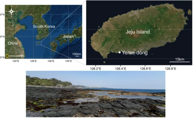
Fig. 1.
Location of the sampling site. Aplysia kurodai, A. juliana, A. parvula, and A.oculifera were collected from tide pools at Yerae-dong in Jeju Island on the south
coast of Korea (33˚14′17.3″N; 126˚23′29.6)
··· 5
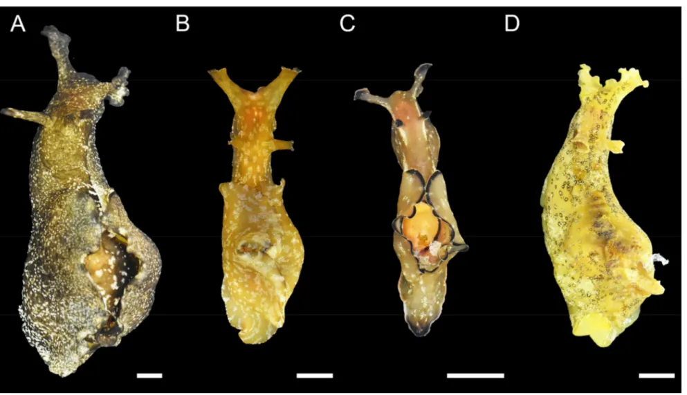
Fig. 2. Photographs showing the
four species of Aplysia. (A) Aplysia kurodai, (B) A.juliana, (C) A. parvula, and (D) A. oculifera. Scale bar = 1 cm.
··· 6
Fig. 3.
Three hemocyte types isolated from Aplysia kurodai, A. juliana, A. parvula, and A.oculifera. The hemocyte types were determined using a combination of flow
cytometry and light microscopy. A total of 10,000 cells were analyzed for individual using flow cytometry. Scale bar = 10 μm.
··· 12
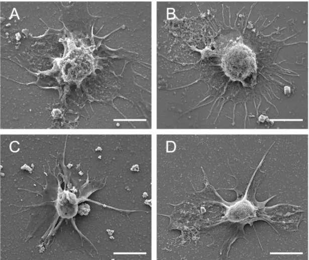
Fig. 4.
Scanning electron micrographs showing the hemocytes of (A) A. kurodai, (B) A.juliana, (C) A. parvula, and (D) A. oculifera. Scale bar = 5 μm.
··· 13
Fig. 5.
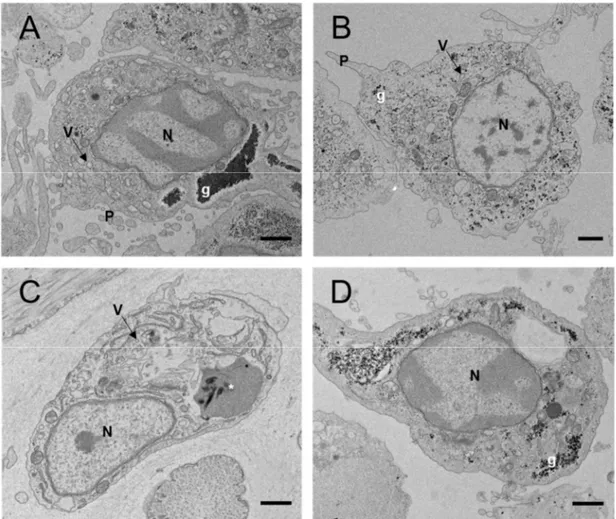
Transmission electron micrographs showing the hyalinocytes of (A) A. kurodai, (B)A. juliana, (C) A. parvula, and (D) A. oculifera. N, nucleus; V, vesicle; P,
pseudopodia; g, glycogen; *, unknown structure. Scale bar = 1 μm.
··· 14
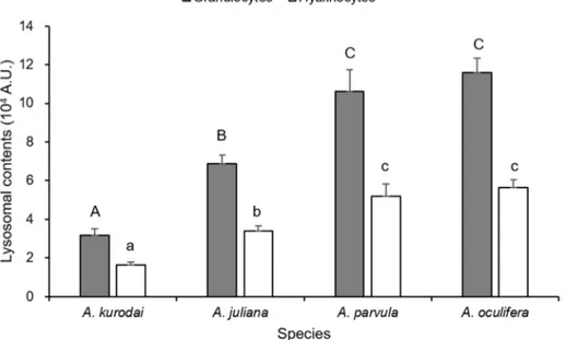
Fig. 6.
Relative quantity of lysosomes in the hemocyte populations of A. kurodai, A. juliana,A. parvula, and A. oculifera.
··· 19
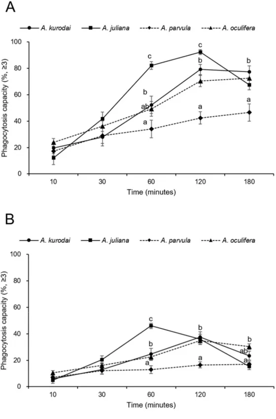
Fig. 7.
Phagocytosis capacity of (A) the granulocytes and (B) hyalinocyte of A. kurodai, A.juliana, A. parvula, and A. oculifera. The percentage of hemocytes having engulfed
more than three fluorescent beads.
··· 21
Fig. 8.
Intracellular oxidative activity of (A) the granulocytes and (B) hyalinocytes of A.List of Tables
Table1.
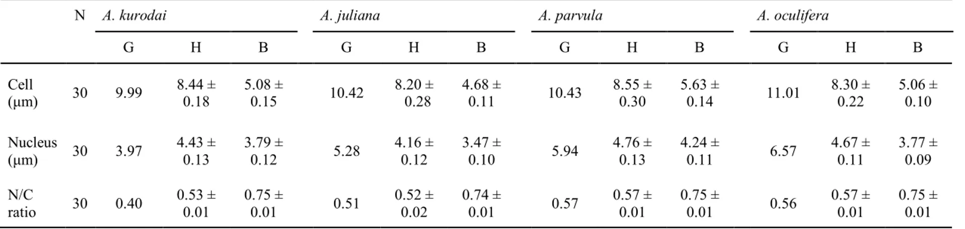
Cell and nucleus diameters and nucleus/cell (N/C) ratio of Aplysia kurodai, A.juliana, A. parvula, and A. oculifer. Values are presented as mean ± standard error.
N: Number of analyzed cells, G: Granulocytes, H: Hyalinocytes, B: Blast-like cells.
··· 16
Table2.
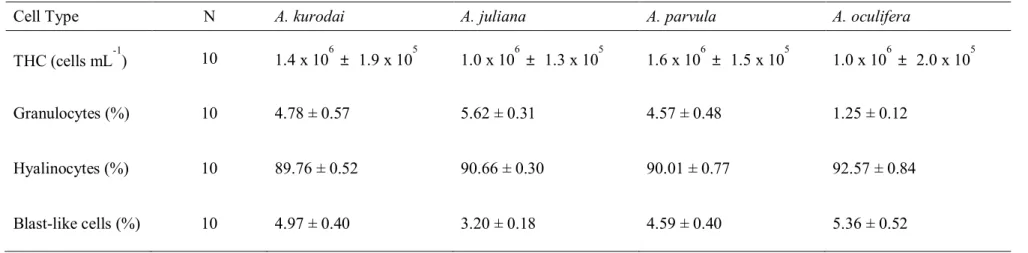
The total hemocyte count (THC) and percentage of each hemocyte types of A.kurodai, A. juliana, A. parvula, and A. oculifera. N: Number of analyzed animals.
··· 18
Table3.
Hemocyte types and total hemocytes count of aquatic gastropod observed in the present and previous studies. THC, Total hemocytes count; G, Granulocytes; H, Hyalinocytes; B, Blast-like cells; FCM, Flow cytometry; LM, Light microscope; SEM, Scanning electron microscope; TEM, Transmission electron microscope.1. Introduction
Like other invertebrates, marine mollusks have an internal cellular response against invading foreign substances (Loker, 2010). The innate immunity of mollusks provides the first line of defense including the physical and chemical barriers, humoral and cellular components with the non-specific response. The cellular immune response in gastropods entirely relies upon cells freely circulating in the hemolymph and infiltrating in tissue, referred to as hemocytes (Donaghy et al., 2010). Hemocytes mediate cellular defense in mollusks through phagocytosis and encapsulation of invading foreign particles, and then secret enzymes and reactive oxygen species (ROS) to destroy the particles. To maintain homeostasis, hemocytes are also involved in various physiological responses such as tissue injury healing, shell formation, nutrient transportation, metabolite excretion, and gas exchange (Cheng, 1984; Hine, 1999; Loker, 2010).
Characterization of hemocytes, such as cell types and functions, under natural and ambient conditions is the first step in understanding the cellular response of mollusks to environmental and biological stresses. In marine mollusks, two main hemocyte types are generally recognized: granulocytes containing numerous intra-cytoplasmic granules and hyalinocytes (or agranulocytes) exhibiting few to no granules. Functionally, both granulocytes and hyalinocytes exhibit pseudopodia, agglutinate, engulf the particles and produce ROS. In marine bivalve mussels (Yang et al., 2015), oyster (Hong et al., 2013), blood cockles (Kim et al., 2020), and marine gastropod abalone (Hong et al., 2019), the types of hemocytes were characterized into the two types mentioned above. The granulocytes are the most active in cellular immune responses compare to hyalinocytes.
Commonly, the classification of molluscan hemocytes have been based on morphological features from light and electron microscopy (Adema et al., 1992; Cheng, 1975; Franchini and Ottaviani, 1990; Mahilini and Rajendran, 2008; Martin et al., 2007; Ottaviani and Franchini, 1988). However, depending on researchers, such a method may lead
to numerous misconceptions (Donaghy et al., 2017) and low percentage of cell types may not be observed under a microscope. Thus, not only morphological analysis but also functional activities of hemocytes should be performed. Among the techniques for identifying cell function, flow cytometry allows fast, accurate, quantitative, and simultaneous analyses of the cell morphology and functions, including immune activities (Donaghy et al., 2009). For this reason, flow cytometry has been used widely in mollusks immunology such as bivalve (Brousseau et al., 1999; Hong et al., 2013, 2020; Lambert et al., 2003; Park et al., 2012) and gastropod (Donaghy et al., 2010; Hong et al., 2019; Ottaviani, 1989; Prokhorova et al., 2018).
The marine gastropods Aplysia, also known as sea hares, belongs to the Family Aplysiidae. Total 44 species have been reported in shallow marine water world-wide (World Register of Marine Species; Carefoot, 1987; Klussmann-Kolb, 2004; Lee et al., 2014). According to Min et al. (2004), only four Aplysia species have been reported in Korea. The Aplysia play a crucial role in grazing macro-phytobenthos in
small tidal pools or shallow
subtidal hard bottoms
. They are an important model species for neurobiology research on memory and learning due to a comparatively simple nervous system with large neurons (Kandel and Schwartz, 1982). Despite their high abundance in the ecosystem and importance as a research model, the immune system on Aplysia has been studied only one species, A.californica (Martin et al., 2007; Pauley et al., 1971, 1972), and internal defense of Aplysia
such as hemocyte types and their functions remain unclear.
According to Martin et al. (2007), A. californica has a single type of circulating hemocyte; hyalinocytes with pseudopodia and no intra-cytoplasmic granules on morphological features. In marine gastropod abalone, H. diversicolor possesses three types of hemocytes, granulocytes, hyalinocytes, and blast-like cells (Hong et al., 2019), while two main hemocyte populations were distinguished in H. discus discus hemolymph: hyalinocytes
same genus, they may have different blood cell types depending on the species. Therefore, in the present study, we characterized the hemocyte of poorly studied four Aplysia species,
Aplysia kurodai, A. juliana, A. parvula, and A. oculifera, as morphology and functional
aspects using a combination of light and electron microscopy and flow cytometry. This result provides baseline information for comparative studies on molluscan hemocytes.
2. Materials and Methods
2.1. Sampling effort
In April and July 2020, Aplysia kurodai, A. juliana, A parvula, and A. oculifera were collected from a rocky intertidal beach in Yerae-dong (33˚14′17.3″N; 126˚23′29.6) in Jeju Island on the south coast of Korea (Fig. 1 and 2). The harvested four Aplysia species were transported to the laboratory and maintained in a tank with aerated seawater (salinity, 30 PSU; water temperature, 20 ℃) over 24 h to acclimate to the laboratory condition. The wet weight of four Aplysia species, A. kurodai, A. juliana, A. parvula, and A. oculifera, used in the experiment ranged 29.9 - 144.3 g, 22.4 - 92.3 g, 2.8 - 11.1 g, and 7.0-43.4 g, respectively.
Fig. 1. Location of the sampling site. Aplysia kurodai, A. juliana, A. parvula, and A. oculifera were collected from tide pools at Yerae-dong in Jeju
2.2. Hemolymph collection
Approximately 500 μL of hemolymph was withdrawn from the hemocoel along the right side of the head of each Aplysia species using a 1-mL syringe fitted with a 22 G x 1 1/4” needle. Collected hemolymph was immediately transferred into a microtube on ice to minimize cell aggregation. Ten individuals of A. kurodai, A. juliana, and A. oculifera were used to characterize hemocyte type and functions using flow cytometry. In A. parvula, hemolymph collection was difficult due to their body size, so hemolymph from two individuals was pooled to obtain a sufficient quantity of hemolymph to be analyzed in flow cytometry.
2.3. Morphology of the hemocytes
2.3.1. Light microscopy
Forty μL of hemolymph from each Aplysia species was placed on glass slides coated with poly-L-Lysine (MAS-11; Matsunami Glass Ind., Ltd., Japan), and the hemocytes were incubated in a humidity chamber at room temperature for 30 min. The adhered hemocytes were stained with Giemsa and Wright stain solution (Sigma, USA). Cell and nucleus diameters of hemocytes were measured from the digitized images using image-analysis software (Zen 2.3 Lite, ZEISS, Germany).
2.3.2. Scanning electron microscopy (SEM)
The hemolymph was attached on a glass slide coated with poly-L-lysine (MAS-11; Matsunami Glass Ind., Ltd., Japan) and incubated at room temperature for 60 min. The incubated hemocytes were fixed with 2% glutaraldehyde (Sigma-Aldrich, USA) at room temperature for 60 min. The fixed hemocytes were washed three times in 1X Phosphate
buffered saline (PBS; 137mM NaCl, 2.7mM KCL, 10mM Na2HPO4, pH 7.3) for 5 min and
dehydrated in graded series of ethanol (50, 70, 90, 95, and 100%). After dehydration, the hemocytes were substituted in graded series of isoamyl acetate (30, 50, 70, and 100%). The hemocytes on slides were coated with platinum using a sputter coater (Q150RS; Quorum Technologies, UK). The surface of hemocytes was observed under the scanning electron microscope (MIRA3; TESCAN, Czech Republic)
2.3.3. Transmission electron microscopy (TEM)
Collected hemocytes were pelleted (6000 rpm, 1 min) and washed three times in 1X PBS for 5 min. The pelleted hemocytes were fixed with 2.5% glutaraldehyde in 0.1 M cacodylate buffer (pH 7.3) at room temperature. After fixation, the hemocytes were treated with 4% OsO4 and 3% potassium ferrocyanide in 0.1 M cacodylate buffer (pH 7.3) for 1 h at 4°C in the dark. After dehydration in an ethanol series (50, 60, 70, 80, 90, and 100%) and propylene oxide, the hemocytes embedded in Epon 812 and polymerized at 70 °C for two days. The embedded hemocytes were cut into 70nm size using an ultramicrotome and then collected on 150 mesh copper grids. The ultrathin sections were double-stained with 2% uranyl acetate (10 min) and lead citrate (5 min). The sections were observed under a transmission electron microscope at 120 kV.
2.4. Cellular immune functions
To identify hemocyte types and cellular immune functions, including phagocytosis capacity, oxidative activity, and lysosomal content, the hemocytes were analyzed using CytoFLEX flow cytometer (Beckman Coulter Life Sciences, USA) equipped with two active lasers (488- and 639-nm) and four channels of fluorescence detectors.
2.4.1. Hemocyte type and count
Total hemocyte count (THC) and the percentage of each hemocyte type were determined using SYBR green I (Invitrogen, USA), a green-fluorescent dye that binds to the double-stranded DNA. THC was calculated as the number of cells per milliliter of hemolymph (cells/mL). Each 50 µL of hemolymph diluted in the equal volume of antiaggregant solution (AASH, 2.5% NaCl and 1.5% EDTA in 0.1 M phosphate buffer, pH 7.4) was incubated with 1,000X SYBR green I (final concentration = 20X) for 30 min in the dark at room temperature. After selecting hemocytes only stained by SYBR green I, hemocyte types were discriminated based upon their relative cell size (forward scatter) and granularity (side scatter).
2.4.2. Lysosomal content
The lysosomal content in the hemocytes was determined using LysoTracker Red (Invitrogen, USA), a red-fluorescent dye that accumulates within lysosomal compartments in the live hemocytes. Each 50 µL of hemolymph from four Aplysia species were mixed with 50 µL of filtered seawater (FSW). Diluted hemolymph was incubated with LysoTracker Red (final concentration = 2 µM) for 60 min in the dark at room temperature. The relative quantity of lysosomal components in the hemocytes was expressed as the intensity of red fluorescence in arbitrary units (A.U.)
2.4.3. Phagocytosis capacity
Phagocytosis was induced using Fluorescent beads (2.0 µm in diameter, Polysciences Inc., USA). Each 150 µL of hemolymph from four species were diluted with 150 µL of FSW. Diluted hemolymph was incubated with beads (final concentration at 2%)
in the dark at room temperature. Phagocytosis capacity of hemocytes was measured at 10, 30, 60, 90, 120, and 180 min reaction time. Phagocytosis capacity was expressed as the percentage of hemocytes that engulfed more than three beads.
2.4.4. Oxidative activity
Oxidative activity was measured using 2’7’-dichlorodihydrofluorescein diacetate (DCFH-DA, Invitrogen, USA), a membrane-permeable, non-fluorescent probe. Inside the hemocytes, DCFH-DA is oxidized to a highly fluorescent 2’7’-dichlorodihydrofluorescein (DCF) by the oxidative activity of hemocytes, including reactive oxygen species (ROS) and reactive nitrogen species (RNS). The oxidative ability was quantified by the green fluorescence detector of the flow cytometer. Each 150 µL of hemolymph diluted in the same volume of FSW was incubated with DCFH-DA (final concentration = 10 µM) in the dark at room temperature and was measured at 10, 30, 60, 90, 120, and 180 min after the reaction. The oxidative activity was expressed as fluorescence arbitrary units (A.U.) provided by the green fluorescence detector.
2.5. Statistical analysis
The hemocyte parameters in the four different Aplysia species were compared using one-way analysis of variance (ANOVA) and Duncan’s range test. All statistical tests were performed using the statistical package for the social sciences (SPSS, IBM, USA). The level of significance was set at P <0.05.
3. Result
3.1. Morphology of the hemocytes
3.1.1. Hemocyte types
Under a light microscope, three types of hemocytes could be observed: granulocytes, hyalinocytes, and blast-like cells. The granulocytes of sea hares exhibited pseudopodia on the cell surface and granules in the cytoplasm. Morphology of the hyalinocyte was similar to that of the granulocytes, while they lack cytoplasmic granules. The blast-like cells were small and round, with very thin cytoplasm. (Fig. 3).
SEM and TEM allowed the characterization of the surface morphology and intracellular ultrastructure of hemocytes, respectively. In SEM and TEM, only the hyalinocytes that accounted for the largest proportion in the THC of the four sea hare species were observed. The hyalinocytes were spherical with many pseudopodia on the cell surface (Fig. 4). The hyalinocytes of four Aplysia species exhibited no cytoplasmic granules with lysosome-like organelle containing glycogen and vesicles in the hemocyte (Fig. 5).
Fig. 3. Three hemocyte types isolated from Aplysia kurodai, A. juliana, A. parvula, and A.
oculifera. The hemocyte types were determined using a combination of flow cytometry and
light microscopy. A total of 10,000 cells were analyzed for the individual using flow cytometry. Scale bar = 10 μm.
Fig. 4. Scanning electron micrographs showing the hyalinocytes of (A) A. kurodai, (B) A.
Fig. 5. Transmission electron micrographs showing the hyalinocytes of (A) A. kurodai, (B) A.
juliana, (C) A. parvula, and (D) A. oculifera. N, nucleus; V, vesicle; P, pseudopodia; g,
3.1.2. Hemocytes size
Table 1 summarizes the size of the cells (C), nucleus (N), and the ratio of nuclei to cells (N/C) of the two types of hemocytes, as determined from the digitized images. Under the light microscope, size data of the granulocytes of the four sea hare species could not sufficiently be collected for analysis because the granulocytes were rarely observed. The mean cell size of hyalinocyte in A. kurodai, A. juliana, A. parvula, and A. oculifera ranged 8.44 µm, 8.20 µm, 8.55 µm, and 8.30 µm, respectively. The cell size of hyalinocyte among each species was no significant difference. However, the mean nucleus size of hyalinocyte of
A. parvula (4.76 µm) was significantly larger (P<0.05) than A. oculifera (4.67 µm), A. kurodai (4.43 µm), and A. juliana (4.16 µm). The mean cell size of blast-like cells in A. kurodai, A. juliana, A. parvula, and A. oculifera ranged 5.08 µm, 4.68 µm, 5.63 µm, and
5.06 µm, respectively. The mean cell size of blast-like cells of A. parvula was significantly larger (P<0.05) than A. oculifera, A. kurodai, and A. juliana. The mean nucleus size of blast-like cells of A. parvula (4.24 µm) was significantly larger (P<0.05) than A. oculifera (3.77 µm), A. kurodai (3.79 µm), and A. juliana (3.47 µm). Consequently, the N/C ratio of blast-like cells among each species was no significant difference. The N/C ratio of hyalinocytes of
Table 1. Cell and nucleus diameters and nucleus/cell (N/C) ratio of Aplysia kurodai, A. juliana, A. parvula, and A. oculifer. Values are presented as
mean ± standard error. N: Number of analyzed cells, G: Granulocytes, H: Hyalinocytes, B: Blast-like cells.
N A. kurodai A. juliana A. parvula A. oculifera
G H B G H B G H B G H B Cell (μm) 30 9.99 8.44 ± 0.18 5.08 ± 0.15 10.42 8.20 ±0.28 4.68 ± 0.11 10.43 8.55 ± 0.30 5.63 ± 0.14 11.01 8.30 ± 0.22 5.06 ± 0.10 Nucleus (μm) 30 3.97 4.43 ± 0.13 3.79 ± 0.12 5.28 4.16 ± 0.12 3.47 ± 0.10 5.94 4.76 ± 0.13 4.24 ± 0.11 6.57 4.67 ± 0.11 3.77 ± 0.09 N/C ratio 30 0.40 0.53 ± 0.01 0.75 ± 0.01 0.51 0.52 ± 0.02 0.74 ± 0.01 0.57 0.57 ± 0.01 0.75 ± 0.01 0.56 0.57 ± 0.01 0.75 ± 0.01
3.2. Cellular immune functions
3.2.1. THC and proportion of each hemocyte types
The flow cytometry analysis also discriminated three types of hemocytes based on the relative cell size and granularity (Fig.3). THC and the proportion of each hemocyte type are summarized in Table 2. Among the four species, the mean THC was highest in A.
parvula (1.6 x 10⁶ cells/mL), followed by A. kurodai (1.4 x 10⁶cells/mL), A. juliana (1.0 x
106 cells/mL), and A. oculifera (9.7 x 10⁵ cells/mL). The mean THC of A. parvula was
significantly higher than that of A. juliana and A. oculifera (Table 2). The hyalinocytes accounted for the largest proportion in the THC of the four sea hare species. The proportion of hyalinocytes in the THC of A. kurodai was 93.7 %, A. juliana was 94.7%, A. parvula was 94.7%, and A. oculifera was 93.7%. For the granulocytes, the proportion in the THC of A.
juliana (5.6 %) was highest, followed by A. kurodai (4.8 %), A. parvula (4.6 %), and A. oculifera (1.3 %). The values of A. oculifera were significantly (P<0.05) lower than that of
Table 2. The total hemocyte count (THC) and percentage of each hemocyte types of A. kurodai, A. juliana, A. parvula and A. oculifera. N: Number of
analyzed animals.
Cell Type N A. kurodai A. juliana A. parvula A. oculifera
THC (cells mL-1) 10 1.4 x 10⁶ ± 1.9 x 10⁵ 1.0 x 10⁶ ± 1.3 x 10⁵ 1.6 x 10⁶ ± 1.5 x 10⁵ 1.0 x 10⁶± 2.0 x 10⁵ Granulocytes (%) 10 4.78 ± 0.57 5.62 ± 0.31 4.57 ± 0.48 1.25 ± 0.12
Hyalinocytes (%) 10 89.76 ± 0.52 90.66 ± 0.30 90.01 ± 0.77 92.57 ± 0.84 Blast-like cells (%) 10 4.97 ± 0.40 3.20 ± 0.18 4.59 ± 0.40 5.36 ± 0.52
3.2.2. Lysosomal content
Figure.6 shows the lysosomal content in the hemocytes of the four sea hares analyzed in this study. ANOVA test indicated that the mean of lysosomal content of granulocytes in A. oculifera (1.2 x 10⁵A.U.) and A. parvula (1.1 x 10⁵A.U.) were significantly (P<0.05) higher than that of A. juliana
(6.9 x 10⁴A.U.), and A. kurodai (3.2 x 10⁴A.U.). Likewise, the lysosomal content of hyalinocytes of
A. oculifera (5.7 x 10⁴A.U.) and A. parvula (5.2 x 10⁴A.U.) were significantly (P<0.05) higher than
that of A. juliana (3.4 x 10⁴A.U.), and A. kurodai (1.6 x 10⁴A.U.).
Fig. 6. Relative quantity of lysosomes in the hemocytes populations of A. kurodai, A. juliana, A.
3.2.3. Phagocytosis capacity
Over the 180 min of the experiment, the phagocytosis activities of the granulocytes of all species tend to increase and were no significant difference until 30 min. The phagocytosis activity of the granulocytes of A. parvula and A. oculifera increased until 180 min of the experiment (46.5 and 72.6 %) and that of A. kurodai and A. juluana increased until 120 min (79.1 and 92.3 %) and then decreased. For A. juliana, the phagocytosis activity showed significantly (P<0.05) higher (1.2-2.4 times) than other species at 60 and 120 min. After 120 min, the phagocytosis activity declined rapidly. Finally, phagocytosis capacities of granulocytes in A. kurodai, A. juliana, A. parvula, and A. oculifera reached 77.4 %, 67.4 %, 46.5 %, and 72.6 % at 180 min, respectively (Fig.7A).
The phagocytosis activity of the hyalinocytes of A. kurodai and A. oculifera increased until 120 min of the experiment (37.5 and 34.8 %) and decreased. The phagocytosis capacity of hyalinocytes of A. parvula showed a similar trend from 10 min to 180 min (7.1, 12.2, 13.0, 16.5, and 16.8 %) and that of A. juluana increased until 60 min (46.0 %) and then decreased. The phagocytosis capacities of hyalinocytes in A. kurodai, A. juliana, A. parvula, and A. oculifera reached 23.4 %, 15.4 %, 16.8 %, and 30.3 % at 180 min, respectively (Fig.7B). The mean phagocytosis capacity of the granulocytes was 2.2-2.6 times higher than that of hyalinocytes. The phagocytosis activity of blast-like cells was excluded from the result due to their negligibly low levels of phagocytosis capacities.
Fig. 7. Phagocytosis capacity of (A) the granulocytes and (B) hyalinocyte of A. kurodai, A. juliana, A.
parvula, and A. oculifera. The percentage of hemocytes having engulfed more than three fluorescent
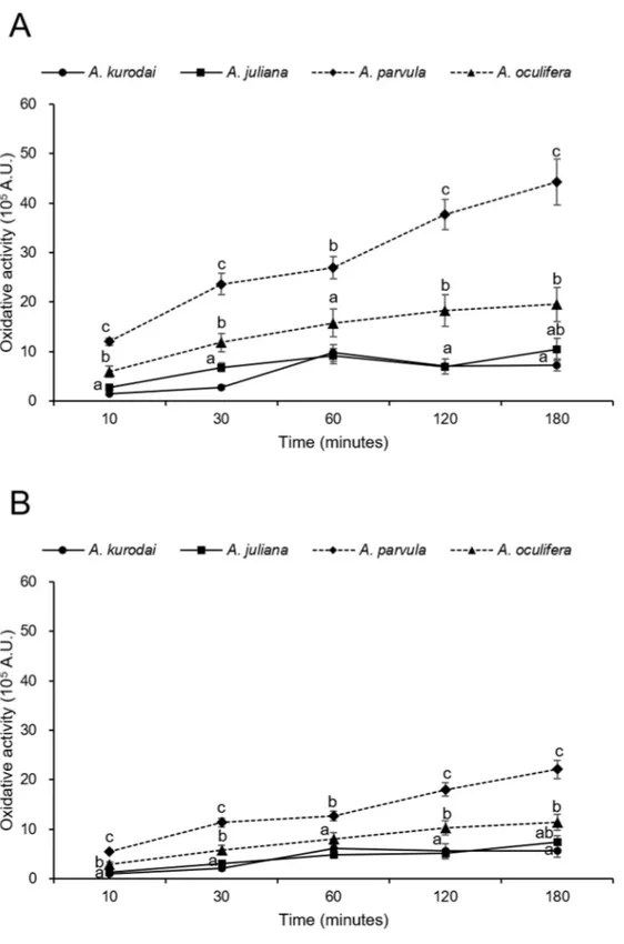
3.2.4. Oxidative activity
Production of ROS and RNS by hemocytes of Aplysias was estimated using the flow cytometry. During the reaction time for 180 min, the oxidative activities of granulocytes and hyalinocytes of all species tend to increase. Over the 180 min of the experiment, the oxidative activities of granulocytes of A. parvula were significantly (P<0.05) higher (approximately 2-5 times) than those of A. kurodai, A. juliana, and A.oculifera. A. juliana (1.0 x 10⁶A.U.), A. parvula (4.4 x 10⁶
A.U.), and A. oculifera (2.0 x 10⁶ A.U.) exhibited the most increased oxidative activities at 180 min
reaction times and A. kurodai (9.8 x 10⁵A.U.) showed the highest activities at 60 min (Fig.8A).
Over the 180 min of the experiment, the oxidative activities of hyalinocytes of A. parvula were significantly (P<0.05) higher (approximately 2-3 times) than those of the other three species. The oxidative activities of hyalinocytes of A. juliana, A. juliana, and, A. oculifera were highest at 180 min and 7.4 x 10⁵ A.U., 2.2 x 10⁶ A.U., and 1.1 x 10⁶ A.U. respectively. For A. kurodai (6.2 x 10⁵
A.U.), the oxidative activities of hyalinocytes showed the highest values at 60 min (Fig.8B). The mean oxidative activities of the granulocytes were 1.4-2.1 times higher than that of hyalinocytes.
Fig. 8. Intracellular oxidative activity of (A) the granulocytes and (B) hyalinocytes of A. kurodai, A.
4. Discussion
In the present study, we first applied both flow cytometry and microscopy to discriminate and characterize different hemocyte types in four species of sea hares, including
Aplysia kurodai, A. juliana, A. parvula, and A. oculifera. In the flow cytometry analysis,
granulocytes and blast-like cells were clearly separated from hyalinocyte populations by cell size and granularity. However, the granulocytes and blast-like cells were hardly observed under the microscope because of the low proportion in the THC (1.3-5.6% in granulocytes and 3.2-5.4% in blast-like cells). Martin et al. (2007) also observed a single type of circulating hemocyte, hyalinocytes without intracellular granules, in A. californica under microscopic observation, probably due to the small number of granulocytes. Microscopic analysis may not allow extensive and quantitative assessment of the hemocyte populations. Alternatively, flow cytometry allows rapid, accurate, and quantitative analyses of hemocyte morphology and functions. Because of these advantages, flow cytometric analysis has been widely performed to characterize the hemocyte types and functions in marine bivalves, Manila clam Ruditapes philippinarum (Park et al., 2012), oyster Crassostrea gigas (Lambert et al., 2013), Saccostrea kegaki (Hong et al., 2020), mussels Mytilus coruscus (Yang et al., 2015) and marine gastropods, abalone Haliotis discus (Donaghy et al., 2010), H. diversicolor (Hong et al., 2019), spiny to shell Turbo cornutus (Donaghy et al., 2010).
The granulocytes of the Aplysia exhibited long pseudopodia from the cell surface with granules in the cytoplasm. Although the granulocytes were a low percentage in the THC, those showed the most active phagocytosis and oxidative activities, so the granulocytes are considered the major hemocyte involved in immune activities. Morphology of the hyalinocyte was similar to that of the granulocytes, while they lack cytoplasmic granules. The hyalinocytes were the most abundant type and were involved in a certain level of immune activities. The blast-like cells of Aplysia are the smallest agranular cells with thin cytoplasm and exhibited an absence of immune activities. Such morphological and
functional features are representative characteristics of undifferentiated cells. However, additional studies are needed to verify that those cells are truly undifferentiated cells by a molecular marker (Cima et al., 2000).
Total hemocyte count (THC) in the hemolymph is changed by hemocyte proliferation or movement between the hemolymph and the tissues (Pipe and Coles, 1995). In gastropods, THC altered depending on environmental conditions such as temperature, pH (Suresh et al., 1994) and can be used as an indicator of physiological conditions. For example, Hong et al. (2019) reported that dry-air exposure stresses decrease THC in the marine gastropod, H. diversicolor. Therefore, we first determined the THC of four Aplysia species to establish the THC baseline of these species using flow cytometry. The mean THC of A. parvula (1.6 x 10⁶cells/mL) was statistically higher than those of A. kurodai (1.4 x 10⁶
cells/mL), A. juliana (1.0 x 10⁶cells/mL), and A. oculifera (9.7 x 10⁵cells/mL). Martin et al.
(2007) also measured THC of A. californica (0.8 x 10⁵cells/mL) by light microscope with a hemocytometer. The mean THC of A. californica was approximately ten times lower than that of four Aplysia species used in the present study and differences in the THC might result from the differential method (Table 3).
Lysosomes are membrane-bound organelles that digest unnecessary materials by releasing various hydrolytic enzymes (Luzio et al., 2007). Lysosomes on mollusks are crucial in detoxification and defense (Moore et al., 2009). Detection of lysosomes in the hemocytes of gastropods was commonly performed using Neutral Red stain and microscopic observation (Martin et al., 2007; Travers et al., 2008). In the present study, flow cytometry allowed that the lysosomal content of granulocytes was twice higher than hyalinocytes in all species. Among the species, A. kurodai showed the least contents of lysosomes in the granulocytes and hyalinocytes. The lysosomal content of the hemocytes of A. oculifera and A.
Phagocytosis is one of the major cellular activities of hemocytes on marine mollusks, closely related to the immune defense against invading foreign materials (Donaghy et al., 2009). In marine mollusks, numerous studies have been investigated the impact of environmental factors and stresses on the immune functions by measuring phagocytic activity (Donaghy et al., 2009; Perez and Fontanetti., 2011). Some studies have reported a decrease in the phagocytosis capacity of hemocytes in gastropod under stressful conditions, including air exposure (Hong et al., 2019), herbicide (Russo et al., 2007). On the other hand, phagocytic activity increased in B. glabrata and Planorbarius corneus exposed to chlorpyrifos (pesticide) (Garate et al., 2020). In Aplysia species, Phagocytosis hemocyte ability in A. californica was reported (Martin et al., 2007; Pauley et al., 1972) using yeast, red blood cell, and foreign materials as phagocytosis inducer. However, a quantitative analysis of phagocytosis was not conducted. In this study, the phagocytosis capacities of the hemocyte against fluorescent beads were demonstrated by flow cytometry. The granulocytes exhibited 2.2-2.6 times higher phagocytic activities than the hyalinocytes, and blast-like cells showed no phagocytosis. The mean phagocytosis activities by granulocytes for all reaction times on A. juliana (59.1 %) was highest, followed by A. kurodai (51.3 %), A. oculifera (50.5 %), and A. parvula (33.8 %). For hyalinocytes, the mean phagocytosis activities in A.
juliana (24.5 %) was highest, followed by A. oculifera (22.9 %), A. kurodai (20.9 %), and A. parvula (13.1 %).
Oxidative activity such as the production of reactive oxygen species (ROS) and reactive nitrogen species (RNS) has been studied in gastropod species (Bhagat et al., 2016; Hahn et al., 2000; Wright et al., 2006; Zelck et al., 2005). Oxidative activity plays a crucial role in non-specific host defense against invading pathogens (Di et al., 2013; Russo et al., 2000). The oxidative activity has been detected in the Aplysia species, Martin et al. (2007) described that the hemocytes of A. californica possess numerous lysosomes and appear to generate ROS after phagocytosing yeast. In the present study, the oxidative activities of the
hemocyte were confirmed by flow cytometry. The granulocytes exhibited 1.4-2.1 times higher oxidative activities than the hyalinocytes. The oxidative capacities of granulocytes on
A. parvula (2.9 x 10⁶A.U.) was most increased over the 180 min of the experiment, followed
by A. oculifera (1.4 x 10⁶A.U.), A. juliana (7.2 x 10⁵A.U.), and A. kurodai (5.1 x 10⁵A.U.).
Also, the mean oxidative activities of hyalinocytes on A. parvula (1.4 x 10⁶ A.U.) was
highest, followed by A. oculifera (7.7 x 10⁵ A.U.), A. juliana (4.4 x 10⁵ A.U.), and A.
kurodai (4.1 x 10⁵A.U.).
Among the four Aplysia species, the THC showed similar values in the range of 106
cells/mL. The lysosomal contents in hemocytes of A. parvula and A. oculifera were significantly (P<0.05) higher than that of other species. The phagocytosis capacities were the highest in A. juliana and were the lowest in A. parvula. The oxidative activities in A. parvula were more active than other species. Such differences in cellular immune activities is thought to be species-specific, which was controlled by a different pathway.
Table 3. Hemocyte types and total hemocytes count of aquatic gastropod observed in the present and previous studies. THC, Total hemocytes count; G,
Granulocytes; H, Hyalinocytes; B, Blast-like cells; FCM, Flow cytometry; LM, Light microscope; SEM, Scanning electron microscope; TEM, Transmission electron microscope.
Family Species THC (cells mL-1) Hemocyte types and proportion (%) Method Reference
Ampullariidae Pila globosa - G (3 types) - H - LM, SEM Mahilini & Rajendran, 2008
Babyloniidae Babylonia areolata 1.2 x 10⁶- 1.5 x 10⁶ G (2 types) 56.8 H 40.0 B 3.2 LM, TEM Di et al., 2011 Pomatiopsidae Oncomelania hupensis 3.5 x 10⁷- 3.6 x 10⁷ Macrophage-like cell 68.8 Lymphocyte-like cell 31.2 LM, SEM, TEM Pengsakul et al., 2013
Potamididae Cerithideopsis californica - G - H - LM, TEM Yoshino, 1976
Viviparidae Viviparus ater 1.8 x 10⁶ G - LM, TEM, FCM Ottaviani, 1989Franchini & Ottaviani., 1990
Aplysiidae Aplysia californica 0.8 x 10⁵ H - LM, SEM, TEM Martin et al., 2007
Aplysia juliana 1.0 x 10⁶ G 5.6 H 90.7 B 3.2 LM, SEM, TEM, FCM present study
Aplysia kurodai 1.4 x 10⁶ G 4.8 H 89.8 B 5.0 LM, SEM, TEM, FCM present study
Aplysia oculifera 1.0 x 10⁶ G 1.3 H 92.6 B 5.4 LM, SEM, TEM, FCM present study
Bulinidae Bulinus truncatus 3.3 x 10⁵ G 97.5 H 2.5 LM Cheng & Guida, 1980
Indoplanorbis exustus - G (3 types) - H - LM, SEM Mahilini & Rajendran, 2008
Lymnaeidae Lymnaea stagnalis - H - TEM Sminia., 1972
Stagnicola palustris 8.4 x 10⁵ G 13.4 H 58.9 Round cell 27.6 LM Russo & Lagadic., 2000
Planorbidae Biomphalaria glabrata 1.4 x 10⁵ G 73.3 H 23.7 LM, TEM, FCM Proknorova et al., 2018
Planorbarius corneus 0.8 x 10⁶- 1.4 x 10⁶ Spreadinghemocytes - RoundHemocytes - LM, TEM Ottaviani., 1983Ottaviani & Franchini, 1988
Neritidae Clithon retropictum - Spreadinghemocytes - RoundHemocytes - TEM Kumazawa et al., 1991
Fissurellidae Megathura crenulata 0.7 x 10⁶ Ovoid hemocyte - LM, SEM, TEM Martin et al., 2007
Haliotidae Haliotis discus discus 2.2 x 10⁶ H 93.3 B 6.8 LM, FCM Donaghy et al., 2010
Haliotis diversicolor 7.1 x 10⁶ G 36.3 H 56.2 B 4.7 LM, FCM Hong et al., 2019
Haliotis tuberculata 6.0 x 10⁶ G - H - B - LM, TEM, FCM Travers et al., 2008
4. Conclusion
In this study, we identified and characterized the hemocytes of four Aplysia
species including Aplysia kurodai, A. juliana, A. parvula, and A. oculifera using flow
cytometry and light and electron microscopy. Flow cytometry identified three types
of circulating hemocytes in the hemolymphs of the four sea hare species;
granulocytes, hyalinocytes, and blast-like cells. The hyalinocytes were the most
abundant cells accounting for 89.6-92.8% of the total hemocytes. Flow cytometry
indicated that the granulocytes and blast-like cells were less than 5.6% and 5.3% of
the total hemocyte populations. The granulocytes of sea hares exhibited pseudopodia
on the cell surface and granules in the cytoplasm. Morphology of the hyalinocyte
was similar to that of the granulocytes, while they lack cytoplasmic granules. The
blast-like cells were small and round, with very thin cytoplasm. Flow cytometry also
revealed that the hemocytes are engaged in cellular defensive activities such as
intra-cellular lysosomal content,phagocytosis, and ROS production. The mean lysosomal
contents of the granulocytes (3.2 x 10
4- 1.2 x 10
5A.U.) was about twice higher than
that of hyalinocytes (1.6 x 10
4- 5.7 x 10
4A.U.). The granulocytes showed
comparatively higher phagocytosis capacity (33.8-51.3%) than that of the
hyalinocytes (13.1-24.5%). Of the four species of sea hares, the ROS production of
the granulocytes (5.7x 10
5– 2.9 x 10
6A.U.) was 1.4-2.1 times higher than that of the
hyalinocytes (4.1 x 10
5- 1.4 x 10
6A.U.). Flow cytometry and microscopy indicated
Reference
Adema, C. M., Harris, R. A., & van Deutekom-Mulder, E. C. (1992). A comparative study of hemocytes from six different snails: morphology and functional aspects. Journal of invertebrate pathology, 59(1), 24-32.
Bhagat, J., Ingole, B. S., & Singh, N. (2016). Glutathione S-transferase, catalase, superoxide dismutase, glutathione peroxidase, and lipid peroxidation as biomarkers of oxidative stress in snails: A review. Invertebrate Survival Journal, 13(1), 336-349.
Brousseau, P., Pellerin, J., Morin, Y., Cyr, D., Blakley, B., Boermans, H., & Fournier, M. (1999). Flow cytometry as a tool to monitor the disturbance of phagocytosis in the clam Mya arenaria hemocytes following in vitro exposure to heavy metals. Toxicology, 142(2), 145-156.
Carefoot, T. H. (1987). Aplysia: its biology and ecology. Oceanography and Marine Biology, 25, 167.
Cheng, T. C. (1975). Functional morphology and biochemistry of molluscan phagocytes. 266(1), 343-379.
Cheng, T. C. (1984). A classification of molluscan hemocytes based on functional evidences. In Invertebrate Blood (pp. 111-146). Springer, Boston, MA.
Cheng, T. C., & Guida, V. G. (1980). Hemocytes of Bulinus truncatus rohlfsi (Mollusca: Gastropoda). Journal of Invertebrate Pathology, 35(2), 158-167.
Cima, F., Matozzo, V., Marin, M. G., & Ballarin, L. (2000). Haemocytes of the clam Tapes philippinarum (Adams & Reeve, 1850): morphofunctional characterisation. Fish & shellfish immunology, 10(8), 677-693.
Di, G. L., Zhang, Z. X., Ke, C. H., Guo, J. R., Xue, M., Ni, J. B., & Wang, D. X. (2011). Morphological characterization of the haemocytes of the ivory snail, Babylonia areolata (Neogastropoda: Buccinidae). Marine Biological Association of the United Kingdom. Journal of the Marine Biological Association of the United
Kingdom, 91(7), 1489.
Di, G., Zhang, Z., & Ke, C. (2013). Phagocytosis and respiratory burst activity of haemocytes from the ivory snail, Babylonia areolata. Fish & shellfish immunology, 35(2), 366-374.
Donaghy, L., Hong, H. K., Park, K. I., Nobuhisa, K., Youn, S. H., Kang, C. K., & Choi, K. S. (2017). Flow cytometric characterization of hemocytes of the solitary ascidian, Halocynthia roretzi. Fish & Shellfish Immunology, 66, 289-299.
Donaghy, L., Hong, H. K., Lambert, C., Park, H. S., Shim, W. J., & Choi, K. S. (2010). First characterisation of the populations and immune-related activities of hemocytes from two edible gastropod species, the disk abalone, Haliotis discus discus and the spiny top shell, Turbo cornutus. Fish & shellfish immunology, 28(1), 87-97.
Donaghy, L., Lambert, C., Choi, K. S., & Soudant, P. (2009). Hemocytes of the carpet shell clam (Ruditapes decussatus) and the Manila clam (Ruditapes philippinarum): current knowledge and future prospects. Aquaculture, 297(1-4), 10-24.
Franchini, A., & Ottaviani, E. (1990). Fine structure and acid phosphatase localization of
hemocytes in the freshwater snail Viviparus ater (Gastropoda,
Prosobranchia). Journal of Invertebrate Pathology, 55(1), 28-34.
Martin, G. G., Oakes, C. T., Tousignant, H. R., Crabtree, H., & Yamakawa, R. (2007). Structure and function of haemocytes in two marine gastropods, Megathura crenulata and Aplysia californica. Journal of Molluscan studies, 73(4), 355-365. Garate, O. F., Gazzaniga, S., & Cochón, A. C. (2020). A comparative study of enzymatic and
immunological parameters in Planorbarius corneus and Biomphalaria glabrata exposed to the organophosphate chlorpyrifos. Aquatic Toxicology, 105544.
Hahn, U. K., Bender, R. C., & Bayne, C. J. (2000). Production of reactive oxygen species by
hemocytes of Biomphalaria glabrata: carbohydrate-specific
Hine, P. M. (1999). The inter-relationships of bivalve haemocytes. Fish & Shellfish Immunology, 9(5), 367-385.
Hong, H. K., & Choi, K. S. (2020). Temporal changes in hemocyte functions of the oyster Saccostrea kegaki (Torigoe & Inaba, 1981) on Jeju Island off the south coast of Korea are closely associated with annual gametogenesis. Marine Pollution Bulletin, 152, 110780.
Hong, H. K., Donaghy, L., & Choi, K. S. (2019). Flow cytometric characterization of hemocytes of the abalone Haliotis diversicolor (Reeve, 1846) and effects of air exposure stresses on hemocyte parameters. Aquaculture, 506, 401-409.
Hong, H. K., Kang, H. S., Le, T. C., & Choi, K. S. (2013). Comparative study on the hemocytes of subtropical oysters Saccostrea kegaki (Torigoe & Inaba, 1981), Ostrea circumpicta (Pilsbry, 1904), and Hyotissa hyotis (Linnaeus, 1758) in Jeju Island, Korea: morphology and functional aspects. Fish & shellfish immunology, 35(6), 2020-2025.
Kandel, E. R., & Schwartz, J. H. (1982). Molecular biology of learning: modulation of transmitter release. Science, 218(4571), 433-443.
Kim, J. H., Lee, H. M., Cho, Y. G., Shin, J. S., You, J. W., Choi, K. S., & Hong, H. K. (2020). Flow cytometric characterization of the hemocytes of blood cockles Anadara broughtonii (Schrenck, 1867), Anadara kagoshimensis (Lischke, 1869), and Tegillarca granosa (Linnaeus, 1758) as a biomarker for coastal environmental monitoring. Marine Pollution Bulletin, 160, 111654.
Klussmann Kolb, A. (2004). Phylogeny of the Aplysiidae (Gastropoda, Opisthobranchia) ‐ with new aspects of the evolution of seahares. Zoologica Scripta, 33(5), 439-462. KUMAZAWA, H. N., TANIGAWA, T., KASAGI, N., & TANAKA, Y. (1991).
Characterization of hemocytes of an estuarine gastropod mollusc, Clithon retropictus, based on lysosomal enzymes. Journal of Veterinary Medical Science, 53(4), 725-726.
Lambert, C., Soudant, P., Choquet, G., & Paillard, C. (2003). Measurement of Crassostrea gigas hemocyte oxidative metabolism by flow cytometry and the inhibiting capacity of pathogenic vibrios. Fish & shellfish immunology, 15(3), 225-240.
Lee, C. H., Kaang, B. K., & Lee, Y. D. (2014). Spawning behavior and egg development of Aplysia kurodai inhabiting the coastal waters of Jeju Island, Korea. Development & Reproduction, 18(1), 25.
Loker, E. S. (2010). Gastropod immunobiology. In Invertebrate immunity (pp. 17-43). Springer, Boston, MA.
Luzio, J. P., Pryor, P. R., & Bright, N. A. (2007). Lysosomes: fusion and function. Nature reviews Molecular cell biology, 8(8), 622-632.
Mahilini, H. M., & Rajendran, A. (2008). Categorization of hemocytes of three gastropod species Trachea vittata (Muller), Pila globosa (Swainson) and Indoplanorbis exustus (Dehays). Journal of invertebrate pathology, 97(1), 20-26.
Min, D.-K., Lee, J.-S., Koh, D.-B., &Je, J.-G. (2004). Mollusks in Korea. Min Molluscan Research Institute.
Moore, M. N., Readman, J. A., Readman, J. W., Lowe, D. M., Frickers, P. E., & Beesley, A. (2009). Lysosomal cytotoxicity of carbon nanoparticles in cells of the molluscan immune system: an in vitro study. Nanotoxicology, 3(1), 40-45.
Ottaviani, E. (1983). The blood cells of the freshwater snail Planorbis corneus (Gastropoda, Pulmonata). Developmental & Comparative Immunology, 7(2), 209-216.
OTTAVIANI, E. (1989). Haemocytes of the freshwater snail Viviparus ater (Gastropoda, Prosobranchia). Journal of molluscan studies, 55(3), 379-382.
Ottaviani, E., & Franchini, A. (1988). Ultrastructural study of haemocytes of the freshwater snail Planorbarius corneus (L.)(Gastropoda, Pulmonata). Acta Zoologica, 69(3), 157-162.
Assessment of immune parameters of manila clam Ruditapes philippinarum in different physiological conditions using flow cytometry. Ocean Science Journal, 47(1), 19-26.
Pauley, G. B., Granger, G. A., & Krassner, S. M. (1971). Characterization of a natural agglutinin present in the hemolymph of the California sea hare, Aplysia californica. Journal of invertebrate pathology, 18(2), 207-218.
Pauley, G. B., & Krassner, S. M. (1972). Cellular defense reactions to particulate materials in the California sea hare, Aplysia californica. Journal of Invertebrate Pathology, 19(1), 18-27.
Pengsakul, T., Suleiman, Y. A., & Cheng, Z. (2013). Morphological and structural
characterization of haemocytes of Oncomelania hupensis (Gastropoda:
Pomatiopsidae). Italian Journal of Zoology, 80(4), 494-502.
Perez, D. G., & Fontanetti, C. S. (2011). Hemocitical responses to environmental stress in invertebrates: a review. Environmental monitoring and assessment, 177(1-4), 437-447.
Pipe, R. K., & Coles, J. A. (1995). Environmental contaminants influencing immunefunction in marine bivalve molluscs. Fish & Shellfish Immunology, 5(8), 581-595.
Prokhorova, E. E., Serebryakova, M. K., Tokmakova, A. S., & Ataev, G. L. (2018).
Hemocytes of mollusc Biomphalaria glabrata (Gastropoda,
Pulmonata). Invertebrate Survival Journal, 15(1), 346-351.
Russo, J., & Lagadic, L. (2000). Effects of parasitism and pesticide exposure on characteristics and functions of hemocyte populations in the freshwater snail Lymnaea palustris (Gastropoda, Pulmonata). Cell biology and toxicology, 16(1), 15. Russo, J., Lefeuvre-Orfila, L., & Lagadic, L. (2007). Hemocyte-specific responses to the
peroxidizing herbicide fomesafen in the pond snail Lymnaea stagnalis (Gastropoda, Pulmonata). Environmental pollution, 146(2), 420-427.
Sminia, T. (1972). Structure and function of blood and connective tissue cells of the fresh water pulmonate Lymnaea stagnalis studied by electron microscopy and enzyme histochemistry. Zeitschrift für Zellforschung und mikroskopische Anatomie, 130(4), 497-526.
Suresh, P. G., Reju, M. K., & Mohandas, A. (1994). Factors influencing total haemocyte counts in freshwater gastropods. Comparative Haematology International, 4(1), 17-24.
Travers, M. A., Da Silva, P. M., Le Goïc, N., Marie, D., Donval, A., Huchette, S., Koken, M. & Paillard, C. (2008). Morphologic, cytometric and functional characterisation of abalone (Haliotis tuberculata) haemocytes. Fish & shellfish immunology, 24(4), 400-411.
Wright, B., Lacchini, A. H., Davies, A. J., & Walker, A. J. (2006). Regulation of nitric oxide production in snail (Lymnaea stagnalis) defence cells: a role for PKC and ERK signalling pathways. Biology of the Cell, 98(5), 265-278.
Yang, H. S., Hong, H. K., Donaghy, L., Noh, C. H., Park, H. S., Kim, D. S., & Choi, K. S. (2015). Morphology and Immune-related activities of hemocytes of the mussel Mytilus coruscus (Gould, 1861) from East Sea of Korea. Ocean Science Journal, 50(1), 77-85.
Yoshino, T. P. (1976). The ultrastructure of circulating hemolymph cells of the marine snail
Cerithidea californica (Gastropoda: Prosobranchiata). Journal of
Morphology, 150(2), 485-493.
Zelck, U. E., Janje, B., & Schneider, O. (2005). Superoxide dismutase expression and H2O2 production by hemocytes of the trematode intermediate host Lymnaea stagnalis (Gastropoda). Developmental & Comparative Immunology, 29(4), 305-314.