저작자표시-비영리-변경금지 2.0 대한민국 이용자는 아래의 조건을 따르는 경우에 한하여 자유롭게 l 이 저작물을 복제, 배포, 전송, 전시, 공연 및 방송할 수 있습니다. 다음과 같은 조건을 따라야 합니다: l 귀하는, 이 저작물의 재이용이나 배포의 경우, 이 저작물에 적용된 이용허락조건 을 명확하게 나타내어야 합니다. l 저작권자로부터 별도의 허가를 받으면 이러한 조건들은 적용되지 않습니다. 저작권법에 따른 이용자의 권리는 위의 내용에 의하여 영향을 받지 않습니다. 이것은 이용허락규약(Legal Code)을 이해하기 쉽게 요약한 것입니다. Disclaimer 저작자표시. 귀하는 원저작자를 표시하여야 합니다. 비영리. 귀하는 이 저작물을 영리 목적으로 이용할 수 없습니다. 변경금지. 귀하는 이 저작물을 개작, 변형 또는 가공할 수 없습니다.
Master's Thesis
Protective effect of myricetin via
activation of antioxidant defense
enzyme against oxidative stressed cell
damage
Department of Medicine
Graduate School
Jeju National University
Zhi-Hong Wang
산화적인 스트레스로 유도된
세포손상에 대하여 항산화 효소
활성을 통한 Myricetin 의 보호
효과
지도교수 현 진 원
왕 지 홍
이 논문을 의학 석사학위 논문으로 제출함
2009 년 8 월
왕지홍의 의학 석사학위 논문을 인준함
심사위원장
위 원
위 원
Protective effect of myricetin via activation of
antioxidant defense enzyme against oxidative
stressed cell damage
Zhi-Hong Wang
(Supervised by Professor Jin-Won Hyun)
A thesis submitted in partial fulfillment of requirement for
the degree of Master of Medicine
2009. 8.
This thesis has been examined and approved.
Department of Medicine
GRADUATE SCHOOL
ABSTRACT
We evaluated the cytoprotective effect of myricetin on oxidative stress damaged cells, it was assessed the scavenging effect of reactive oxygen species (ROS) and activities of antioxidant enzymes. Myricetin showed the scavenging effect of 1,1-diphenyl 2-picrylhydrazyl (DPPH ) radicals, and intracellular ROS. In addition, myricetin restored the activity and protein expression of cellular antioxidant enzymes such as superoxide dismutase (SOD), catalase (CAT), and glutathione peroxidase (GPx) reduced by hydrogen peroxide (H2O2) treatment. H2O2 induced cellular DNA and lipid damages, and myricetin was found to
prevent the DNA damage shown by inhibition of DNA tail, and decrease of nuclear phospho-histone H2A.X expression, which are markers for DNA strand breakage and to inhibit membrane lipid peroxidation shown by inhibition of thiobarbituric acid reactive substance(TBARS) formation and of fluorescence intensity of diphenyl-1-pyrenylphosphine (DPPP). These results suggest that myricetin protects cells against H2O2 induced cell damage
via inhibition of ROS generation and activation of antioxidant enzymes.
Key words: myricetin; reactive oxygen species; antioxidant enzyme; lipid peroxidation; DNA strand breakage.
CONTENTS
ABSTRACT···ⅰ CONTENTS···ⅱ LIST OF FIGURES···ⅳ
Ⅰ. INTRODUCTION···1
Ⅱ. MATERIALS AND METHODS···3 1. Reagents
2. Cell culture
3. DPPH radical scavenging activity 4. Intracellular ROS measurement 5. Western blot
6. SOD activity 7. CAT activity 8. GPx activity 9. Comet assay
10. Lipid peroxidation detection 11. Statistical analysis
Ⅲ.RESULT···10
1. Radical scavenging activity of myricetin 2. Effect of myricetin on antioxidant enzymes 3. Effect of myricetin on cellular DNA damage induced by H2O2 4. Effect of myricetin on H2O2 induced lipid peroxidation Ⅳ. DISCUSSION···20
Ⅴ. REFERENCES···22
Ⅵ. ABSTRACT IN KOREAN···28
LIST OF FIGURES
Figure 1. Chemical structure of myricetin···3
Figure 2. Effect of myricetin on DPPH radicals and intracellular ROS···11
Figure 3. Effects of myricetin on the protein expression and activity of antioxidant enzymes···12
Figure 4. Effect of myricetin on DNA damage induced by H2O2 treatment···17
Figure 5. Effect of myricetin on inhibition of lipid peroxidation induced by H2O2
Ⅰ. INTRODUCTION
The univalent reduction of molecular oxygen results in the generation of ROS including superoxide anions, hydroxyl radicals as well as hydrogen peroxide [1-3]. Excessive ROS can damage sugars, proteins, polyunsaturated lipid, and DNA, thus leading to degenerative processes and diseases [4-11]. However, aerobic organisms have antioxidant and antigenotoxic defense systems, protecting against oxidative and genotoxic damage. This enzymatic defense mechanisms included SOD, which catalyses the dismutation of the superoxide anion to H2O2; CAT, which converts H2O2 to water and an oxygen molecule; and
seleno-dependent GPx, which catalyses the degradation of H2O2 and hydroperoxide through
the utilization of reduced glutathione.
Flavonoids are structurally heterogenous polyphenolic compounds, which are widely distributed in plant foods, and which may exert beneficial effects, including protection from cardiovascular disease, cancer, diabetes, and neurodegenerative disorders [12-16]. Most of these beneficial effects originate from their potent antioxidant and free radical scavenging properties, as well as their ability to modulate many cellular enzyme functions [17-20]. Flavonoids can provide both short and long-term protection against oxidative stress via a variety of mechanism including acting as antioxidants themselves, directly neutralizing toxic ROS through the donation of hydrogen ions, inducing antioxidant enzymes, or modulating cell signaling pathways [21].
fruits,vegetables, herbs, and other plants. Recently, it has been reported that myricetin is high effective with respect to scavenging ROS and exhibits a cytoprotective effect against oxidative stress [22-26], anti-inflammatory effect [27-29], and anti-mutagenic effect [30-31]. The present study focused on investigating the cytoprotective effect of myricetin via activation of antioxidant defense enzyme.
Ⅱ. MATERIALS AND METHODS
1 . Reagents
Myricetin (Figure 1), 1,1-diphenyl-2-picrylhydrazyl (DPPH) radical, and 2',7'-dichlorodihydrofluorescein diacetate (DCF-DA) were purchased from Sigma (St. Louis, MO, USA), and the thiobarbituric acid (TBA) was purchased from BDH laboratories (Dorset, UK). Anti-Cu/Zn SOD and CAT antibodies were purchased from Biodesign International Company (Saco, Maine, USA). Anti-Mn SOD antibody was purchased from Stressgen Corporation (Ann Arbor, MI, USA), and anti-GPx antibody was purchased from Santa Cruz Biotechnology (Delaware Avenue, CA, USA). Diphenyl-1-pyrenylphosphine (DPPP) was purchased from Molecular Probes (Eugene, Oregon, USA).
Figure 1. Chemical structure of myricetin
A
C
2 . Cell culture
Chinese hamster lung fibroblasts (V79-4) cells from the American type culture collection were maintained in an incubator at 37 °C with a humidified atmosphere of 5% CO2 and
cultured in Dulbecco’s modified Eagle’s medium containing 10% heat-inactivated fetal calf serum, streptomycin (100 mg/ml) and penicillin (100 unit/ml).
3 . DPPH radical scavenging activity
Various concentrations of myricetin were added to a 1 × 10⁻⁴ M solution of DPPH in
methanol, and the reaction mixture was shaken vigorously. After 1 h, the amount of DPPH remaining was determined at 520 nm [32]. The DPPH radical scavenging activity (%) was calculated as [(optical density of DPPH radical)-(optical density of DPPH radical with myricetin treatment)]/(optical density of DPPH radical) × 100.
4 . Intracellular ROS measurement
To detect intracellular ROS, the DCF-DA method was used. DCF-DA diffuses into cells, where it is hydrolyzed by intracellular esterase to polar 2',7'-dichlorodihydrofluorescein. This non-fluorescent fluorescein analog is trapped in cells and can be oxidized by intracellular oxidants to the highly fluorescent 2',7'-dichlorofluorescein [33]. The V79-4 cells were seeded in a 96 well plate at a concentration of 1 × 10⁵cells/ml. Sixteen hours after plating, cells were treated with various concentrations of myricetin and 30 min later 1 mM H2O2 was added to
2',7'-dichlorofluorescein was detected at 485 nm excitation and at 535 nm emission, using a PerkinElmer LS-5B spectrofluorometer. The intracellular ROS scavenging activity (%) was calculated as [(optical density of H2O2)-(optical density of H2O2 with myricetin
treatment)]/(optical density of H2O2) × 100.
5 . Western blot
The V79-4 cells were placed in a plate at 1 ´ 105 cells/ml. Sixteen hours after plating, the
cells were treated with 10 mg/ml of myricetin. The cells were harvested at 24 h and washed twice with PBS. The harvested cells were then lysed on ice for 30 min in 100 ml of lysis buffer [120 mM NaCl, 40 mM Tris (pH 8), 0.1% NP 40] and centrifuged at 13,000 ´ g for 15 min. Supernatants were collected from the lysates and protein concentrations were determined. Aliquots of the lysates (40 mg of protein) were boiled for 5 min and electrophoresed in 10% SDS-polyacrylamide gel. Blots were transferred onto nitrocellulose membranes (Bio-Rad, CA, USA), which were then incubated with primary antibody. The membranes were further incubated with secondary immunoglobulin G-horseradish peroxidase conjugates (Pierce, IL, USA), and then exposed to X-ray film. Protein bands were detected using an enhanced chemiluminescence Western blotting detection kit (Amersham, Buckinghamshire, UK).
6 . SOD activity
16 h after plating, were treated with myricetin at 10 mg/ml. After 1 h, 1 mM H2O2 was added
to the plate, which then was incubated for an additional 24 h. The cells were then washed with cold PBS, and scraped. The harvested cells were suspended in 10 mM phosphate buffer (pH 7.5) and then lysed on ice by sonicating twice for 15 sec. Triton X-100 (1%) was then added to the lysates and incubated for 10 min on ice. The lysates were clarified by centrifugation at 5,000 ´ g for 10 min at 4 °C to remove cellular debris. The protein content of the supernatant was determined using the Bradford method. The total SOD activity was used to detect the level of epinephrine auto-oxidation inhibition [34]. Fifty microgram of protein was added to 500 mM phosphate buffer (pH 10.2) and 1mM epinephrine. Epinephrine rapidly undergoes auto-oxidation at pH 10 to produce adrenochrome, a pink colored product, which was assayed at 480 nm using a UV/VIS spectrophotometer in the kinetic mode. SOD inhibits the auto-oxidation of epinephrine. The rate of inhibition was monitored at 480 nm and the amount of enzyme required to produce 50% inhibition was defined as one unit of enzyme activity. The total SOD activity was expressed as units/mg protein.
7 . CAT activity
Fifty microgram of protein was added to 50 mM phosphate buffer (pH 7.0) and 100 mM H2O2 and this was subsequently incubated for 2 min at 37 °C. The absorbance of the mixture
8 . GPx activity
Fifty micrograms of the protein was added to 25 mM phosphate buffer (pH 7.5) containing 1 mM EDTA, 1 mM NaN3, 1 mM glutathione (GSH), 0.25 unit of glutathione
reductase, and 0.1 mM NADPH. After incubation for 10 min at 37 °C, H2O2 was added to
the reaction mixture at a final concentration of 1 mM. The absorbance was monitored at 340 nm for 5 min. The GPx activity was measured as the rate of NADPH oxidation by changes in absorbance at 340 nm [36]. The GPx activity was expressed as units/mg protein and one unit of enzyme activity was defined as the amount of enzyme required to oxidize 1 mM NADPH.
9 . Comet assay
A Comet assay was performed to assess oxidative DNA damage [37,38]. The cell pellet (1.5 × 10⁵ cells) was mixed with 100 ml of 0.5% low melting agarose (LMA) at 39 °C and spread on a fully frosted microscopic slide that had been pre-coated with 200 ml of 1% normal melting agarose (NMA). After solidification of the agarose, the slide was covered with another 75 ml of 0.5% LMA and then immersed in lysis solution (2.5 M NaCl, 100 mM Na–EDTA, 10 mM Tris, 1% Trion X-100, and 10% DMSO, pH 10) for 1 h at 4 °C. The slides were then placed in a gel electrophoresis apparatus containing 300 mM NaOH and 10 mM Na–EDTA (pH 13) for 40 min to allow for DNA unwinding and the expression of the alkali labile damage. An electrical field was applied (300 mA, 25 V) for 20 min at 4 °C to draw negatively charged DNA toward an anode. After electrophoresis, the slides were
washed three times for 5 min at 4 °C in a neutralizing buffer (0.4 M Tris, pH 7.5) and then stained with 75 ml of ethidium bromide (20 mg/ml). The slides were observed using a fluorescence microscope combined with image analysis (Kinetic Imaging, Komet 5.5, UK). The percentage of total fluorescence in the tail and the tail length of the 50 cells per slide were recorded.
10 . Lipid peroxidation detection
Lipid peroxidation was assayed by measuring related substances that react with thiobarbituric acid (TBARS) [39]. The V79-4 cells were seeded in a culture dish at a concentration of 1 ´ 105 cells/ml, and 16 h after plating, were treated with myricetin at 10
mg/ml. After 1 h, 1 mM H2O2 was added to the plate, which was incubated for a further 1 h.
The cells were then washed with cold PBS, scraped, and homogenized in ice-cold 1.15% KCl. About 100 ml of cell lysate was combined with 0.2 ml of 8.1% SDS, 1.5 ml of 20% acetic acid (adjusted to pH 3.5) and 1.5 ml of 0.8% thiobarbituric acid (TBA). The mixture was adjusted to a final volume of 4 ml with distilled water and heated to 95 °C for 2 h. After cooling to room temperature, 5 ml of a mixture of n-butanol and pyridine (15:1, v/v) was added to each sample, and the mixture was shaken vigorously. After centrifugation at 1,000 ´ g for 10 min, the supernatant fraction was isolated, and the absorbance measured at 532 nm. Lipid peroxidation was also estimated by using fluorescent probe, DPPP. After cells were incubated with 5 mM DPPP for 15 min in the dark, cells were treated with H2O2. The DPPP
excitation wavelength of 351 nm and an emission wavelength of 380 nm.
11 . Statistical analysis
Results are represented as the mean ± standard error of the mean (SEM). The results were subjected to an analysis of the variance (ANOVA) using the Tukey test to analyze the difference. p<0.05 were considered significantly.
Ⅲ. RESULT
1 . Radical scavenging activity of myricetin
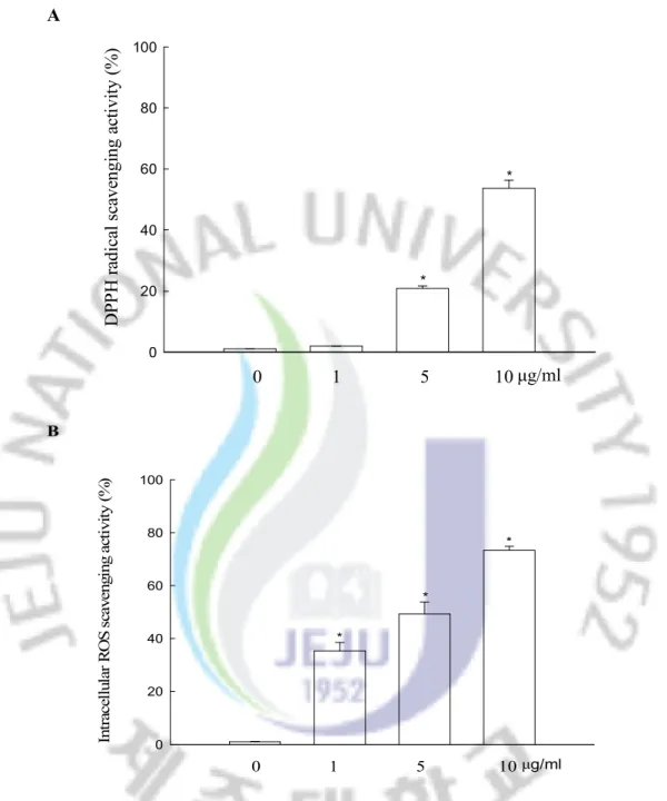
The scavenging ability of myricetin on DPPH radicals, and intracellular ROS in V79-4 cells was measured. With respect to DPPH radicals, myricetin was able to scavenge 21% at 5 mg/ml, and 54% at 10 mg/ml, respectively (Figure 2A). The intracellular ROS scavenging activity of myricetin was 35% at 1 mg/ml, 49% at 5 mg/ml, and 73% at 10 mg/ml, respectively (Figure 2B). Taken together, these results suggest that myricetin has reactive radical scavenging effects.
2 . Effect of myricetin on antioxidant enzymes
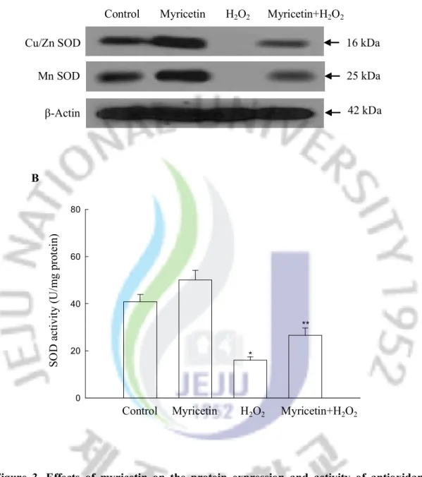
To investigate the effect of myricetin on protein expression of SOD, the Western blot analysis was performed. As shown in Figure 3A, myricetin increased the protein expression of both Cu/Zn SOD and Mn SOD. H2O2 treatment resulted in a decrease of both types of
SOD protein expressions; however, treatment with myricetin reversed this effect. To determine whether the effect of myricetin on SOD protein expression resulted in increased SOD activity, its activity was assessed. In fact, myricetin increased the activity of SOD, showing 50 U/mg protein at 10 mg/ml of myricetin compared to 40 U/mg protein in the control (Figure 3B). H2O2 treatment resulted in a decrease in SOD activity to 16 U/mg
A 0 20 40 60 80 100 0 1 5 10 D PP H r ad ic al s ca ve ng in g ac tiv ity ( % ) μg/ml * * B 0 20 40 60 80 100 0 1 5 10μg/ml In tr ac el lu la r R O S s ca ve ng in g ac ti vi ty ( % ) * * *
Figure 2. Effect of myricetin on DPPH radicals and intracellular ROS. (A) The amount of DPPH radicals was determined spectrophotometrically at 520 nm. (B) The intracellular ROS levels were detected using the DCF-DA method. The measurements were made in triplicate and values are expressed as the mean ± SEM. *Significantly different from control (p<0.05).
A
Control Myricetin H2O2 Myricetin+H2O2
Cu/Zn SOD Mn SOD β-Actin 25 kDa 42 kDa 16 kDa 0 20 40 60 80 B
Control Myricetin H2O2 Myricetin+H2O2
* ** SO D a ct iv ity ( U /m g pr ot ei n)
Figure 3. Effects of myricetin on the protein expression and activity of antioxidant enzymes. (A) Western blot analysis was performed using anti-Cu/Zn SOD, Mn SOD antibodies. (B) The enzyme activities are expressed as average enzyme unit per mg protein ± SEM. *Significantly different from control (p<0.05), and **significantly different from H2O2
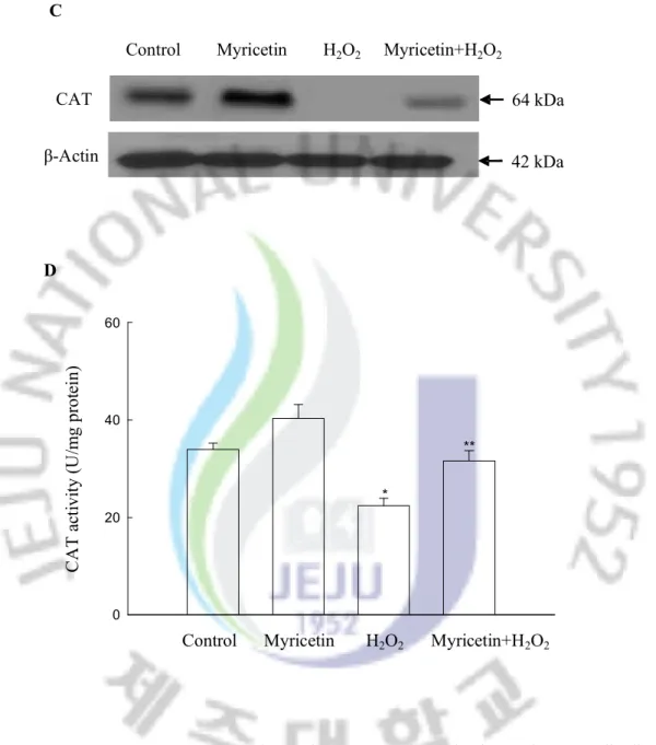
however, treatment with myricetin was able to restore this decrease (Figure 3C). Myricetin increased the activity of CAT, exhibiting 40 U/mg protein at 10 mg/ml of myricetin compared to 34 U/mg protein in the control (Figure 3D). Treated cells with H2O2 decreased the CAT
activity to 22 U/mg protein; however, treatment with myricetin restored the activity to a level of 32 U/mg. With respect to GPx, H2O2 treatment resulted in a decrease of GPx protein
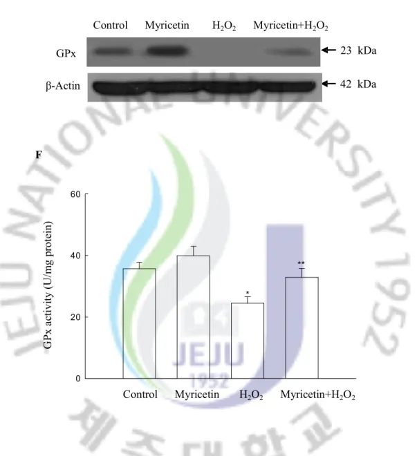
expressions; however, treatment with myricetin restored it (Figure 3E), a finding that is consistent to GPx protein activity (Figure 3F).
C
CAT
Control Myricetin H2O2 Myricetin+H2O2
64 kDa 42 kDa β-Actin 0 20 40 60 D C A T a ct iv ity ( U /m g pr ot ei n)
Control Myricetin H2O2 Myricetin+H2O2
*
**
Figure 3. Continued. (C) Western blot analysis was performed using anti-CAT antibodies. (D) The enzyme activities are expressed as average enzyme unit per mg protein ± SEM. *Significantly different from control (p<0.05), and **significantly different from H2O2
Control Myricetin H2O2 Myricetin+H2O2 GPx β-Actin 23 kDa E 42 kDa 0 20 40 60 F G Px a ct iv ity ( U /m g pr ot ei n) * **
Control Myricetin H2O2 Myricetin+H2O2
Figure 3. Continued. (E) Western blot analysis was performed using anti-GPx antibodies. (F) The enzyme activities are expressed as average enzyme unit per mg protein ± SEM. *Significantly different from control (p<0.05), and **significantly different from H2O2
3 . Effect of myricetin on cellular DNA damage induced by H2O2
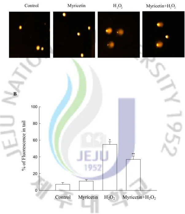
The treatment of cells with H2O2 increased the comet parameters of tail length and
percentage of DNA in the tails of the cells. Figures 4A and B illustrate the increase in the percentage of DNA in the tail upon H2O2 treatment. Treatment with myricetin decreased the
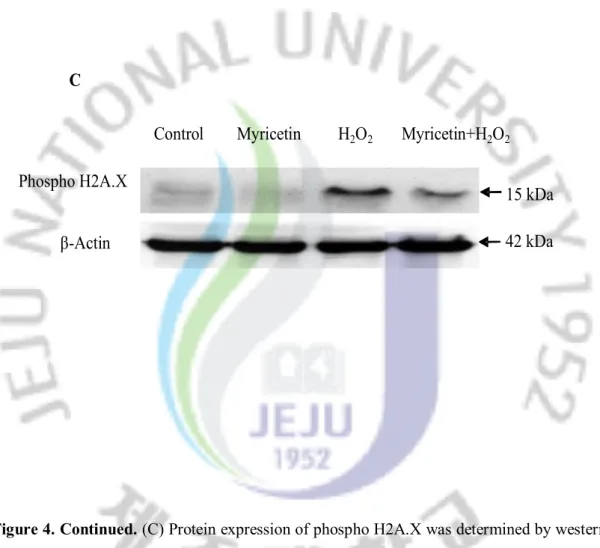
comet tail length and resulted in a decrease in percentage of DNA in the tail. The phosphorylation of nuclear histone H2A.X, a sensitive marker for breaks in double stranded DNA [40], increased in the H2O2-treated cells, as demonstrated by Western blot (Figure 4C).
However, the treatment of myricetin decreased the expression of phospho histone H2A.X, indicating a protective effect of myricetin on H2O2-induced DNA damage.
4 . Effect of myricetin on H2O2 induced lipid peroxidation
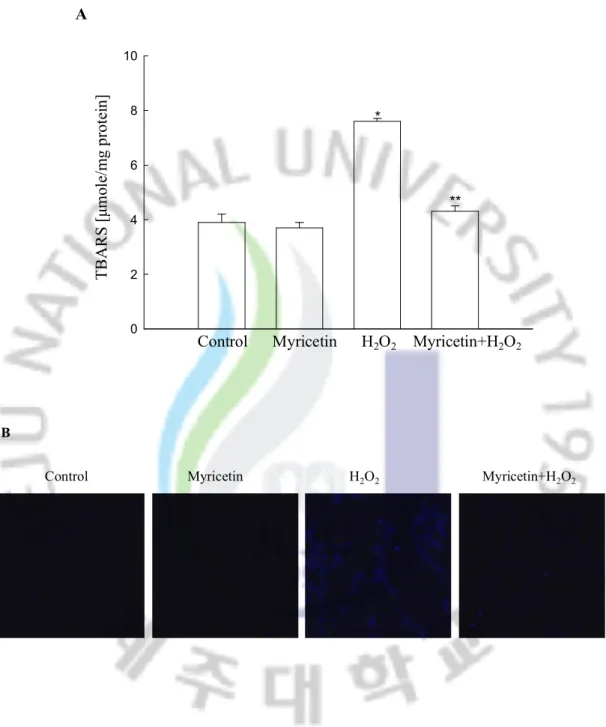
Cells treated with H2O2 exhibited an increase in the level of lipid peroxidation, as
monitored by the generation of TBARS. However, myricetin prevented this H2O2-induced
peroxidation of lipids as compared to H2O2 treated cells (Figure 5A). This pattern was also
confirmed by detection of DPPP fluorescence intensity. DPPP reacts with lipid hydroperoxides to produce a fluorescent product, DPPP oxide [41]. H2O2 treatment increased
the fluorescence intensity of DPPP and myricetin reduced it in H2O2-treated cells (Figure
B 0 20 40 60 80 100 % o f F lu or es ce nc e in ta il * **
Control Myricetin H2O2 Myricetin+H2O2
Figure 4. Effect of myricetin on DNA damage induced by H2O2 treatment. (A)
Representative images and (B) percentage of cellular DNA damage were detected using an alkaline comet assay.
Control Myricetin H 2 O 2 Myricetin+H 2 O 2
Phospho H2A.X
Control Myricetin H
2O
2Myricetin+H
2O
215 kDa
C
β-Actin
42 kDa
Figure 4. Continued. (C) Protein expression of phospho H2A.X was determined by western blot analysis.
0 2 4 6 8 10 A T B A R S [μ m ol e/ m g pr ot ei n]
Control Myricetin H2O2 Myricetin+H2O2
*
**
Control Myricetin H2O2 Myricetin+H2O2
B
Figure 5. Effect of myricetin on inhibition of lipid peroxidation induced by H2O2
treatment. (A) Lipid peroxidation was assayed by measuring the amount of TBARS. (B) Lipid peroxidation was detected using confocal microscopy after DPPP staining.
Ⅳ. DISCUSSION
In our system, we propose that the antioxidant effect of myricetin may involve two
mechanisms of action: (1) a direct scavenging effect on free radicals, as illustrated by the DPPH data, and (2) an indirect effect via the induction of antioxidant enzymes activities. Structurally, myricetin is 3¢,4-dihydroxy catechol in a B ring. The coplanarity of the molecule, and the presence of 2, 3 unsaturated and together an oxo functional group at position 4 in the C ring are the one recognized as important for antioxidant scavenging activity and therefore, giving myricetin protective properties against oxidative stress [42]. The cytoprotective mechanism of myricetin against oxidative stress was also investigated by assessing the status of various antioxidant enzymes including SOD, CAT, and GPx [43,44]. SOD represents the first line of defense against free radicals by dismutating toxic superoxide into the less reactive H2O2. In our study, the protein expressions of mitochondrial Mn SOD
and cytosolic Cu/Zn SOD were significantly decreased in H2O2 treated cells, leading to
corresponding decrease in SOD activity. Myricetin treatment recovered the decreased SOD protein expression and activity. CAT is also a major primary antioxidant defense component that works primary to catalyze the decomposition of H2O2 to water, sharing this function
with GPx. Therefore, both of these enzymes detoxify H2O2 derived from SOD activity. At
low concentration, H2O2 is the preferred substrate for GPx because it has low Km value
GPx were significantly decreased in H2O2 treated cells, resulting in corresponding decrease
in the activity of both enzymes. However, myricetin treatment recovered the decreased CAT and GPx protein expression and activity. Taken together we have shown that, myricetin can inhibit the adverse effects of oxidative stress and may protect the cellular environments from free radical damage by indicating the cellular antioxidant enzyme defense system, allowing for the repair of damaged DNA and lipid.
Ⅴ. REFERENCES
1 Halliwell B, Gutteridge JM. Free radicals, lipid peroxidation, and cell damage. Lancet 1984; 2:1095.
2 Halliwell B, Gutteridge JM. Oxygen toxicity, oxygen radicals, transition metals and disease. Biochem J 1984; 219:1-14.
3 Gutteridge JM, Halliwell B. Free radicals and antioxidants in the year 2000. A historical look to the future. Ann N Y Acad Sci 2000; 899:136-47.
4 Halliwell B, Gutteridge JM. Free radicals and antioxidant protection: mechanisms and significance in toxicology and disease. Hum Toxicol 1988; 7:7-13.
5 Sawada M. Neuroprotective and toxic changes in microglia in neurodegenerative disease. Parkinsonism Relat Disord 2009; 1:39-41.
6 Wakamatsu TH, Dogru M, Tsubota K. Tearful relations: oxidative stress, inflammation and eye diseases. Arq Bras Oftalmol 2008; 71:72-9.
7 Eleuteri E, Magno F, Gnemmi I, Carbone M, Colombo M, La Rocca G, et al. Role of oxidative and nitrosative stress biomarkers in chronic heart failure. Front Biosci 2009; 14:2230-7.
8 Mirshafiey A, Mohsenzadegan M. The role of reactive oxygen species in immunopathogenesis of rheumatoid arthritis. Iran J Allergy Asthma Immunol 2008; 7:195-202.
9 Hori M, Nishida K. Oxidative stress and left ventricular remodelling after myocardial infarction. Cardiovasc Res 2009; 81:457-64.
10 Du W, Adam Z, Rani R, Zhang X, Pang Q. Oxidative stress in Fanconi anemia hematopoiesis and disease progression. Antioxid Redox Signal 2008; 10:1909-21.
11 Roessner A, Kuester D, Malfertheiner P, Schneider-Stock R. Oxidative stress in ulcerativecolitis-associated carcinogenesis. Pathol Res Pract 2008; 204:511-24.
12 Steinmetz KA, Potter JD. Vegetables, fruit, and cancer. I. Epidemiology. Cancer Causes Control 1991; 2:325-57.
13 Grassi D, Desideri G, Croce G, Tiberti S, Aggio A, Ferri C. Flavonoids, vascular functionand cardiovascular protection. Curr Pharm Des 2009; 15:1072-84.
14 Mandel SA, Amit T, Weinreb O, Reznichenko L, Youdim MB. Simultaneous manipulation of multiple brain targets by green tea catechins: a potential neuroprotective strategy for Alzheimer and Parkinson diseases. CNS Neurosci Ther 2008; 4:352-65.
15 Lu MP, Wang R, Song X, Chibbar R, Wang X, Wu L, et al. Dietary soy isoflavones increase insulin secretion and prevent the development of diabetic cataracts in streptozotocin-induced diabetic rats. Nutr Res 2008; 28:464-71.
16 Richter M, Ebermann R, Marian B. Quercetin-induced apoptosis in colorectal tumor cells: possible role of EGF receptor signaling. Nutr Cancer 1999; 34:88-99.
17 Abdel-Raheem IT, Abdel-Ghany AA, Mohamed GA. Protective effect of quercetin against gentamicin-induced nephrotoxicity in rats. Biol Pharm Bull 2009; 32:61-7.
18 Zhang R, Kang KA, Piao MJ, Ko DO, Wang ZH, Chang WY, et al. Preventive effect of 7,8-dihydroxyflavone against oxidative stress induced genotoxicity. Biol Pharm Bull 2009; 32:166-71.
19 Piao MJ, Kang KA, Zhang R, Ko DO, Wang ZH, You HJ, et al. Hyperoside prevents oxidative damage induced by hydrogen peroxide in lung fibroblast cells via an antioxidant effect. Biochim Biophys Acta 2008; 1780:1448-57.
20 Middleton E Jr, Kandaswami C, Theoharides TC. The effects of plant flavonoids on mammalian cells: implications for inflammation, heart disease, and cancer. Pharmacol Rev 2000; 52:673-751.
21 Williams RJ, Spencer JP, Rice-Evans C. Flavonoids: antioxidants or signalling molecules? Free Radic Biol Med 2004; 36:838-49.
22 Shimmyo Y, Kihara T, Akaike A, Niidome T, Sugimoto H. Three distinct neuroprotective functions of myricetin against glutamate-induced neuronal cell death: involvement of direct inhibition of caspase-3. J Neurosci Res 2008; 86:1836-45.
23 Dajas F, Rivera F, Blasina F, Arredondo F, Echeverry C, Lafon L, et al. Cell culture protection and in vivo neuroprotective capacity of flavonoids. Neurotox Res 2003; 5:425-32. 24 Fawcett JR, Bordayo EZ, Jackson K, Liu H, Peterson J, Svitak A, et al. Inactivation of the human brain muscarinic acetylcholine receptor by oxidative damage catalyzedby a low molecular weight endogenous inhibitor from Alzheimer's brain is prevented by pyrophosphate analogs, bioflavonoids and other antioxidants. Brain Res 2002; 950:10-20.
25 Molina-Jiménez MF, Sánchez-Reus MI, Andres D, Cascales M, Benedi J. Neuroprotective effect of fraxetin and myricetin against rotenone-induced apoptosis in neuroblastoma cells. Brain Res 2004; 1009:9-16.
26 Molina-Jiménez MF, Sánchez-Reus MI, Cascales M, Andrés D, Benedí J. Effect of fraxetin on antioxidant defense and stress proteins in human neuroblastoma cell model of rotenone neurotoxicity. Comparative study with myricetin and N-acetylcysteine. Toxicol Appl Pharmacol 2005; 209:214-25.
27 Park HH, Lee S, Son HY, Park SB, Kim MS, Choi EJ, et al. Flavonoids inhibit histamine release and expression of proinflammatory cytokines in mast cells. Arch Pharm Res 2008; 31:1303-11.
28 Wang J, Mazza G. Effects of anthocyanins and other phenolic compounds on the production of tumor necrosis factor alpha in LPS/IFN-gamma-activated RAW 264.7 macrophages. J Agric Food Chem 2002; 50:4183-9.
29 Alexandrakis M, Letourneau R, Kempuraj D, Kandere-Grzybowska K, Huang M, Christodoulou S, et al. Flavones inhibit proliferation and increase mediator content in human leukemic mast cells (HMC-1). Eur J Haematol 2003; 71:448-54.
30 Hayder N, Bouhlel I, Skandrani I, Kadri M, Steiman R, Guiraud P, et al. In vitro antioxidant and antigenotoxic potentials of myricetin-3-o-galactoside and myricetin-3-o-rhamnoside from Myrtus communis: modulation of expression of genes involved in cell defence system using cDNA microarray. Toxicol In Vitro 2008; 22:567-81.
31 Duthie SJ, Dobson VL. Dietary flavonoids protect human colonocyte DNA from oxidative attack in vitro. Eur J Nutr 1999; 38:28-34.
32 Lo SF, Nalawade SM, Mulabagal V, Matthew S, Chen CL, Kuo CL, et al. In Vitro propagation by asymbiotic seed germination and 1,1-Diphenyl-2-picrylhydrazyl (DPPH) radical scavenging activity studies of tissue culture raised plants of three medicinally important species of Dendrobium. Biol Pharm Bull 2004; 27:731-5.
33 Rosenkranz AR, Schmaldienst S, Stuhlmeier KM, Chen W, Knapp W, Zlabinger GJ. A microplate assay for the detection of oxidative products using 2',7'-dichlorofluorescin-diacetate. J Immunol Meth 1992; 156:39-45.
34 Misra HP, Fridovich I. The role of superoxide anion in the autoxidation of epinephrine and a simple assay for superoxide dismutase. J Biol Chem 1972; 247:3170-5.
35 Carrillo MC, Kanai S, Nokubo M, Kitani K. (-) deprenyl induces activities of both superoxide dismutase and catalase but not of glutathione peroxidase in the striatum of young male rats. Life Sci 1991; 48:517-521.
36 Paglia DE, Valentine, WN. Studies on the quantitative and qualitative characterization of erythrocyte glutathione peroxidase. J Lab Clin Med 1967; 70:158–69.
37 Rajagopalan R, Ranjan S, Nair C. Effect of vinblastine sulfate on gamma-radiation-induced DNA single-strand breaks in murine tissues. Mutat Res 2003; 536:15-25.
39 Ohkawa H, Ohishi N, Yagi K. Assay for lipid peroxides in animal tissues by thiobarbituric acid reaction. Anal Biochem 1979; 95:351-8.
40 Rogakou EP, Pilch DR, Orr AH, Ivanova VS, Bonner WM. DNA double-stranded breaks induce histone H2A.X phosphorylation on serine 139, J. Biol. Chem 1988; 273:5858–68.
41 Okimotoa Y, Watanabea A, Nikia E, Yamashitab T, Noguchia N. A novel fluorescent probe diphenyl-1-pyrenylphosphine to follow lipid peroxidation in cell membranes. FEBS Lett 2000; 474:137–40.
42 Rice-Evans C. Flavonoid antioxidants. Curr Med Chem 2001; 8:797-807.
43 Liochev SI, Fridovich I. The effects of superoxide dismutase on H2O2 formation. Free
Radic Biol Med 2007; 42:1465-9.
44 Goldstone AB, Liochev SI, Fridovich I. Inactivation of copper, zinc superoxide dismutase by H2O2: mechanism of protection. Free Radic Biol Med 2006; 41:1860-3.
45 Yu BP. Cellular defenses against damage from reactive oxygen species. Physiol Rev 1994; 74:139-62.
VI. ABSTRACT IN KOREAN
이 실험은 myricetin 의 세포에서의 산화적인 손상으로부터 보호효과를 ROS 소거능력과 항 산화 효소 활성 변화를 통하여 평가하였다. Myricetin 은 DPPH radicals 소거능력과 세포 내에서의 ROS 소거능력을 확인함으로써 자체의 ROS 소거능 및 세포 내에서의 ROS 소거능을 확인할 수 있었다. 그리고 myricetin 은 세포 내의 항 산화 효소인 SOD, Catalase, GPx 와 같은 항 산화효소의 활성과 발현을 H2O2 에 의한 항 산화 효소 활성과 발현의 감소로부터
회복하였다. 그리고 H2O2에 의해 세포의 DNA 손상과 지질의 산화를 일으켰으며,
myricetin 이 Comet assay 와 DNA 손상 marker 인 H2A.X 의 발현을 통하여 DNA 손상보호를 확인하였고, DPPP 와 MDA assay 를 통하여 세포의 지질 과산화로부터 보호 효과가 있음을 확인할 수 있었다. 이러한 결과로부터 myricetin 이 H2O2 에
의한 세포손상으로부터 ROS 생성의 방지와 항 산화 효소 활성 증가를 통한
.
Ⅶ 감사의 글
짧다면 짧은 2 년의 석사 학위 과정을 마쳤습니다. 이제 곧 한국을 떠나야 할 즈음에 내 가슴은 뜨거운 감격과 무한한 감개로 샘솟음을 느끼고 있습니다. 2 년의 학습 생활은 충실하면서도 아름다웠지만 모든 일이 순풍에 돛 단 격은 아니었습니다. 생활에서 여러 가지 어려움을 겪게 되었지만 매 번 삶의 진창에 빠져 허우적대고 있을 때마다 주변의 사람들은 마치 암흑 속에서 밝혀지는 등불처럼, 혼돈 속에서 가려내는 맑음처럼 헤쳐나올 수 있게끔 수많은 도움을 주었습니다. 이 모든 것은 우선 저의 지도 교수님이신 현진원 교수님의 배려와 갈라 놓을 수 없습니다. 언제나 어머니처럼 자상한 보살핌과 끊임없는 가르침으로 풍부한 전문 지식을 습득하게 하였고 진정한 사람의 도리를 깨닫게 하였습니다. 교수님 그 동안 참으로 수고가 많으셨습니다. 진심으로 감사 드립니다. 그리고 저에게 가르침과 도움을 주신 고영상 교수님, 강희경 교수님을 비롯한 여러 교수님들께도 모두모두 감사 드립니다. 이 분들은 제주대에서의 2 년이란 세월에서 저에게 있어서 가장 존경하는 선생님이셨으며, 가장 자상한 가장이기도 하셨습니다. 실험실의 형님 누나들, 특히 경아 누나, 장예 누나, 미경 누나, 동옥 형님,기천 형님은 생활과 학습에서 사심없이 저를 도와주셨고 저 또한 형님 누나들을 한 가족처럼 여기면서 지냈습니다. 형님 누나들 앞으로도 나날이 발전하기를 바라고 천리만리 떨어져 있어도 꼭 연락을 자주 하시길 바랍니다. 너무너무 보고 싶을 테니깐요. 다시 한번 감사 드립니다. 중국에 있는 저의 부모님, 그리고 친구들, 당신들에 대한 나의 그리움은 한 시각도 멈추지 않았습니다. 이번에 순조롭게 졸업을 하게 된 것도 나를 지지해준 최고의 보답이라고 생각합니다. 그리고 타 실험실에 있는 선배님과 친구들, 당신들의 도움이 없었더라면 저 오늘 여기까지 오지도 못했을 것입니다. 항상 행운이 동반하시길 바랍니다. 끝으로, 몸을 담고 있는 아름다운 이 섬에게 감사 드립니다. 아름다운 섬, 아름다운 사람들, 아름다운 시작이 있으면 아름다운 끝도 있을 것입니다. “안녕히!” 라는 말 조차도 잔인하다고 느껴지지만 현실은 또 반드시 새로운 여정을 시작해야 합니다. 감사합니다! 아름다운 제주; 감사합니다! 아름다운 한국. 당신들을 사랑합니다! 우리 꼭 다시 만날 수 있겠죠?