서 론
근상피종이 최초로 기술된 것은 1943년으로 침샘에서 발생한 것 이었다.1) 근상피세포는 일반적으로 샘상피 조직에서 발견되며 췌 장을 제외한 땀샘, 젖샘 및 침샘의 외분비선에 위치하는 특징을 가진다. 그러므로 근상피종은 주로 침샘의 종양으로 발병하며 이 외에도 유방,2) 코인두,3) 후두,4) 피부,5) 폐,6) 골,7,8) 연부 조직11-18) 등 에서도 발생된 것이 보고되고 있다. 연부 조직에 발생하는 14.4% 로 매우 드물다.11) 연부 조직 악성 근상피종은 굉장히 희귀한 질환으로 주로 증례 보고로 발표되고 있다. 처음으로 보고된 원발성 연부 조직 악성사지에 발생한 연부 조직 악성 근상피종
Soft Tissue Malignant Myoepithelioma in the Extremities
공창배 • 이정욱 • 고재수* • 송원석 • 조완형 • 전대근 • 이수용
원자력병원 정형외과학교실, *병리학교실 목적: 사지에 발생한 연부 조직 악성 근상피종의 진단, 치료 및 예후에 대하여 알아보고자 하였다. 대상 및 방법: 2008년 1월부터 2014년 10월까지 연부 조직 악성 근상피종으로 진단받고 본원에서 치료받은 6명의 환자를 대상으로 하였다. 2명의 환자는 타원에서 조직 검사 없이 무계획 절제술(unplanned excision)을 시행받은 이후 본원으로 전원되었으며, 나머지 4명의 환자는 모 두 본원에서 조직 검사 및 광범위 절제술을 시행받았다. 결과: 평균 연령은 41세(33-54)였고, 남자가 3예, 여자가 3예였으며 평균 추시 기간은 28개월(9-45)이었다. 1명의 환자에서만 술 후 항암요 법을 시행하였다. 연구 대상 환자 중 4명만이 악성 연부 조직 종양 의심하에 조직 검사 후 광범위 절제술을 시행하였다. 본원에서 광범위 절제 술을 시행한 환자 4명은 모두 절제연에 종양 세포가 관찰되지 않았다. 무계획 절제술 이후 전원된 환자 2명 모두 본원에서 재절제술 시행받았 으며, 이중 한명은 재절제술 병리 조직에서 잔존하는 종양 세포는 관찰되지 않았다. 6명의 환자 모두에게서 수술 후 평균 6개월(3-29)에 국소 재발 소견이 관찰되었고 4명의 환자에서는 수술 후 평균 7개월(3-14)에 원격 전이도 관찰되었다. 원격 전이가 발생한 4명의 환자는 모두 질 병으로 인하여 사망하였고, 국소 재발만 발생한 2명의 환자중 1명은 환자는 재발하여 추시 관찰 중이며 나머지 1명의 환자는 재수술후 2년간 재발이나 전이 없이 경과 관찰 중이다. 결론: 연부 조직 악성 근상피종은 극히 드물게 발병하는 질환으로 재발과 전이를 잘 하는 공격적인 악성 연부 조직 종양으로 적절한 치료법으 로는 광범위 절제술이 권장되며, 국소 재발을 줄이기 위해서는 악성 연부 조직 종양일 가능성을 염두에 두고 반드시 술 전 조직 검사를 하여 악성임을 확인하고 이후 계획된 광범위 절제술을 시행하는 것이 중요하다고 생각된다. 색인단어: 악성 근상피종, 연부 조직, 사지 근상피종은 1995년에 Burke 등에 의해 보고된 후복막 종양이었 다.9) 이후 1997년 Kilpatrick 등이 19예의 연부 조직 근상피종 환자 에 대해 보고하였는데 이중 2예에서 전이가 일어났다고 하였다.18) 2002년 악성 근상피종은 세계보건기구(WHO) 분류에 등재되었고 2003년 Hornick 등은 101예의 근상피종을 연구하여 악성 근상피 종 진단 기준을 제시하였다.11) 저자들은 사지에 발생한 연부 조직 악성 근상피종 환자 6예를 진단하고 치료한 경험이 있기에 이 환자들에 대한 치료 결과를 문 헌 고찰과 함께 보고하자고 한다.대상 및 방법
본 연구는 2008년부터 2014년 10월까지 본원에서 사지의 연부 조 직 악성 근상피종으로 확진된 후 치료 받은 6명의 환자를 대상으 로 하였다. 2명의 환자는 타원에서 조직 검사 없이 무계획 절제술 (unplanned excision)을 시행받은 이후 본원으로 전원되었으며, 나 머지 4명의 환자는 모두 본원에서 조직 검사 및 광범위 절제술을 접수일 2014년 10월 30일 심사수정일 2014년 11월 26일 게재확정일 2014년 11월 30일 교신저자 조완형 서울시 노원구 노원로 75, 원자력병원 정형외과 TEL 02-970-1243, FAX 02-970-2403 E-mail chowanda@naver.comCopyrights © 2014 by The Korean Bone and Joint Tumor Society
“This is an Open Access article distributed under the terms of the Creative Commons Attribution Non-Commercial License (http://creativecommons.org/licenses/by-nc/3.0/) which permits unrestricted non-commercial use, distribution, and reproduction in any medium, provided the original work is properly cited.”
시행받았다(Table 1). 이들의 임상 기록, 단순 방사선 사진 및 전산 화 단층촬영, 자기공명영상, 양전자 단층 촬영(Positron emission tomography, PET), 골스캔, 병리 소견을 후향적으로 분석하였다. 환 자의 임상 양상, 종양의 해부학적 위치, 크기, 치료 방법, 수술적 절 제연과 병리학적 등급, 국소 재발, 원격 전이 여부를 확인하였다.
결 과
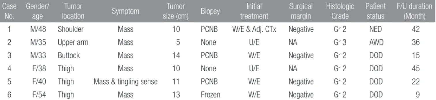
남자가 3예, 여자가 3예였으며, 평균 연령은 41세(33-54)였다. 증 상 발현후 본원에서 확진을 받기까지의 기간은 평균 7개월(1-48) 이었으며, 평균 추시 기간은 28개월(9-45)이었다. 원발 병변 부위 는 대퇴부 3명, 견갑부 1명, 상완부 1명, 둔부 1명이었다. 초기 증 상은 통증을 동반하지 않은 종괴가 5명, 저린감을 동반한 종괴가 1명이었으며, 조직학적으로 5명의 환자가 2등급, 1명의 환자가 3 등급으로 확인되었다. 연구 대상 환자 중 4명만이 악성 연부 조직 종양 의심하에 조직 검사 후 광범위 절제술을 시행하였다(Fig. 1). 본원에서 광범위 절제술을 시행받은 환자 4명은 모두 절제연에 종양 세포가 관찰되지 않았다(Table 1). 무계획 절제술 이후 전원Figure 1. 40-year-old woman suffered from tingling sensation for two weeks (Case 5). (A) Initial T2-weighted axial MR image shows a large deep-seated soft tissue mass at right posterior thigh. (B) T2-weighted axial MR image which was taken 14 months after the index operation shows a local recurrence. (C) The tumor cells show variegated appearance from chondromyxoid to spindle cell morphology with hypercellularity and nuclear atypism (H&E stain, ×100). (D) High power field view reveals atypical tumor cells showing prominent nucleoli and frequent mitotic figures (H&E stain, ×400). (E) Immunostaining for keratin (PAN-K) is positive (×100).
A B
C D E
Table 1. Demographic and Clinico-pathologic Data
Case No. Gender/ age Tumor location Symptom Tumor size (cm) Biopsy Initial treatment Surgical margin Histologic Grade Patient status F/U duration (Month)
1 M/48 Shoulder Mass 10 PCNB W/E & Adj. CTx Negative Gr 2 NED 42
2 M/35 Upper arm Mass 5 None U/E NA Gr 3 AWD 36
3 M/33 Buttock Mass 14 PCNB W/E Negative Gr 2 DOD 15
4 F/38 Thigh Mass 10 None U/E NA Gr 2 DOD 45
5 F/40 Thigh Mass & tingling sense 11 PCNB W/E Negative Gr 2 DOD 22
6 F/54 Thigh Mass 13 Frozen W/E Negative Gr 2 DOD 9
PCNB, percutaneous core needle biopsy; W/E, wide excision; U/E, unplanned excision; Adj. CTx, adjuvant chemotherapy; NED, no evidence of disease; AWD, alive with disease; DOD, dead of disease; NA, not assessed.
된 환자 2명은 본원에서 재절제술 시행받았으며, 이중 한 명은 재 절제술 병리 조직에서 잔존하는 종양 세포는 관찰되지 않았다(증 례 2). 국소 재발은 6명 환자 모두에서 수술 후 평균 6개월(3-29)에 발 생하였다(Table 2). 재발 병변에 대해서는 광범위 절제술을 기본 으로 하였으며, 재발 병변에 대한 수술적 치료 이후 방사선 치료 를 같이 시행한 경우가 1명이었다(증례 1). 이 환자는 유일하게 수 술 후 항암 치료를 시행한 환자로 항암 치료 2달 후 국소 재발 관 찰되었고, 재절제술 시행한 절제연에서 종양 세포 관찰되어 수술 후 방사선 치료를 시행하였는데, 이후 다시 국소 재발 발생하였 다. 두번째로 재발한 병변에 대해 또 다른 광범위 절제술 후 환자 는 현재 2년간 재발이나 원격 전이 없이 추시 관찰중이다. 증례 2
Table 2. Treatments for Local Recurrence and/or Metastasis
Case No.
Time to relapse from index surgery
(months)
Treatment for local recurrence
Surgical margin of operation for local
recurrence
Time to metastasis from index surgery
(months)
Metastasis location Treatment for metastasis
1 20 W/E & postop. RT Positive None -
-2 29 W/E Negative None -
-3 6 None - 6 Lung
-4 5 W/E Negative 14 Abdomen, chest wall, pelvis Excision
5 14 Preop. CTx & W/E Negative 3 Lung Wedge resection
6 3 W/E Negative 5 Lung, Brain CTx, brain RT
W/E, wide excision; RT, radiation therapy; CTx, chemotherapy.
Figure 2. 35-year-old man complained about his right arm mass which is developed 3 months ago (Case 2). (A) Initial T2-weighted axial MR image shows a soft tissue mass at right posterior upper arm. (B) After unplanned excision, T2-weighted axial MR image which was taken after referral shows a localized mass lesion. Re-excision was performed and the pathologic specimen revealed that there was no residual tumor cell. However, local recurrence developed at 29 months after re-excision. (C) High power field view reveals nuclear atypism and mitosis (H&E stain, ×400). (D) Immunostaining for keratin (PAN-K) is positive (×200). (E) Immunostaining for smooth muscle actin is also positive (×100).
A B
는 외부에서 무계획적 절제술 이후에 내원하여 재절제술을 시행 받은 환자로 재절제술 병리 조직에서 잔존하는 종양 세포는 관찰 되지 않았다. 하지만, 재절제술을 시행받은지 29개월후에 다시 국 소 재발 소견 확인되어 재수술 시행하였고, 최근 국소 재발이 다 시 확인되어 광범위 절제술을 받을 계획이다(Fig. 2). 원격 전이는 4명의 환자에서 수술 후 평균 7개월(3-14)에 관찰 되었다(Table 2). 전이 병변의 경우 수술적 치료가 가능한 경우 수 술을 시행하였고, 수술이 어려운 경우 방사선 치료를 시행하였다. 전이 부위는 폐 전이만 있는 경우가 2명, 폐 전이와 뇌 전이가 동 반된 경우가 1명, 다발성 전이가 1명이었다. 원격 전이가 발생한 4 명의 환자 모두 질병으로 인하여 사망하였고 첫 수술 후 질병으로 인한 사망까지의 평균 기간은 23개월(9-45)이었다.
고 찰
악성 근상피종은 타액선 종양의 1% 이하를 차지하며, 중등도 혹은 고도의 악성을 보이는 드문 종양으로 60-70%에서 이하선을 침범 한다.19) 이 종양은 이전에 존재한 근상피종이나 다형성 선종에서 발생하고 드물게 de novo로도 발생하나, de novo로 발생하는 경우 악성도가 더 높은 것으로 알려져 있다.20) 남녀간의 발병 빈도의 차 이는 없으며 발병 시기는 넓은 분포를 보이지만 삼십대와 오십대 에 특히 발병률이 높다.10,11) 대개 사지와 지대(limb girdle)에 발생하 며 관절을 침범한 경우는 없다고 한다. 증상은 주로 통증을 동반하 지 않은 종괴이며 종괴의 크기는 평균 4.7 cm (1-20)이다.10-12) 본 연구 결과 사지에 발생한 악성 근상피종의 국소 재발율은 100%, 원격 전이율은 67%로 확인되었다. 증례수가 적어서 이 수 치를 일반화하기는 어렵지만, 악성 연부 조직 종양중에서도 상당 히 공격적인 종양이라 판단된다. Hornick 등은 전 세계 병리 의사 들에게 의뢰하여 근상피종 환자 101명의 결과를 보고한 바 있으 며, 61명은 양성, 40명은 악성 근상피종 이었고, 악성 근상피종 환 자중 임상 결과가 확인 가능한 환자는 31명이었다.11) 이들중 13명 의 환자에서 국소 재발이, 10명의 환자에서 원격 전이가 관찰되어 국소 재발율은 42%, 원격 전이율은 32%이었다. 같은 그룹에서 4 년뒤 다시 전 세계 병리 의사들에게 의뢰하여 소아 악성 근상피종 환자 29명에 대한 임상 결과를 보고하였다.12) 추시 관찰이 가능하 였던 23명의 환자중 9명의 환자에서 국소 재발이, 12명의 환자에 서 원격 전이가 발생하여 국소 재발율은 53%, 원격 전이율은 43% 라 하였다. 본원에서 치료받은 환자들의 예후가 이 두 논문의 결 과에 비해 더 안좋은 부분에 대해 명쾌한 설명을 하기는 어렵지 만, 종양 크기가 크고 병리 등급이 높다는 것과 무계획 절제술 환 자군이 포함되어 있다는 점 등을 생각해 볼 수 있겠다. 육안 소견상으로 악성 근상피종은 결정형이나 소엽형이며 흰 색, 노란색, 황갈색을 띄고 점액성이나 아교성으로 관찰된다.12) 현 미경적 소견상으로는 주로 충실성으로 성장하며 낭성 변화 혹 은 괴사를 동반하기도 한다. 구성하는 세포는 근상피종에서 볼수 있는 세포들로서, 유상피 세포, 형질세포양 세포, 방추형 세포 그 리고 투명 세포로 나눌 수 있다.21) 악성 근상피종의 진단에 필요한 면역조직화학적 표지자는 cytokeratin, vimentin, MSA (muscle specific actin), SMA (smooth muscle actin), EMA (epithelial mem-brane antigen), S-100 protein, GFAP (glial fibrillary acidic protein), calponin 및 desmin 등이다. 악성 근상피종에서 이들 표지자에 대 한 양성율은 보고자마다 차이가 있지만 cytokeratin (AE1:AE3) 90-100%, cytokeratin (CAM5.2) 90%, EMA 21-100%, SMA 50-80%, MSA 50-80%, desmin 10%, calponin 75%, vimentin 100%, S-100 protein 100%, GFAP 50%로 보고되고 있다.10-12) 새로운 표지자로 p63, CK14가 보고되고 있으나 양성율이 20-30%로 민감도가 낮아 2차 표지자로서만 유용성이 있다고 한다.10-12,16) 침샘에서 발생한 악성 근상피종의 경우 침윤성 성장 방식이 악 성 진단의 최고의 예측인자이며 세포학적 비정형성은 악성 근상 피종의 진단에 필요하지 않은 것으로 알려져 있는 반면 연부 조 직 근상피종은 절반 가까이 침윤성 경계를 보이며 이러한 침윤 성 성장 방식이 임상적인 종양의 예후와 관련이 없다고 한다.11) Hornick 등에 따르면 적어도 중등도 이상의 세포학적 비정형성 이 있어야 국소 재발이나 원격 전이가 보고 된다고 하여 중등 도 이상의 세포학적 비정형성이 연부조직 악성 근상피종의 유 일한 악성 진단의 지표라 기술하였다.11) 연부조직 악성 근상피종 의 감별 진단으로는 고등급 골외성 점액성 연골육종(High-grade extraskeletal myxoid chondrosarcoma), 상피양 악성 말초 신경초종 (Epitheloid MPNST), 근위형 상피양 육종(Proximal-type epitheloid sarcoma) 등이며, 앞서 기술한 종양 표지자를 이용하여 감별 진단 한다.8,11,12,14,16) 연부 조직에 발생한 악성 근상피종의 적절한 치료법으로는 광 범위 절제술이 권장되며, 항암 요법이나 방사선 치료의 효과는 아 직 불확실하다.10) Gleason 등은 국소 재발율이 변연부 절제술에서 는 80%, 광범위 절제술에서는 17%로 절제연이 국소 재발과 유의 한 상관 관계가 있다고 하였는데,11) 국소 재발을 줄이기 위해서는 악성 연부 조직 종양일 가능성을 염두에 두고 반드시 술 전 조직 검사를 하여 악성임을 확인하고, 계획된 광범위 절제술을 시행하 는 것이 중요하다고 생각된다.
결 론
연부 조직 악성 근상피종은 극히 드물게 발병하는 질환으로 재발 과 전이를 잘 하는 공격적인 악성 연부 조직 종양으로 적절한 치 료법으로는 광범위 절제술이 권장되며, 국소 재발을 줄이기 위해 서는 악성 연부 조직 종양일 가능성을 염두에 두고 반드시 술 전 조직 검사를 하여 악성임을 확인하고 이후 계획된 광범위 절제술 을 시행하는 것이 중요하다고 생각된다. 항암 치료 및 방사선 치료의 효과는 아직 밝혀진 바 없으며 이에 대한 추가적인 임상적 연구가 필요하다.
참고문헌
1. Sheldon W. So-called mixed tumors of the salivary glands. Arch Pathol. 1943;35:1-20.
2. Bigotti G, Di Giorgio CG. Myoepithelioma of the breast: his-tologic, immunologic, and electromicroscopic appearance. J Surg Oncol. 1986;32:58-64.
3. Tuncel U, Ergul G, Ozlugedik S, Unal A. Myoepithelial car-cinoma in the nasopharynx: an unusual localization. Yonsei Med J. 2004;45:161-5.
4. Martínez-Madrigal F, Santiago Payán H, Meneses A, Domín-guez Malagón H, Rojas ME. Plasmacytoid myoepithelioma of the laryngeal region: a case report. Hum Pathol. 1995;26:802-4.
5. Hornick JL, Fletcher CD. Cutaneous myoepithelioma: a clini-copathologic and immunohistochemical study of 14 cases. Hum Pathol. 2004;35:14-24.
6. Veeramachaneni R, Gulick J, Halldorsson AO, Van TT, Zhang PL, Herrera GA. Benign myoepithelioma of the lung: a case report and review of the literature. Arch Pathol Lab Med. 2001;125:1494-6.
7. Alberghini M, Pasquinelli G, Zanella L, et al. Primary malig-nant myoepithelioma of the distal femur. APMIS. 2007;115: 376-80.
8. Park JS, Ryu KN, Han CS, Park YK. Malignant myoepithe-lioma of the humerus with a satellite lesion: a case report and literature review. Br J Radiol. 2010;83:e161-4.
9. Burke T, Sahin A, Johnson DE, Ordóñez NG, Mackay B. Myo-epithelioma of the retroperitoneum. Ultrastruct Pathol. 1995; 19:269-74.
10. Lee JR, Georgi DE, Wang BY. Malignant myoepithelial tumor of soft tissue: a report of two cases of the lower extremity and a review of the literature. Ann Diagn Pathol. 2007;11:190-8. 11. Hornick JL, Fletcher CD. Myoepithelial tumors of soft tissue:
a clinicopathologic and immunohistochemical study of 101
cases with evaluation of prognostic parameters. Am J Surg Pathol. 2003;27:1183-96.
12. Gleason BC, Fletcher CD. Myoepithelial carcinoma of soft tis-sue in children: an aggressive neoplasm analyzed in a series of 29 cases. Am J Surg Pathol. 2007;31:1813-24.
13. Mahdi Y, Zouaidia F, Zouhair A, et al. Combined myoepithe-lial carcinoma and myoepithelioma in soft tissue: a case report and review of the literature. J Med Case Rep. 2014;8:317. 14. Harada O, Ota H, Nakayama J. Malignant myoepithelioma
(myoepithelial carcinoma) of soft tissue. Pathol Int. 2005;55: 510-3.
15. Combalía A, Marco V, Seijas R, Domínguez R. Rare presenta-tion of a soft-tissue myoepithelial carcinoma. J Orthop Sci. 2013. [Epub ahead of print]
16. Neto AG, Pineda-Daboin K, Luna MA. Myoepithelioma of the soft tissue of the head and neck: a case report and review of the literature. Head Neck. 2004;26:470-3.
17. Rastrelli M, Passuello N, Cecchin D, Basso U, Tosi AL, Rossi CR. Metastatic malignant soft tissue myoepithelioma: a case report showing complete response after locoregional and sys-temic therapy. J Surg Case Rep. 2013;2013. pii: rjt109.
18. Kilpatrick SE, Hitchcock MG, Kraus MD, Calonje E, Fletcher CD. Mixed tumors and myoepitheliomas of soft tissue: a clini-copathologic study of 19 cases with a unifying concept. Am J Surg Pathol. 1997;21:13-22.
19. Nagao T, Sugano I, Ishida Y, et al. Salivary gland malignant myoepithelioma: a clinicopathologic and immunohistochemi-cal study of ten cases. Cancer. 1998;83:1292-9.
20. Bombí JA, Alós L, Rey MJ, et al. Myoepithelial carcinoma aris-ing in a benign myoepithelioma: immunohistochemical, ul-trastructural, and flow-cytometrical study. Ultrastruct Pathol. 1996;20:145-54.
21. Savera AT, Zarbo RJ. Defining the role of myoepithelium in salivary gland neoplasia. Adv Anat Pathol. 2004;11:69-85. 22. Noronha V, Cooper DL, Higgins SA, Murren JR, Kluger HM.
Metastatic myoepithelial carcinoma of the vulva treated with carboplatin and paclitaxel. Lancet Oncol. 2006;7:270-1.
Soft Tissue Malignant Myoepithelioma in the Extremities
Chang-Bae Kong, Jung-Wook Lee, Jae-Soo Koh*, Won Seok Song,
Wan Hyeong Cho, Dae-Geun Jeon, and Soo-Yong Lee
Departments of Orthopedic Surgery and *Pathology, Korea Cancer Center Hospital, Seoul, Korea
Purpose: We report the diagnosis, treatment outcomes and prognosis of the patients with soft tissue malignant myoepithelioma in the extremities.
Materials and Methods: We retrospectively reviewed 6 patients with soft tissue malignant myoepithelioma in the extremities who were treated at our institution between 2008 and 2014. Two patients received unplanned excision at another hospital and remaining 4 patients underwent the biopsy procedures and received wide excision at our hospital.
Results: There were 3 men and 3 women with mean age of 41 (33−54) years. The average follow up was 28 (9−45) months. Among the 6 patients, only 4 patients underwent biopsy procedures under the impression of malignant soft tissue sarcoma. Surgical margins for these 4 patients were negative. Two patients who had unplanned excision received another re-excision and one of them showed no residual tumor in the resected specimen. Local recurrences were developed in all patients and distant metastasis in 4 patients. All 4 patients who developed distant metastasis died due to disease progression. Among the 2 patients who developed local recurrence only, one patient has another local recurrence after re-operation and remaining one patient is no evidence of disease for 2 years after resection of locally recurred mass.
Conclusion: Soft tissue malignant myoepithelioma in the extremities is a rare disease and shows an aggressive behavior. Appropri-ate biopsy under the impression of soft tissue malignancy is necessary and complete surgical resection with wide margins is the recommended treatment of choice.
Key words: malignant myoepithelioma, soft tissue, extremities
Received October 30, 2014 Revised November 26, 2014 Accepted November 30, 2014 Correspondence to: Wan Hyeong Cho
Department of Orthopedic Surgery, Korea Cancer Center Hospital, 75, Nowon-ro, Nowon-gu, Seoul 139-706, Korea
TEL: +82-2-970-1243 FAX: +82-2-970-2403 E-mail: chowanda@naver.com
Copyrights © 2014 by The Korean Bone and Joint Tumor Society
“This is an Open Access article distributed under the terms of the Creative Commons Attribution Non-Commercial License (http://creativecommons.org/licenses/by-nc/3.0/) which permits unrestricted non-commercial use, distribution, and reproduction in any medium, provided the original work is properly cited.”