Expression of HCNl subunits is age-dependently regulated in developing hippocampus of rats
Yoon-SiI Yang
,
Moon-Seok Kang,
Seon-Hee Kim,
Su-Yong Eun,
Sung-Cherl Jung Departmenl01PhySÎology‘80h∞
I 01Medicine‘Jeju NalionalUniversity‘Jeju.KoreaAbstract
Hyperpolarization-activated,cyclic nucleotide-gated cation channels (HCN) encode hyperþolarization-activated calion current (ι),playing regulatory roles in resting membrane properties. synaplic integration,and intrinsic ex이tabilities of hippocampal neurons. Expression patterns 01 HCN sublypes seem 10 be Quietty variable 10 excital0ry conditions 01 neurons,even though it has been previously observed that expression levels 01 HCN1,2 and 4 are differentially re9u
’
ated during developmental period In the presenl study. we investigaled expression patterns 01 HCNl during an ear1y devetopmental period within postnatal 3 weeks by using Western btol analysis. Inconsistenl with previous reperts,HCNl 'expression was highesl in neonatal and initial postnatal perlods,while neurons in poslnalal third week revealed lower levels. This result indicates that HCNl proleins are age-dependently decreased during firsl postnatal 3 weeks. This finding suggesls that CA1 neurons of early developmental hippocampi may have specific neuroprotective mechanisms correlated with HCNl subunit against hyperexcilabilities or excil이。xic damages. (J Med Ufe 8012012;9:64-63)Key Words : j
,
Channel,HCNl,hippecampus,excitotoxici앙Introduction
It is well known that expression levels and gating properties of ion channels such as voltage-dependent K'’
Na’,and hyperpolari7..ation-activated caüon currents (Ih) are dynamically and systemicalIy changed in deveIopmental marnma1ian neurons (F떠k et 떠,2003: Cing이ani et al., 2002: Maletic-Savatic et ai.,1995: Perney et ai. ,1992 Scheinman et al., 1989: Beckh et al., 1989). The hyperpolarization-activat.ed. cYclic nucIeotide-gated cation (HCN) channels encoding 1h contribute 1.0 detennine the resting membrane potentials (RMPs),input resistance (Rι) and synaptic integra디。n,and limits hyperpoIarization 와ld
depol밍ization of cytosolic membranes in neurons (Magee,
1998; Poolos et a1.. 2002; Surges et 외,2004,Pape,1996 WiIIiams and Stuart,2000). In addition,recent studies
。
bseπed that HCN subunils p뻐y very important roles in regulation of overexcit8.tion,which can induce neuronaI disease such as seizures (Ql때<louz et a1.. 2010 여 hrfjeld-Johnsen et 떠,2α)8). In studies on molecuIar composition,Address1α correspOndence: Sung
→
::;her1JungDepartmenl01마harmacologι Je씨NalionalUniversitySch。이 。! Medicine,102 Jejudaehakno,690-756,Jeju,Korea
E-mai1:jungsC@jejunu.ac.kr
-‘1
“
HCN have four members of gene family (HCNI-4) and proteins encoded by each of these homomeric-form channels. The each subunit of HCN channels differently displays according to spatial and temporal developmentaI patterns of brains αasilyev and Barish. 2002; Bender et 외, 2001‘Surgeß et al.,2006). HCNl subunils Qui이씨 activate and deactivate by hyperpolari7..ation‘compared 'With other subunits (Ludwig et 외 .1998; Santoro et a1.. 1998; Je잉a et
외, 1999: seαeπ et 외 ‘1999) and strongly expressed in principal neurons and interneurons of hippocampi.
m 야lis study. we tested the expression Ievel of HCNl during early postnata1 3 weeks in hippocampa1neurons of SD rals. We c1assified neurons in this period in1.o early developmental (ED) and late developmental (LD),neurons based on postnatai days (ED neurons: P6-8; LD neuron PlS .•..19). Consequently , HCNl expression is age-dependentlý decreased at least within first 3 posmatal weeks, suggesting that ED neurons are non-sensitive t
。
overexcitation than LD neuronsMethods 1.Tissue preparatlon
HippOCBmpiwere prepared from embryo (n = 2) and pups (n = 20) during the f1rst 3 postnatal weeks of
-YoonSilYang.moonSeokKang,않。n HeeK.im.Su YongEun.SungCherlJung
D.wley (SD) rats. Experiments were .pproved by the Animal Care and Use Committee of J~u Naüonal University. Brains were quickly exu-acted from decapitated rats and then hippocampus were rapidly dissected on ice in Ca2+-free nonnal 앙Tode solution containing (in mM): 140 N.CI. 5.4 KCI.2.3 MgCb. 10 HEPES. 5 glucose (pH 7.4)
2. Western blot analysis
Hippocampiwere homogenized on ice in lySis buffer (120 mM N.CI. 40 mM까is pH 8.0. 0.1% NP 40)없d Iysed for 3αnin on ice η18 Iysate was then centrifuged at 13.α)()X g for 15 rnin at 4 t. The superna떠nts were collected from
the Iysates and the protein concen뼈üons were detennined
by using a protein잃say kit (Bio-Rad. Hercules. CA. USA) An equal amôunt of protein was electrophoresed in 8% SDS-polyacrγlamide gel and σ'ansfeπed onw nitrocellulose membranes (Bio-Rad,Hercules. CA,USA>,'I'he membranes subsequenUy inununostained wlth primary antibodys. HCNl antib어y (1:1α)(). Millipore.Bedford. MA) for 24 뼈 at 4
t
andβ .cOO(1:1αJOO.Cen signaling. Laborawries. USA)for 2 hrs. at room temperature (RT),and tl1en the membranes incubated with horseradish peroxidase (HRP)-coniugated anti-rabbit or mouse inununoglobutinG (IgG) (1:5αm‘Cen
signa1ing,Laboratories,USA) respectively,fol1owed by exposure
ω
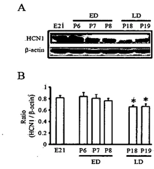
X-ray rùm. The protein bands were det.ect.ed using an enhanced chemiluminescence western blotting detection kit (Amersham. μtUe Ch외fonι Buck:in링1amshire, UK)tested using Western blot analysi~ .and measured by norma1izationwith correspondirig levels of ß-actin. Figure 1 shows the HCNl expression levels in neonata1 and postnatal hippocampus of rats. In ED hippocampus. the expression levels of HCNl of p6 were similar 、씨th E21 but age-dependent decreas in HCNl protein levels was revealed (Fi밍tre 1. E21 = 0.82 :!: 0.07. n = 2: p6 = 0.84 :!: 0.07. n = 4: p7 = 0.81 :!: 0.07. n = 4: p8 = 0.77 :!: 0.04. n = 4) Moreover,HCNl levels. in consistent with previous report (Bender et 외 2001). was significantJy decreased in LD hippocampus (Figure 1. P18 = 0.66 :!: 0.02. n = 4: P19 = 0.65 :!: 0.03. n = 4. p(0.05l πlis resulOOdicatesthat the expression level of HCNl is age-dependentiy decreased and this reflects that ED hippocampus is protected from cell damages by overexcitationin developmenta1stage
Discussion
。
ur results indicate ti1at ED hippocampimay be stable to excitatory stirnulalion because more HCNl proteins express in ED hipp∞뼈lpi than in LD hippocampi. In the previous reports,expression levels of HCN 1,2 and 4 subtypes areA
ED E2Ì P6 P7 P8 HCN11!'.IiIIi.l' p;;ocnnI
를를롤룸플
3. Statistical analysisD.ts ana!ysis and ststistic.1 sign피cance were peñonned by using Excel (Microsoft) software and then data were expressed as mean va1ue:tstandard error of mean (SEM) The Student' 5 t-test was used,and the significance between grcups was indicated for p values of(0.05
Results
B
훌훌::
? ••••*
*
...•
,
It has been reported that the expression levels of HCN1 age-dependentiy change in developing neurons within frrst 3 postnata1 weeks. In some previous papers,the expression levels of HCNl are increased during this developmental period (surges et al.. 2006‘Vasilyev and B양ish. 2002)
However,Bender et a1. reported that expression levels of HCN1 in ED neurons were higher than in LD neurons (2001). We. therefoTe. confirmed the changes of HCNl expression levels in this period. HCNl protein levels were
E21 P6 :P7 P8 P18 P19 ED LD
Figure 1. Age-dependent decrease in expression leveIs of HCNl is observed in the developing hippocampus of rats. π1e expression levels of HCNl were decreased with age,
as shown in the film(A) and quantitative analysis(B) Proteins of HCNl iso!ated from embrγ'O(n=2)and postnatal rat hippocampus (n=4/age) and the expression levels of HCNl were measured using Westem blot analysis. Optical dcnsities of individual bands were normalized to corresponding levels ofß-actin
70-Expression of HCN1 subunits is age-dependent1y regulated in developing hippoc와npus of rats
differently regulated during deve10pmental periods in m밍nm외ian brains. Generally,HCN2 subtypes are increased γhile HCN4 subtypes are decreased (Surges et al.,2006; Brewster et al.,2007). However ,it is not ensured whether expression levels of HCN1 are increased (Surges et al., 2006) or decreased (Bender et al.,2001) in this period. In the present study,we tested this issue by using western blot analysis to confrrm cbange of HCN1 proteins duringthe first 3 postnata1 weeks. Consequently ,age-dependent increase in expression levels of HCN1 was obseπed during this period and HCN1 protein 1eve1s were not different between embrio-21 day and ED hippocampus (Fig. 1)
The HCN1 subtype p1ays important ro1es in the determina1ion of membrane properties such as IDJPs and Rn and p따↑icipates in re밍1ation of excitability of neurons Recent stu이es reported that deficiency or downregl니la1ion of
HCNl subunits induces enhanced excitabilities and epileptogenesis because of the increased neuronal Rn and input summa1ion (Huang et al.,2009; Jung et 머,2007; Kole et al,.2007; Sb삶1 et a1.,2(04). Moreover ,ac1ivation of Th charmels encoded by HCN1 induces the decreased neurona1 excitabili1ies due to the decreased Rn (Fan et a1.,2005; Poolos et al.,2002; Johnston ,2006). Therefore ,our result suggests that ED hippocampi may be regu1ated by neurons but 1ess in LD hippocampi
Reference
1) Beckb ,S ,Noda ,M,Lubbert ,H ,and Numa ,S Differential regulation of three sodium channel messenger RNAs in the rat central nervous system during development. Embo J 1989;8:3611-3616
2) Bender ,RA,Brewster ,A,Santoro ,B,1udwig ,A, Hofmann,F,Biel,M,and Baram ,TZ. Differential and age-dependent expression of hyperpolarization-activated , cyc1ic nuc1eotide-gated cation channel isoforms 1
→
suggests eγolving roles in the developing rat hippocampus. Neuroscience 2001;106:689-6983) Brewster,A1,Chen ,Y,Bender ,RA,Yeh ,A,Shigemot
。’
R,and Baram ,1'2. Q니an1itative ana1ysis and subcellulardistribution of mRNA and protein expression of 야1e hyperpo1arization-activated cyclic nuc1eotide-gated 야larmels 랴lToughout development in .rat hippocampus Cereb Cortex 2007;17:702-712
4) Cingolani,1A,Gymnopou1os,M,Boccaccio,A,Stocker , M,and Pedarzani ,P. Developmental regulation of small conductance 않2十 activated K+ charmel expression and function in rat Purkinje neurons. J Neurosci 2002;22
4456-4467
5) Dybrfjeld-Jobnsen ,J ,Morgan ,RJ,Foldy,C,and S이tesz, 1. Upregulated H-current in hνperexcitable CA1 dendrites after febrile seizures 싼ont Cell Neurosci 2008;2:2 6) F머k,T,Kilani,RK,8trazdas ,LA,Borders ,RS,Steidl ,
JV,γ.001,AJ,and Sherman ,SJ. Developmental re밍ula1ion
。
f the A-type potassiurn-channel current in hippocampal neurons: role of the Kvbeta 1.1 subunit. Neuroscience 2003;120:387-4047) Fan,Y,Fricker ,D,Brager ,DH,Chen ,X,1u ,HC, C버twood,RA,and Johnston ,D. Activity-dependent decrease of excitabi1ity in rat hippocampal neurons throu양1 increases in l(h). Nat Neurosci 2005;8:1542-1551 8) Huang,Z,Walker ,MC,and Sbab ,MM μ ss of dendritic
HCN1 subunits enhances cortical excitability and epileptogenesis. J Neurosci 2009;29:10979-10988
9) Jung,S,Jones ,TD,Lugo,JN ,Jr. ,Sheerin ,AH,뻐ller, ,JW,D'Ambrosio ,R,Anderson ,AE,and Poolos ,NP
Progressive dendritic HCN channe10pathy during epileptogenesis in the rat pilocarpine model of epilepsy J Neurosci 2007;27:13012-13021
10) Kole,빠I,Brauer ,AU,and Stuart ,GJ. 1nherited cortical HCN1 channel loss amp1ifies dendritic calcium electrogenesis and burst frring in a rat absence epilepsy model. J Pbysiol 2007;578:507-525
11) Ludwig,A,Zong ,X,Jegliiscb ,M,Hofm밍m,F ,and Biel, M. A family of byperpol없ization-ac1ivated mamma1ian
cation cbannels. Nature 1998;393:587-591
12) Magee,JC. Dendritic hyperpolarization-activated currents modify the iritegrative properties of hippocampal CA1 pyramidal neurons. J Neurosci 1998;18:7613-7624 13) Maletic-Savatic ,M,1enn ,NJ ,and Trimmer ,JS
Differential spatiotemporal expression of K+ channel polypep1ides in rat hippocampal neurons developing in situ and in vitro. J Neurosci 1995;15:3840-3851
14)
α
니ardouz,M,Lema ,P ,Awad ,PN,Di Cristo ,G,andCarmant,1. N-methyl-D-aspartate ,hyperpolarization activated ca1ion cuπent (Ih) and gamma-aminobutyric acid conductances govern the risk of epileptogenesis following febrile seizures in rat hippocampus. Eur J Neurosci 201O;31:1252-126C
15) Pape ,HC. Queer current and pacemaker: the hyperpolarization-ac1ivated ca1ion current in neurons Annu Rev Pbysiol 1996;58:299-327
16) Poolos,NP,Migliore,M,and Jobnston ,D. Pbarrna
∞
logical upregu1ation of h-charmels reduces the excitabi1ity of pyramida1 neuron dendrites. Nat Neurosci 2002;5:767← 774Yoon Sil Yang,moon Seok K잉19,Seon Hee Kim,Su Yong Eun,Sung Cherl Jung
17) Rosenkranz ,JA ,and Johnston ,D. Dopaminergie regulation of neuronal excitability throt띔h modulation of Ih in layer V entorhinal cortex. J Neurosci 2006;26:3229-3244
18) Santoro,B,μu ,DT,γ"ao,H,B않tseh,D,Kandel ,ER,
Siegelbaum ,SA,and Tibbs ,GR Identifieation of a gene encoding a hyperpolarization-activated paeemaker ehannel of bram. Cen 1998;93:717-729
19) Seheinm밍1,RI,Auld,VJ,Goldin ,AL,Davidson ,N,Dunn ,
RJ,and Catteral1 ,WA. Developmental regl띠ation of sodium channel expression in the- rat forebrain. J BiolChem 1989;264:10660-10666
20) Seifert,R,Scholten ,A,Gauss ,R,Mincheva ,A,1ichter , P,and Kaupp ,00. Molecular characterization of a slowly gating human hyperpolarization-activated channel predominantly expressed in thalamus ,heart ,and testis ProeNatlAeadSei U S A 1999;96:9391-9396
21) Shah ,MM,Anderson ,AE ,Leung ,V,Lin ,X,and Johnston ,D. Seizme-induced plasticity of h ehannels in
entorhina1 cortieal layer III pyramidal nemons. Neuron 2004;44:495-508
22) Surges ,R ,Brewster ,AL ,Bender ,RA ,Beck ,H , Feuerstein ,TJ ,and Baram ,'IZ. Regl나lated expression of
HCN charmels and eA1v1Plevels shape the propertîes of the h eurrent in developîng rat hippocampus. Eur J Neurosei 2006;24:94-104
23).Surges ,R ,Freiman ,TM,and Feuerstein ,TJ. Input resistance is voltage dependent due to activation of Ih channels in rat CAl pyramidal eells. J Neurosci Res 2004;76:475-480
24) Vasîlyev,DV,and Barish ,I\IlE. Postnatal development of
야1e hyperpo1arization-activated excitato:ry CUlTent Ih in mouse hippoeampal pyramidal neurons. J Neurosci 2002;22:8992-9 따4
25) Williams,SR,and Stu않t,GJ. Site independence of EPSP
time course is mediated by dendritic I(h) in neocortical pyramidal neurons. J Neurophysiol 2000;83:3177-3182