저작자표시-비영리-변경금지 2.0 대한민국 이용자는 아래의 조건을 따르는 경우에 한하여 자유롭게 l 이 저작물을 복제, 배포, 전송, 전시, 공연 및 방송할 수 있습니다. 다음과 같은 조건을 따라야 합니다: l 귀하는, 이 저작물의 재이용이나 배포의 경우, 이 저작물에 적용된 이용허락조건 을 명확하게 나타내어야 합니다. l 저작권자로부터 별도의 허가를 받으면 이러한 조건들은 적용되지 않습니다. 저작권법에 따른 이용자의 권리는 위의 내용에 의하여 영향을 받지 않습니다. 이것은 이용허락규약(Legal Code)을 이해하기 쉽게 요약한 것입니다. Disclaimer 저작자표시. 귀하는 원저작자를 표시하여야 합니다. 비영리. 귀하는 이 저작물을 영리 목적으로 이용할 수 없습니다. 변경금지. 귀하는 이 저작물을 개작, 변형 또는 가공할 수 없습니다.
A THESIS
FOR THE DEGREE OF MASTER OF SCIENCE
Isolation of BEMB from Polysiphonia japonica inhibit LPS-induced pro
-inflammatory responses by preventing ROS production and down-reg
ulating NF-kB in in vitro and in vivo Zebrafish models
EUN-YI KO
Department of Marine Life Sciences
GRADUATE SCHOOL
JEJU NATIONAL UNIVERSITY
August, 2016
i CONTENTS 국문초록 ... iii LIST OF FIGURES ... vi ABSTRACT ... 1 INTRODUCTION ... 2
MATRERIALS AND METHODS ... 5
Materials ... 5
Isolation of BEMB from Polysiphonia japonica ... 6
Cell culture ... 8
Determination of NO production and cell viability ... 8
Determination of PGE2 and cytokine production ... 8
Western blot analysis in RAW264.7 macrophages ... 9
Measurement of ROS of intracellular reactive oxygen species in RAW264.7 cells ... 10
Detection of NF-kB in RAW264.7 cells by immunofluorescence and confocal Microscopy ... 10
Maintenance of zebrafish ... 11
ii
Western blot analysis in zebrafish ... 12
RNA extraction and quantitative RT-PCR (qRT-PCR) ... 12
Statistical analysis ... 13
RESULTS AND DISCUSSIONGS ... 15
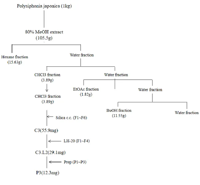
Extraction and isolation of BEMB from Polysiphonia japonica ... 15
Effect of BEMB form P.japonica on production of NO and PGE2 in LPS-stimulated RAW 264.7 cells ... 19
Effects of BEMB on iNOS and COX-2 protein in RAW264.7 cells ... 21
BEMB inhibit LPS induced cytokine production ... 23
Effect of BEMB on LPS-induced NF-kB activity in RAW264.7 cells ... 25
Effect of BEMB on LPS-induced ROS generation in RAW264.7 cells ... 28
Effect of BEMB against LPS-induced NO production and iNOS and COX-2 protein in zebrafish ... 31
Effects of BEMB and NAC against LPS-induced ROS production in zebrafish ... 34
Effect of BEMB on LPS-induced NF-kB activity in zebrafish ... 37
DISCUSSION ... 39
REFERENCES ... 42
iii 국문초록 염증은 생체 또는 조직에 물리적 자극, 화학적 물질, 세균감염 등의 변화를 가져오는 자극이 가해질 때 손상부위를 복구하려는 생체반응으로서 자극이 가해지면 국소적으로 염증성 성분이 증가되면서 염증이 유발이 된다. 하지만, 염증반응이 만성화되면 류마티스 관절염, 동맥 경화증, 위염, 천식, 당뇨병 및 심혈관 질환 등의 각종 질병의 원인이 되며, 염증성 질환이 유발된다 염증반응에 관여하는 대표적인 세포 중 하나인 대식세포는 염증성 매개물질인 interleukin (IL)-6, IL-1β, tumor necrosis factor-α (TNF-α) lipopolysaccharide (LPS)와 같은 자극에 의해 염증반응의 전사인자인 nuclear factor-κB (NF-κB)를 활성화시키며 pro-inflammatory cytokine과 nitric oxide (NO), prostaglandin E2 (PGE2), inducible nitric oxide synthase (iNOS), cyclooxygenase-2 (COX-2) 등의 염증유발인자들을 생성한다.
이러한 염증 치료제로 천연자원을 이용한 연구들이 많이 하고 있으며, 자원들 중 하나로 해조류에 대한 관심이 많아 지고 있다. 해조류는 칼륨, 요오드, 칼슘 등의 각종 무기염류와 비타민 A,C 등의 함량이 높은 자원이며, laminaran 및 fucoidan 등과 같이 인체에 유용한 다당류뿐만 아니 라 여러 생리활성물질을 함유하고 있다. 해조류의 다양 한 생리활성에 대한 연구들이 증가되고 있으나, 홍조류의 떨기나무꼴폴리시포니아 (Polysiphonia japonica) 에 대한 연구는 미비한 편이다. 특히 떨기나무꼴폴리시포니아 추출물에서 분리한 물질들에 대한 항염증 실험, 세포실험 및 동물실험에 대한 연구는 보고되어 있지 않다. 그래서 이 연구에서는 떨기나무꼴폴리시포니카에서 분리한
3-bromo-5-(ethoxymethyl)-1,2-iv
benzenediol (BEMB)에 항염효과를 세포실험, 대표적인 대식세포인 RAW264.7 세포를 이용하여 수행하였다.
먼저, 떨기나무꼴폴리시포니카를 80% 메탄올 추출을 하고 그 후 클로포름 분획 물에서 3-bromo-5-(ethoxymethyl)-1,2-benzenediol (BEMB)를 분리하였다. 그리고 BEMB를 LPS로 자극된 RAW264.7 세포에서 독성과 NO, PGE2 생성양을 확인하였다. 그 결과, BEMB 12.5, 25, 50 μM에서 독성이 없었으며, 농도 의존적으로 NO와 PGE2양이 감소함을 확인하였다. 그 후, western blot 분석방법을 통해 염증유발인자 인 iNOS, COX-2 단백질 발현양을 확인한 결과, 가장 높은 농도인 50μM 에서 iNOS, COX-2 단백질 발현양이 매우 감소함을 확인 할 수 있었으며, ELISA kit를 이용하여 cytokine (IL-6, IL-1β)의 양 또한 농도의존적으로 감소함을 확인할 수 있었다. 그리고 다시, western blot 분석방법을 통해 염증반응 전사인자인 NF-κB의 활성화를 확인하였다. 그결과, 인산화된 P65, P105가 BEMB의 50 μM에서 발현양이 줄어듬을 확인하였다. 세포 내의 활성산소종 (Reactive oxygen species ROS) 생성 역시 염증 반응에서 중요한 인자이다. ROS의 조절로 염증이 억제되는지 확인 하고자 LPS로 자극된 RAW264.7 세포에 BEMB 와 대표적인 ROS 억제제인 N-acetylcysteine (NAC)을 처리 하여 ROS의 발생양을 확인하였다. 그 결과, BEMB가 처리 된 부분에서는 ROS의 양이 억제되었으며, 억제된 양은 NAC이 처리된 군과 비슷하였다. NAC이 처리된 비교군 또한 LPS가 처리된 군 보다 ROS, NO양이 줄어들었으며, western blot 분석방법을 통해 iNOS, COX-2의 발현양 또한 줄어듬을 확인하였다.
v 기관을 가지고 있는 척추동물로 최근 실험동물 모델로 주목받고 있다. 체외수정을 하며 난을 대량 확보 가능하고, 난의 투명하여 형태적 관찰이 용이한다. 이러한 장점을 이용하여 최근 염증연구에서도 동물모델로서 많이 이용되어 지고 있다. BEMB가 RAW264.7 대식세포 상에서 항염증효과를 보여 동물실험으로 항염증 효과를 확인하기 위해 LPS로 자극된 제브라 피쉬에서 염증효과를 확인하였다.
그 결과 LPS가 자극된 제브라피쉬에서 BEMB가 농도 의존적으로 NO, ROS 생성 양과 iNOS, COX-2 단백질 발현양이 억제됨을 확인하였다. 또한 LPS가 자극된 제브라피쉬에서도 NAC을 처리한 결과, NO, ROS 생성양과 iNOS, COX-2 단백질 발현양이 억제됨을 확인하였다. 또한, 제브라피쉬에서 RNA를 추출하여 PCR 결과 BMEM 50μM에서 NF-κB의 관련 유전자의 발현이 줄어듬을 확인 하였다. 이 모든 결과를 종합해 볼 때, 떨기나무꼴폴리시포니카에서 분리한 BEMB는 RAW264.7 세포에서 ROS 발생양과 NF-kB 활성화를 조절하여 항염증 효과를 나타내었으며, 더 나아가 동물모델인 제브라피쉬에서도 ROS 발생양과 NF-kB 유전자를 조절하여 항염증 효과를 나타내었다. 이로써 떨기나오꼴폴리시포니카는 천연 염증치료제로 사용할 수 있음을 사료되어진다.
vi
LIST OF FIGURES
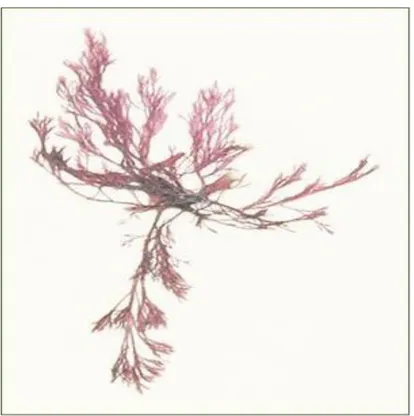
Fig. 1. The photography of the alga, Polysiphonia japonica
Fig. 2. Isolation scheme of BEMB from the alga Polysiphonia japonica. Fig. 3. HPLC fingerprinting analysis of P.japonica chloroform extract. Fig. 4. Carbon NMR spectrum and structure of BEMB.
Fig. 5. Effect of BEMB on production of NO and PGE2 production in LPS-stimulated RAW 264.7 cells. RAW264.7 cells were simultaneously stimulated with LPS (1 μg/ml) and
BEMB (12.5, 25, 50 μM). (A) Measured of NO production by Griess reagent. (B) Supernatants were collected PGE-2 concentration in the supernatants were determined by ELISA kit. (C) Cell viability was indicated by MTT assay. All the results were expressed as means ± S.E. of over three individual experiments; *P < 0.05.
Fig. 6. Effects of BEMB on LPS-induced iNOS and COX-2 protein expressions in
RAW 264.7 cells. Cells stimulated with LPS (1 μg/ml) in the presence of BEMB (12.5, 25,
and 50 μM) for 24 h at 37°C. iNOS and COX-2 protein level were determined via Western blotting.
Fig. 7. Inhibitory effects of BEMB on the pro-inflammatory cytokine production in
LPS-stimulated RAW264.7 cells. The production of (A) IL-1β, and (B) IL-6 were assayed
in the culture medium of cells stimulated with LPS (1 μg/ml) for 24 h in the presence of BEMB (12.5, 25, and 50 μM). Supernatants were collected, and the IL-1β, and IL-6 concentration in the supernatants were determined by ELISA kit. All the results were expressed as means ± S.E. of over three individual experiments; *P < 0.05.
Fig. 8. Inhibitory effects of BEMB on LPS-induced phosphorylation of NF-kB. (A)Cells
vii
different concentrations (12.5, 25, and 50 μM) of BEMB. Cell lysates were extracted, and protein levels of p-p65, p-p105 were analyzed by Western blot. (B) The analysis of NF-kB translocation on RAW264.7 the nucleus induced by 30 minutes of LPS treatment was performed by confocal microscopy (x 1000).
Fig. 9. Effect of BEMB and NAC on the generation of ROS in LPS-stimulated
RAW264.7cells. RAW264.7 cells were seeded into 96-well plates at a starting density of
5×10⁴ cell/well and 18h cultured and pre-incubated with florescence dye DCF-DA for 1 h. Then, cell were pre-treated PSC14 (12.5, 25, 50 μM/ml), N-acetylcysteine (NAC) and treated with LPS (1 μg/ml) at after 2hour. Fluorescence was measured at 485nm excitation and 555nm emission wavelengths using Fluostar Optima (BMG Labtechnologies) fluorescent microplate reader. All the results were expressed as means ± S.E. of over three individual experiments; *P < 0.05.
Fig. 10. Effect of BEMB and NAC on the production of NO , expression of iNOS and
COX-2 in LPS-stimulated RAW264.7cells. Cells were pretreated with BEMB (12.5 25 and
50 μM) and NAC (2 mM) for 2 h and then incubated with stimulation with 1 μg/ml LPS. (A) Supernatants were collected, (A) NO production in the supernatants were determined by Griess reagent.(B) iNOS and COX-2 protein level were determined via Western blotting. All the results were expressed as means ± S.E. of over three individual experiments; *P < 0.05.
Fig. 11. Effect of BEMB on LPS-induced NO production and iNOS and COX-2 protein
expressions in zebrafish. Zebrafish embryos stimulated with LPS (10 μg/ml) in the presence
of BEMB 3 dpf. (A) NO levels were measured by image analysis and fluorescence microscope. The fluorescence intensity was quantified using image J program. (B) iNOS and COX-2 protein level were determined via Western blotting. All the results were expressed as means ± S.E. of over three individual experiments; *P < 0.05.
viii
Fig. 12. Effects of BEMB and NAC on LPS-induced ROS and NO Production in
Zebrafish. Zebrafish embryos stimulated with LPS (10 μg/ml) in the presence of BEMB and
NAC (250 μM) 3 dpf. (A), BEMB ROS level (B) NAC ROS level (C) NAC NO level. Assessments were measured by image analysis and fluorescence microscope. The fluorescence intensity was quantified using image J program. (D) iNOS and COX-2 protein level were determined via Western blotting. All the results were expressed as means ± S.E. of over three individual experiments; *P < 0.05.
Fig. 13. Effect of BEMB on LPS-induced NF-kB activity in zebrafish. Gene expre
ssion (mRNA) of P65, P100/P52 and IκbaA evaluated by RT-PCR. Zebrafish embryos stimulated with LPS (10 μg/ml) in the presence of BEMB (50 μM) 3 dpf. Bars indi cate mean±standard error of three independent experiments.
ix
LIST OF TABLES
Table. 1 Quantitative PCR primer sequences used for the analysis of genes involved in
1 ABSTRACT
The anti-inflammatory effects of 3-bromo-5-(ethoxymethyl)-1,2-benzenediol (BEMB) from Polysiphonia japonica were evaluated in lipopolysaccharide LPS induced in RAW264.7 macrophages and Zebrafish models. BEMB had anti-inflammatory effects, inhibiting nitric oxide (NO), prostaglandin E2 (PGE2), inducible nitric oxide synthase (iNOS),
cyclooxygenase-2 (COX-2), interleukin (IL)-1β, and interleukin (IL)-6 productions in LPS-activated RAW264.7 cells, without inducing cytotoxicity. Furthermore, BEMB inhibited LPS-induced NF-κB activation and reactive oxygen species (ROS) production in RAW 264.7 cells. Moreover, in this study investigated the effect of anti-inflammatory through NO and ROS production in zebrafish embryos. BEMB significantly inhibited LPS-induced NO and ROS generation similar to the ROS inhibitors, N-acetylcysteine (NAC). BEMB and NAC also suppressed the expressions of iNOS and COX-2 in LPS-stimulated zebrafish embryos. Moreover, BEMB suppressed mRNA expression of NF-kB (p65, p100/52 and ikbA) in LPS-induced zebrafish. Collectively, in this study data indicate that BEMB suppresses the production of pro-inflammatory mediators such as iNOS and COX-2 as well as their regulatory in LPS-induced RAW264.7 cells and zebrafish embryos by inhibiting the ROS and NF-kB. Therefore, this study suggest that BEMB should be utilized as potential anti-inflammatory agents for the treatment of anti-inflammatory diseases.
2 1. Introduction
Inflammatory reaction, typically characterized by redness, swelling, heat, and pain, is a common physiologic response to defend the host from injurious stimuli such as pathogens, toxins and local injuries (Li, et al, 2011). LPS, the major component of the Gram-negative bacterial cell wall, can trigger and accelerate an inflammatory cascade. LPS has been linked to a wide variety of human diseases, including gastro-intestinal illness (Mariana et al, 2010). Thus, LPS has been widely used to mimic features of inflammatory diseases (Kempe S et al, 2005). LPS can activate mononuclear phagocytes (monocytes and macrophages) and other types of cells to secrete inflammatory cytokine (IL-6, IL-1β and TNF-α) and pro-inflamamtory mediators (iNOS and COX-2) (Lanan et al, 2012, Kim et al, 2007). IL-1β is expelled primarily by monocytes and macrophages as well as by nonimmune cells, such as fibroblasts, during cell injury, endothelial cells, infection, invasion, and inflammation. IL-6 is a cytokine produced by a number of normal and transformed cells and known to be an endogenous mediator of LPS activated fever(Zhang et al, 2007., Bartold et al, 1991). Thus, inhibition of the production of these inflammatory cytokine and mediators is an important target in the development of anti-inflammatory agents.
Reactive oxygen species (ROS), particularly generated from NADPH oxidase by immune cells, such as macrophages, is an important innate immune response to LPS. Reactive oxygen species (ROS) are key signaling molecules that play an important role in the progression of inflammatory disorders and excessive ROS has strong pro-inflammatory effects and can serve as secondary messenger to further amplify LPS-induced inflammatory. (Fan et al, 2014, Tak et al, 2001).
3
the development of anti-inflammatory drugs targeting NF-κB (Karin et al. 2004, Toby et al, 2009). In immune cells NF-κB contain a heteromeric complex of two components, p65 (rel A) , C-rel, RelB, NF-B1(p50/105), NF-B2 (p52/100). (Lee et al. 2006) The p65 composition include in the main trans activating domain responsible for NF-κB transcription factor function. (Mahdad et al. 2008). Many studies have suggested that the expressions of inducible nitric oxide synthase (iNOS) and cyclooxygenase-2 (COX-2) which are enzymes involved in the generation of inflammatory mediators can be regulated by several transcription factors, one of them is nuclear factor-kappa B (NF-κB). (Bubici C et al. 2006).
As an animal model, the zebrafish has been widely used in studies on molecular genetics and development biology, drug discovery, and toxicology because of their physiological similarity to mammals (Allan et al. 2014, Craig et al. 2007). The zebrafish has numerous advantages as a toxicological model species. For example, their comparatively small size, fecundity, large clutches, low cost, and rapid embryonic development in vivo (Eisen et al. 1996, fishman et al. 1999). In addition, the zebrafish has a very short development, plan of the basic body is laid out 24 h post- fertilization (hpf), embryos hatch approximately 2-3 days post- fertilization (dpf) and they complete maturity at about a 3 months. Furthermore, one female can spawn about 100 eggs per day, which are fertilized by sperm released into the water by males. (kim ea al. 2014). Furthermore, zebrafish have recently been used in toxicology and drug discovery studies investigating oxidative stress and inflammation, because the embryos and juveniles are transparent. This property allows the visualization of specific cells, tissues, and organs under the microscope (Park et al. 2011, Zhang et al. 2013, Kang et al. 2014, Ko et al. 2014). However, many studies have anti-inflammatory experiments in zebrafish, there is not much paper, the experimental in inflammation by
4
controlling the ROS. N-acetyl-L-cysteine (NAC) is commonly used to identify and test ROS (reactive oxygen species) inducers, and to inhibit ROS (Halasi et al, 2013). Therefore, in this study identified regulation of ROS and inhibition of inflammatory by NAC in LPS-induced zebrafish inflammation model.
Marine algae are attracting attention as a material for multi-functional physiological regulatory functions in various whitening, anti-inflammatory, anti-cancer, and anti-obesity effects (El Gamal et al. 2010, Wijesekara et al. 2011). Marine algae have attracted a range of uses in the food, cosmetic and pharmaceutical industries and in biotechnology (Briggs et al. 2002). Among the algae, Polysiphonia japonica (Phylum Rhodophyta, Class Florideophyceae, Order Ceramiales, Family Rhodomelaceae, Fig 1) is found throughout south Coast of the Korea. (Kudo et al. 1986). Many papers have a lot of Polysiphonia algae in the research, reported effect of Polysiphonia is antioxidant, anti-fungi anti-inflammatory (Ke et al. 2007, Gwak et al. 2006 , Heo et al. 2006). But, P. japonicawas not much research.
Therefore, in this study for the first time, isolated of 3-bromo-5-(ethoxymethyl)-1,2-benzenediol (BEMB) from P. japonica and examined for anti-inflammatory activities in in vitro RAW264.7 cells and in vivo zebrafish model.
5
2. Material and method
2.1. Materials
The red alga P. japonica was collected from along the coast of Jeju Island, Korea. The sample was washed thrice with tap water to remove salt, sand, and epiphytes attached to its surface, followed by careful rinsing with fresh water and freezing in a medical refrigerator at -20°C. Thereafter, the frozen sample was lyophilized and homogenized with a grinder prior to extraction. All chemicals and reagents used were of analytical and sourced from trusted commercial sources. Dulbecco’s Modified Eagle’s Medium (DMEM), fetal vobine serum (FBS), penicillin-streptomycin, trypsin-EDTA were purchased from Gibco.(Grand Island,NY). Dimethyl sulfoxide (DMSO), lipopolysaccharide (LPS; from Escherichia coli strain) and phosphate buffered saline (PBS) were purchased from Sigma Chemical Co(St. Louis, MO, USA). The ELISA kits for PGE2, TNF-α, IL-1β, and IL-6 were purchased from R&D Systems, Inc. (St. Louis, MO, USA) and BD Biosciences (San Diego, CA, USA). Antibodies were from the following sources: iNOS, COX-2, IκB-α, P-P65, P-P105 and β-actin were purchased from Cell Signaling Technology (Beverly, MA, USA). The secondary antibodies were obtained from Cell Signaling Technology. 2,7-dichlorofluorescein diacetate (DCF-DA), diaminofluorophore4-amino-5-methylamino-2′7′-difluorofluorescein diacetate (DAF-FM DA), acridine orange were purchased from Sigma (St. Louis, MO, USA). High-performance liquid chromatography (HPLC) grade solvents were purchased from Burdick & Jackson (Muskegon, MI, USA). The other chemicals and reagents used were of analytical grade.
6 2.2. Extraction and isolation
The dried Polysiphonia japonica powder was extracted thrice with 80% aqueous methanol at the room temperature. The liquid layer was obtained via filtration, and the filtrate was concentrated by using an evaporator under reduced pressure. The extract was suspended in water, and the aqueous layer was partitioned with chloroform. Then, the chloroform fraction was fractionated by silica column chromatography with stepwise elution of chloroform– methanol mixture (30:1→1:1) to separate active fractions in chloroform extract. A combined active fraction was further subjected to a Sephadex LH-20 column saturated with 100% methanol, and then purified by reversed phase high performance liquid chromatography (HPLC) using a Waters HPLC system (Alliance 2690; Waters Corp., Milford, MA, USA) equipped with a Waters 996 photodiode array detector and C18 column (J’sphere ODS-H80, 250 × 4.6 mm, 4 μm; YMC Co., Kyoto, Japan) by stepwise elution with methanol–water gradient (UV range, 290 nm; flow rate, 1 ml/min). Finally, the purified compounds were identified by comparing its 1H and 13C NMR data with literature (Mikami et al., 2013). The compound was dissolved in DMSO and employed in experiments in which the final concentration of DMSO in culture medium was adjusted to <0.01%.
7
8 2.3. Cell culture
RAW 264.7 murine macrophages were obtained from American Type Culture Collection (ATCC, Rockville, MD, USA). These cells were cultured in DMEM supplemented with 10% FBS, 100 μ/mL penicillin, and 100 μg/mL streptomycin at 37℃under 5% CO2-humidified air.
2.4. Determination of NO production and cell viability
After pre-incubation of RAW 264.7 cells (1.5 × 10⁵ cells/ml) with LPS (1 μg/ml) plus samples at 37 ℃ for 24 h, the quantity of nitrite accumulated in the culture medium was
measured as an indicator of NO production. Briefly, a 100 μl of cell culture medium was mixed with 100 μl of Griess reagent (1% sulfanilamide and 0.1% naphthylethylenediamine dihydro chloride in 2.5% phosphoric acid), the mixture was incubated at room temperature for 10 min, and the absorbance at 540 nm was measured in a microplate reader (Thermo Max, CA, USA).
Cytotoxicity was assessed using an MTT assay, which is a test of metabolic competence predicated upon the assessment of mitochondrial performance. The cells were seeded into 24-well plates at a concentration of 1.5 x 10⁵ cells/ml. After 70% confluent of cultures, the cells were treated with isolated BEMB (12.5, 25, 50 μM) from P. japonica. Then, the cells were incubated for an additional 24 h at 37ºC. MTT stock solution (100 μl; 2 mg/ml in DPBS) was then applied to each of the wells, to a total reaction volume of 500 μl. After 4 h of incubation, and the supernatants were aspirated. The formazan was dissolved in DMSO and absorbances was measured via microplate reader at a wavelength of 540 nm.
9
2.5. Determination of PGE2 and cytokine production.
Serum levels of PGE2, IL-1β, and IL-6 were determined using a commercially available
enzyme linked immunosorbent assay (ELISA) kit (Biosource International Inc,R&D Systems) according to the manufacturer's instruction. PGE2, IL-1β, and IL-6 were determined from a
standard curve and measured via microplate reader at a wavelength of 450 nm.
2.6. Western blot analysis in RAW264.7 macrophages
RAW264.7 cells (1.5 x 10⁵ cells/mL) were pre- treated with BEMB (12.5, 25, 50 μM) and LPS (1 μg/ml) for 24h. The cells were washed twice PBS, and the cells were resuspended in RIPA buffer (sigma) for 10 min on ice. Cell lysates were centrifuged at 14,000 x g for 20 min at 4ºC. And then protein contents in the supernatant was measured using Bio-Rad protein assay kit (Bio-Rad, CA, USA) with bovine serum albumin (BSA) as a standard. The lysate containing 30 μg of protein was subjected to electrophoresis on 10% sodium dodecyl sulfate-polyacrlyamide gel, and the gel was transferred onto a nitrocellulose membrane (Bio-Rad, Hercules, CA). The membranes were blocked with 5% milk and 3% BSA in TBS buffer containing 0.2% Tween 20 (TBST) for 2h at room temperature. Then the membrane was incubated with specific primary rabbit polyclonal anti-rabbit : iNOS, P-P65, P-P105 and β-actin (1:1000, Cell signaling, Inc.), or mouse monoclonal anti-mouse COX-2 (1:1000, Cell signaling, Inc.) at 4ºC for overnight. Membranes were three times washed with TBST and incubated with goat anti-rabbit or anti-mouse IgG HRP conjugated secondary antibody (1:3,000 dilution, Cell signaling, Inc) in TBST that contained 5% skimmilk and 3% BSA for 2h at room temperature. After three times washing with TBST for 10 min. Following the addition, signals were developed using ECL western blotting detection kit exposed to Biorad
10 Chemidac system.
2.7. Detection of NF-kB in RAW264.7 cells by Immunofluorescence and Confocal Microscopy
Cells cultured confocal chamber slides (5x10⁴ cells/well) for 18h after, pre-treated BEMB then treated with LPS and incubated for 24h. Cells were fixed with 4% paraformaldehyde for 10 minutes at room temperature, after by incubation in methanol for 10 minutes at 4 ºC. After three times washed PBS and further blocked with 1% BSA for 1h.After three times washes PBS then primary antibody p65 (1:100, Cell signaling) incubated for overnight at 4 ºC. After three times washed PBS, Secondary antibodies were incubated for 1h 30min with Alexa Fluor488–labeled Rabbit IgG (1:500 dilution, cell signaling). After 3 times washed PBS then cell nuclei were stained with DAPI using Vectashield mounting medium (Vector, Burlingame, CA). Fluorescence was analyzed using a Zeiss confocal microscope system and camera (ZEISS Instruments).
2.8. Measurement of ROS of intracellular reactive oxygen species in RAW264.7 cells
Intracellular ROS were measured using the oxidation DCF-DA fluorescent dye. RAW264.7 cells were seeded into 96-well plates at a starting density of 5×10⁴ cell/well and 18h cultured and pre-incubated with florescence dye DCF-DA for 1 h. Then, cells were pre-treated BEMB (12.5, 25, 50 μM/ml), N-acetylcysteine (NAC) and treated with LPS (1 μg/ml) at after 2hour. Fluorescence was measured at 485nm excitation and 555nm emission wavelengths using Fluostar Optima (BMG Labtechnologies) fluorescent microplate reader. All experiments were
11
performed in at least 6 parallels and repeated three times.
2.9. Maintenance of zebrafish
Zebrafish were maintained in a temperature-controlled room at 28°C with a 13:11 hour day/night cycle. Zebrafish were fed two times a day. The day before, zebrafish were randomly selected for interbreeding in the male-to-female ratio of 2:1. The embryos were collected from the breeding case and washed to remove any debris lying at the bottom of the tank. The collection of embryos obtained post-spawning were staged, dispensed in embryo media. Embryo media contained deionized water with 60 mg/liter Instant Ocean red salts (Spectrum Brands, Mentor, OH).
2.10. Measurement of ROS and NO production in zebrafish by image analysis
Generation of NO in inflammatory zebrafish model was estimated using a fluorescent probe dye, DAF-FM DA and Production of intracellular ROS in zebrafish embryos were detected using an oxidation-sensitive fluorescent probe dye, DCF-DA. Embryos were treated with 12.5, 25, and 50 μM BEMB 2 h later, 10 μg mL-1 LPS was added to the plate. Embryos selected at 3dpf, transferred into 24 well plate and treat embryo medium containing 10 μM DAF-FM DA and 20 μg/ml DCF-DA. ROS incubated for 1 h and NO incubated for 2h in the dark at 28°C. After incubation, the embryos were washed in embryo media and anesthetized before visualization. The images of stained embryos were observed using a fluorescent microscope, which was equipped with a CoolSNAP-Pro color digital camera (Olympus, Japan).
12 2.11. Western blot analysis in zebrafish
At 3dpf, harvest embryos were washed PBS buffer and treated RIPA buffer. The homogenates were centrifuged at 12,000 g for 15 min in 4°C, and the supernatants were collected, after that protein contents in the supernatant was measured. The lysate containing 30 μg of protein was subjected to electrophoresis on 10% sodium dodecyl sulfate-polyacrlyamide gel, and the gel was transferred onto a nitrocellulose membrane (Bio-Rad, Hercules, CA). The membranes were blocked with 5% skim milk in TBS buffer containing 0.2% Tween 20 (TTBS) for 3H at room temperature. Then the membrane was three times washed TTBS buffer, and incubated with specific primary rabbit iNOS, COX- Ab (1:1000, Cell signaling) at 4ºC for overnight. After, membranes were washed with TTBS and incubated with peroxidase-conjugated secondary antibodies in 5% skimmilk in TBST (diluted 1:5000; Cell signaling) for 2 h at room temperature. After three times washing with TBST for 10 min. Following the addition, signals were developed using ECL western blotting detection kit exposed to Biorad Chemidac system.
2.12. RNA extraction and quantitative RT-PCR (qRT-PCR)
Zebrafish and isolated gonads were homogenized and Invitrogen RNA elution kit. RNA was isolated according to manufacturer’s instruction. cDNA synthesis was performed using qScript cDNA synthesis kit (Quanta Biosciences, USA). Primers were designed for listed genes (Table. 1). SYBR Green (Takara) was used to determine the expression levels of all
13
genes. Takara PCR conditions for SYBR Green consisted of a denaturation step for 5 minutes at 95º C followed by 40 cycles of 95º C for 2 seconds and 60º C for 30 seconds. Data analysis was performed using standard curve method and ΔΔCt method .
2.13. Statistical analysis
All data in this study are expressed as means ± S.D. Significant differences among the groups were determined using the unpaired Student’s t-test. A value of *p < 0.05 was considered to be statistically significant.
14
Table. 1 Quantitative PCR primer sequences used for the analysis of genes involved in
zebrafish NF-κB
Gene Symbol Oligo Primer Sequence
Annealing Temperature (˚C) Amplification product (bp) p65 Forward tccctggagagaagagcaac 60 200 Reverse cagtcttttcccaccagctc 60 p100/p52 Forward gagccctttgtgcaagagac 60 216 Reverse tgtgtgtgtcaccagctgaa 60
ikbA Forward tttcggaggagatggagaga 60 184
Reverse ctgttcaggtacgggtcgtt 60
b-actin
Forward ggttttgctggagatgatgc 60
171
15
3. Result
3.1. Extraction and isolation of BEMB from Polysiphonia japonica
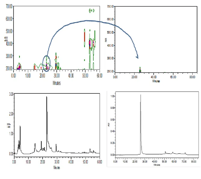
The isolation of the BEMB compounds from P. japonica was carried out as Fig. 2. The dried P. japonica (1 kg) was extracted twice with 80% aqueous methanol at room temperature. The methanol extract was filtered and concentrated under reduced pressure. The residue was subjected to successive extraction with organic solvents of n- hexane, chloroform, ethyl acetate and n-buthanol. The chloroform layer was concentrated under vacuum and the crude residue (3.89 g) was applied to a silica gel column and eluted with CHCl3 and CHCl3-MeOH mixture (100:1-1:1) with increasing proportion of methanol. The HPLC and NMR data of the compound in fraction were reference to reported by Heo et al. (2008). (Fig. 3, 4)
16
17
18
19
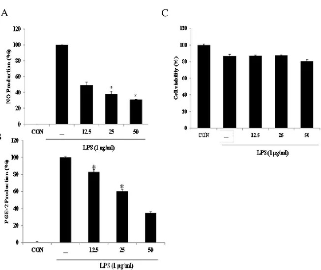
3.2. Effect of BEMB on production of NO and PGE2 in LPS-stimulated RAW 264.7 cells
To identify the potential anti-inflammatory activity of BEMB on LPS-induced the production of NO and PGE2 were measured by griess reagents and ELISA assays, respectively. BEMB significantly inhibited (49% and 30%) LPS-induced NO and PGE2 production at 50 μM respectively (Fig 5 A and B). Moreover, BEMB markedly reduced LPS-induced NO and PGE productions in a dose-dependent manner (Fig. 5 A and B). BEMB did not have any cytotoxic effect on RAW264.7 cells at the employed concentrations (12.5, 25, and 50 μM, Fig 5 C). Therefore, the anti-inflammatory effect of BEMB were deemed not to be attributable to cytotoxic effect.
20
Fig. 5. Effect of BEMB on production of NO and PGE2 production in LPS-stimulated
RAW 264.7 cells. RAW264.7 cells were simultaneously stimulated with LPS (1 μg/ml) and
BEMB (12.5, 25, 50 μM) for 24 h. Supernatants were collected, and (A) NO and (B) PGE2 production in the supernatants were determined by Griess reagent and ELISA kit, respectively. (C) Cell viability was indicated by MTT assay. All the results were expressed as means ± S.E. of over three individual experiments; *P < 0.05.
A
B
21
3.3. Effects of BEMB on expression of iNOS and COX-2 protein in LPS-stimulated RAW264.7 cells
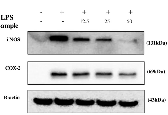
Western blot analysis was carried out to investigate to whether the inhibitory effect of BEMB on LPS-induced NO and PGE2 production were related to modulation of iNOS and COX-2 expression. In these experiments, treatment with LPS stimulated significantly iNOS and COX-2 protein expression. However, BEMB was inhibited iNOS and COX-2 in a dose-dependent manner. (Fig. 6.). Therefore, inhibition of NO and PGE2 production in LPS-induced RAW 264.7 cells is related to the inhibition of the iNOS and COX-2 protein.
22
Fig. 6. Effects of BEMB on LPS-induced iNOS and COX-2 protein expressions in RAW 264.7 cells. Cells stimulated with LPS (1 μg/ml) in the presence of BEMB (12.5, 25,
and 50 μM) for 24 h at 37°C. iNOS and COX-2 protein level were determined via Western blotting. (131kDa) (69kDa) (43kDa) i NOS COX-2 Β-actin
LPS
Sample
-
+ + + +
-
-
12.5 25 5023
3.4. Effects of BEMB on pro-inflammatory cytokines production in LPS-stimulated RAW 264.7 cells
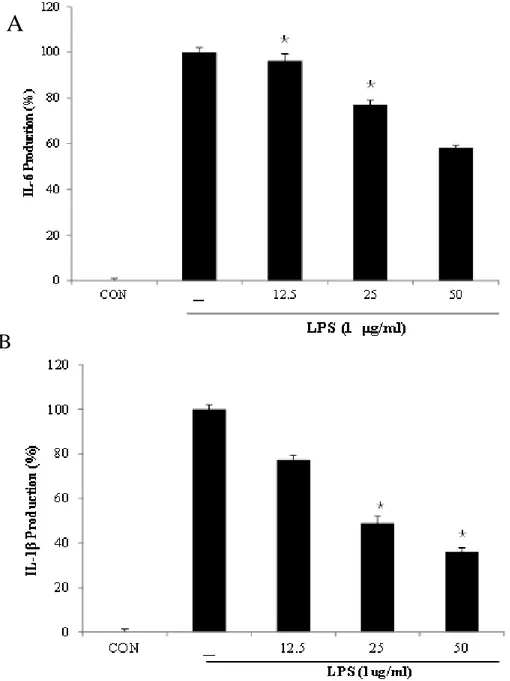
In this study, then further, evaluated the inhibitory activity of BEMB against pro-inflammatory cytokine (IL-1β, and IL-6) production in LPS-stimulated RAW 264.7 cells. The production of these cytokines was significantly increased by LPS than that seen. In contrast, pre-treatment with BEMB decreased the LPS-induced IL-1β and IL-6 production in a concentration-dependent manner. (Fig. 7.)
24
Fig. 7. Inhibitory effects of BEMB on the pro-inflammatory cytokine production in LPS-stimulated RAW264.7 cells. The production of (A) IL-1β, and (B) IL-6 were assayed
in the culture medium of cells stimulated with LPS (1 μg/ml) for 24 h in the presence of BEMB (12.5, 25, and 50 μM). Supernatants were collected, and the IL-1β, and IL-6 concentration in the supernatants were determined by ELISA kit. All the results were expressed as means ± S.E. of over three individual experiments; *P < 0.05.
A
25
3.5. Effect of BEMB on LPS-induced NF-kB activity in RAW264.7 cells
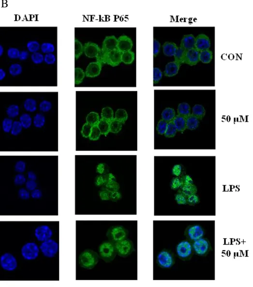
Nuclear factor-kB (NF-kB) is important transcriptional factors that may regulate a variety of inflammatory and immune responses. (Jiang XH et al, 2001) Accordingly, we were performed western blot to assay the effect of BEMB on the phosphorylation of NF-κB on in LPS-indcued RAW264.7 cell. BEMB was significantly blocked the phosphorylation P65 and p105 NF-κB activation induced by LPS. (Fig. 8A). In addition, the analysis of NF-kB translocation on RAW264.7 the nucleus induced by 30 minutes of LPS treatment was performed by confocal microscopy. Consequently, show in Fig. 8 B untreated cells expressing low levels of p65, cytoplasmically located p65 fluorescence were observed and LPS-induced cells showed the accumulation of p65 mainly in nuclear. Nuclear translocation of NF-κB p65 by LPS was markedly reduce in cultures treated with BEMB. (Fig. 8B) Thus, the result proved that anti-inflammatory effect of BEMB with LPS-induced RAW 264.7 cells involves the NF-κB pathway.
26 (65 kDa) p -p105 p-p65 (120 kDa) (43 kDa) Β-actin LPS Sample -
-
+
+
- 50 - 50A
27
Fig. 8. Inhibitory effects of BEMB on LPS-induced phosphorylation of NF-kB. (A)Cells
were treated for 30 min with LPS (1 μg/ml) alone or with LPS (1 μg/ml) coupled with different concentrations (12.5, 25, and 50 μM) of BEMB. Cell lysates were extracted, and protein levels of p-p65, p-p105 were analyzed by Western blot. (B) The analysis of NF-kB translocation on RAW264.7 the nucleus induced by 30 minutes of LPS treatment was performed by confocal microscopy (x 1000).
28
3.6. Effect of BEMB on LPS-induced ROS generation in RAW264.7 cells
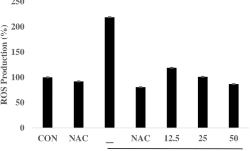
LPS induces ROS production in RAW264.7cells, important underlying LPS-elicited inflammatory responses. LPS-induced ROS production can be explained by several inflammatory mechanisms, for example iNOS, COX-2.(Dilshara MG et al, 2014) Therefore, we determined the effect of BEMB on ROS production with LPS-induced RAW macrophages using an oxidation sensitive fluorescent dye DCF-DA. BEMB inhibited LPS induced ROS production in a concentration-dependent manner and similar to the activity of antioxidants such as NAC. (Fig. 9). In addition, in this study investigated the roles of ROS in LPS-induced iNOS and COX-2 protein expression level by western blot. The result, showed in Fig. 10 A and B, decreased production of NO, and PGE2 and expression of iNOS and COX-2 protein, in the existence of the ROS inhibitors NAC. Accordingly, these results suggest that the inhibitory effects of BEMB on LPS-induced NO, PGE2, iNOS and COX-2 production are at least partially correlated with a decrease in ROS generation.
29
Fig. 9. Effect of BEMB and NAC on the generation of ROS in LPS-stimulated RAW264.7cells. RAW264.7 cells were seeded into 96-well plates at a starting density of
5×10⁴ cell/well and 18h cultured and pre-incubated with florescence dye DCF-DA for 1 h. Then, cells were pre-treated PSC14 (12.5, 25, 50 μM/ml), N-acetylcysteine (NAC) and treated with LPS (1 μg/ml) at after 2hour. Fluorescence was measured at 485nm excitation and 555nm emission wavelengths using fluorescent microplate reader. All the results were expressed as means ± S.E. of over three individual experiments; *P < 0.05.
0 50 100 150 200 250
CON NAC ㅡ NAC 12.5 25 50
ROS P rod u ction (% ) LPS 1 μg/ml
30
Fig. 10. Effect of BEMB and NAC on the production of NO, expression of iNOS and COX-2 in LPS-stimulated RAW264.7cells. Cells were pretreated with BEMB (12.5 25 and
50 μM) and NAC (2 mM) for 2 h and then incubated with stimulation with 1 μg/ml LPS. (A) Supernatants were collected, (A) NO production in the supernatants were determined by Griess reagent. (B) iNOS and COX-2 protein level were determined via Western blotting. All the results were expressed as means ± S.E. of over three individual experiments; *P < 0.05.
0 20 40 60 80 100 120
CON NAC LPS LPS+NAC
N O Pr o d u ct io n ( % )
C
B
A
31
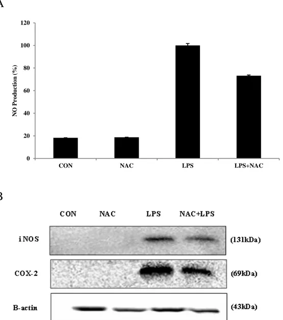
3.7. Effect of BEMB against LPS-induced NO production and expression of iNOS and COX-2 protein in zebrafish
In the study, we demonstrated BEMB inhibit LPS-induced inflammation by ROS and NF-κB of macrophages in vitro. Therefore, next, we investigated the in vivo anti-inflammatory effect of BEMB using the induced zebrafish model. The production of NO in the LPS-induced inflammatory zebrafish model was analyzed using DAF-FM DA. Fig. 11A illustrates the NO levels in zebrafish with or without LPS and or BEMB. These data show that, the NO level in LPS-stimulated zebrafish increased to 143.2% compared with the negative control group. However, the NO productions in zebrafish treated with different concentrations of BEMB (12.5, 25, and 50 μM) were reduced, and a significant reduction was observed at 50 μM.
iNOS and COX-2 are major inflammatory mediators. To determine the mechanism by which BEMB reduces LPS-induced NO production, we investigated the ability of BEMB (12.5, 25, and 50 μM) to influence the LPS-induced production of iNOS and COX-2. Fig. 11B shows the effect of BEMB on iNOS and COX-2 protein expression in zebrafish embryos by western blot analysis. The iNOS and COX-2 protein expressions were significantly increased in LPS-treated (10 μg/mL). However, BEMB significantly suppressed the protein expression of iNOS in a concentration-dependent manner and slightly inhibited COX-2 expression at 50 μM.
32
A
33
Fig. 11. Effect of BEMB on LPS-induced NO production and iNOS and COX-2 protein expressions in zebrafish. Zebrafish embryos stimulated with LPS (10 μg/ml) in the presence
of BEMB 3 dpf. (A) NO levels were measured by image analysis and fluorescence microscope. The fluorescence intensity was quantified using image J program. (B) iNOS and COX-2 protein level were determined via Western blotting. All the results were expressed as means ± S.E. of over three individual experiments; *P < 0.05.
(131kDa) (69kDa) (43kDa)
LPS
Sample
-
+ + + +
i NOS COX-2 Β-actin-
-
12.5 25 50B
34
3.8. Effects of BEMB and NAC against LPS-induced ROS production in zebrafish embryos
The production of ROS in the LPS-induced inflammatory zebrafish model was analyzed using DCF-DA. In this study assessed the inhibitory activity of BEMB on LPS-stimulated ROS production in zebrafish. The level of ROS production in the positive group (only LPS treated) reached to 153% of that observed in controls. However, zebrafish treated with 50 μM BEMB had significantly reduced ROS levels. Also, BEMB inhibited LPS induced ROS production in a concentration-dependent manner and similar to the activity of antioxidants such as NAC. (Fig. 12 A,B). In order to identify of ROS involved in inflammatory that treated NAC in zebrafish induced by LPS. The result, NAC inhibited NO production and iNOS, COX-2 protein expression in LPS-stimulated zebrafish embryos. (Fig. 12 C,D). These data indicate that the inhibition of LPS-induced iNOS and COX-2 expression through the modulation of ROS generation in zebrafish embryos. In this study first identified in ROS role in inducing pro-inflammatory mediator (NO, iNOS and COX-2) by LPS in zebrafish embryos.
35
A
36
Fig. 12. Effects of BEMB and NAC on LPS-induced ROS and NO Production in Zebrafish. Zebrafish embryos stimulated with LPS (10 μg/ml) in the presence of BEMB and
NAC (250 μM) 3 dpf. (A), BEMB ROS level (B) NAC ROS level (C) NAC NO level. Assessments were measured by image analysis and fluorescence microscope. The fluorescence intensity was quantified using image J program. (D) iNOS and COX-2 protein level were determined via Western blotting. All the results were expressed as means ± S.E. of over three individual experiments; *P < 0.05.
37
3.9. Effect of BEMB on LPS-induced NF-kB activity in zebrafish.
In this study performed realtime-PCR to assay the effect of BEMB of NF-κB on induced LPS in zebrafish (3 dpf). After 3dpf exposure in vivo, p65, p100/p52 mRNA level in zebrafish was detected by Quantitative RT-PCR. As shown in Fig. 13, the relative content of p65 was highest at LPS, and the level of p65 and p100/p52 at this exposure concentration was 8-fold of its level in the control. However, zebrafish treated with BEMB dependence on concentration had reduced p65 and p100/p52 NF-κB gene expression. The inhibitory effect of ikbaA levels was slightly but not significantly reduced by BEMB in LPS-induced zebrafish models. As a result, BEMB can be seen that there are anti-inflammatory effects by controlling the NF-kb in in vivo model zebrafish.
38
Fig. 13. Effect of BEMB on LPS-induced NF-kB activity in zebrafish. Gene expression
(mRNA) of P65, P100/P52 and IκbaA evaluated by RT-PCR. Zebrafish embryos stimulated with LPS (10 μg/ml) in the presence of BEMB (50 μM) 3 dpf. Bars indicate mean±standard error of three independent experiments.
39 4. Discussion
In this study, we first demonstrated the anti-inflammatory activity of BEMB from P. japonica in LPS-induced in in vitro RAW264.7 cells and in vitro zebrafish embryos. The LPS-induced expression of iNOS and COX-2 in microphages plays a key role in the pathogenesis of inflammatory conditions (Jung et al. 2013). Especially, the regulation between iNOS and COX-2 is involved in inflammatory diseases such as sepsis and arthritis (Gehrmann et al. 1995). Overproduction of NO by iNOS can connect to cytotoxicity, inflammation, and the development of autoimmune disorders (Liu ea al. 1995, Shin et al 2008). PGE2 is strongest inflammatory mediators in inflammatory response. And, it was transformed from COX2 (Chun et al. 2006). Therefore, we examined the inhibitory activity of BEMB on NO and PGE2 production in RAW264.7 cells via the suppression of iNOS and COX-2 expression. We found that BEMB significantly inhibits the NO and PGE2 production and iNOS and COX-2 expression in LPS-induced RAW264.7 cells.
In addition, BEMB also significantly inhibited the LPS-induced production of IL-6 and IL-1 β. IL-1β can stimulate the production of a host of other cytokine, they have earned a position of prominence at the head of the inflammatory cytokine cascade (Jian Wang et al. 2015). Moreover, IL-1β increases the expression of adhesion factors on endothelial cells to enable transmigration of leukocytes (Huo M et al. 2013).
NF-κB-mediated regulation of pro-inflammatory gene expression is controlled by interactions with the IκB family (Kempe et al. 2005). NF-κB is a transcription factor that regulates the expression of pro-inflammatory mediators, including COX-2, iNOS, IL-1 and IL-6 showed that degradation and phosphorylation of IκB, a cytoplasmic inhibitor of NF-κB was induced by inflammatory stimuli, resulting in the translocation of the activated p65 the
40
cytoplasm to the nucleus (Parul.R et 2005). Our data indicated that BEMB suppression by LPS induced IκB-α phosphorylation and degradation, and the activation of NF-κB p105. (Kim et al. 2010, Rhee et al. 2007. Henkel et al. 1993). In this study, we found that BEMB inhibited LPS-induced NF-κB activation and nuclear translocation of NF-κB p65. These results revealed that the effects of BEMB on inflammatory mediator and cytokine production are mediated via NF-κB pathway inhibition.
ROS production is central to the progression of many inflammatory diseases. Many studies have shown that the ROS is dual role a signaling molecule and a mediator of inflammation. (Zhang et al. 2014). Increased ROS generation can damage biological molecules and lead to cells or tissue injury (Rezaie et al. 2007, Ziegler et al. 2011) as well as induces inflammation by increasing the level of cytokines such as IL-1β, IL-6, and TNF-α (Geronikaki et al. 2006). BEMB inhibited LPS induced ROS production in a concentration-dependent manner. Therefore, the BEMB has an effect in anti-inflammation regulate as both a NF-kB signaling and ROS production.
Recently, zebrafish model have been use to rapidly and simply assess anti-inflammatory activity on LPS-stimulated inflammation in vivo (Park et al. 2011). The advantages of zebrafish xenotransplantation have been demonstrate in several studies in which in vivo fluorescent imaging was used to evaluate tumorigenesis, inflammatory, and metastatic phenotype. (Lee et al. 2015) Transformation of DCF-DA by ROS in the presence of dioxygen generates highly fluorescent triazole derivatives (Itoh et al. 2000, Bölck et al. 2014). Production of intracellular ROS can be detection using the oxidation sensitive fluorescent dye, DCF-DA, emits fluoresce upon interaction with ROS (Handa et al. 2006). We assessed the inhibitory activity of BEMB on LPS-stimulated NO production in zebrafish using a
41
fluorescent probe dye, DAF-FM DA. Transformation of DAF-FM DA by NO in the presence of dioxygen generates highly fluorescent triazole derivatives (Itoh et al. 2000, Bölck et al. 2014). Accordingly, ROS and NO generation were assessed in LPS-induced inflammation zebrafish model. In the present study, ROS and NO production in zebrafish were increased with the LPS treatment. However, BEMB pre- treatment significantly reduced the LPS-induced ROS and NO production to the same extent as the anti-oxidant NAC, which alone inhibited the LPS-induced expression of iNOS and COX-2 in the zebrafish embryos. Moreover, BEMB attenuated the LPS-induced NF-κB p65 and p100/p52 in the zebrafish embryos.
In conclusion, our results demonstrate that BEMB significantly inhibited the levels of pro-inflammatory mediators, including NO, PGE2, iNOS, COX-2, IL-6, and IL-1β in LPS-induced RAW264.7 cells. Also, BEMB suppressed the production of NO and the expression of iNOS and COX-2 in LPS-stimulated zebrafish embryos. Moreover, BEMB reduced ROS generation and NF-κB activation in LPS-induced RAW 264.7 cells and zebrafish embryos. Taken together, these results indicate that BEMB exert its anti-inflammatory action in vitro and in vivo through attenuation of ROS and NF-κB signaling pathways. Therefore, we suggest that BEMB should be utilized as potential anti-inflammatory agents for the treatment of inflammatory diseases.
42
REFERENCES
Allan V. Kalueff, Adam Michael Stewart, and Robert Gerlai. (2014). Zebrafish as an emerging model for studying complex brain disorders. Trends Pharmacol Sci. 35(2): 63–75)
Briggs JP. (2002). The zebrafish: a new model organism for integrative physiology. Am J Physiol Regul Integr Comp Physiol 282: R3–R9.
Bubici C, Papa S, Pham CG, Zazzeroni F, Franzoso G.(2006). The NF-kappaB-mediated control of ROS and JNK signaling. Histol Histopathol.21(1). 69-80.
Couture R, Harrisson M, Vianna RM, Cloutier F. Kinin receptors in pain and inflammation Eur J Pharmacol 2001;429:161–76.
Chun Seung-Chu,Jee Seon Young , Lee Sang Gon, Park Sook Jahr, Lee Jong Rok, Kim Sang Chan. (2007). Anti-Inflammatory Activity of the Methanol Extract of Moutan Cortex in LPS-Activated Raw264.7 Cells. Evidence-Based Complementary and Alternative Medicine. 4(3)327–333
Craig PM, Wood CM, McClelland GB. (2007). Oxidative stress response and gene expression with acute copper exposure in zebrafish (Danio rerio).Am J Physiol Regul Integr Comp Physiol. 293(5):R1882-92.
Dilshara MG, Lee KT, Choi YH, Moon DO, Lee HJ, Yun SG, Kim GY. (2014).Potential chemoprevention of LPS-stimulated nitric oxide and prostaglandin E2 production by a-L-rhamnopyranosyl-(1-6)-ß-D-glucopyranosyl-3-indolecarbonate in BV2 microglial cells through suppression of the ROS/PI3K/Akt/NF-kB pathway. Neurochem Int.
43
El Gamal, A. A. (2010) .Saudi pharmaceutical journal : SPJ : the official publication of the Saudi Pharmaceutical Society. Biological importance of marine algae, 8, 1-25.
Fan SH, Zhang ZF, Zheng YL, Lu J, Wu DM, Shan Q, Hu B, Wang YY. (2009). Troxerutin protects the mouse kidney from d-galactose-caused injury through anti-inflammation and anti-oxidation. Int Immunopharmacol.9.1,91-96
.
Hao Tang, Hui Ge,Zhi-Bin Chen, Xiong-Ming Luo, Feng-Juan Su, Yan-Bing Liang, Zhen-Yu Li, Jing-Guo Wu,Qing Yang, Li-Jin Zeng, Zhong-Fu Ma. (2015). Micrometam C Protects against Oxidative Stress in Inflammation Models in Zebrafish and RAW264.7 Macrophages. Marine Drugs.13(9), 5593-5605
Huo M, Gao R, Jiang L, Cui X, Duan L, Deng X, Guan S, Wei J, Soromou LW, Feng H, Chi G. (2013). Suppression of LPS-induced inflammatory responses by gossypol in RAW 264.7 cells and mouse models. Int Immunopharmacol. 15(2):442-9.
Jiang XH, Wong BC, Lin MC, Zhu GH ,Kung HF, Jiang SH, Yang D, Lam SK.(2001).Functional p53 is required for triptolide-induced apoptosis and AP-1 and nuclear factor-kappaB activation in gastric cancer cells.20(55):8009-18.
Jung HA, Jin SE, Ahn BR, Lee CM, Choi JS.(2013). Anti-inflammatory activity of edible brown alga Eisenia bicyclis and its constituents fucosterol and phlorotannins in LPS-stimulated RAW264.7 macrophages.Food Chem Toxicol. 59:199-206
Kang, M. -C., Kim, K. -N., Wijesinghe, W. A. J. P., Yang, X., Ahn,G. & Jeon, Y. -J. (2014). Protective effect of polyphenol extracted from Ecklonia cava against ethanol induced oxidative damage in vitro and in zebrafish model. J. Funct.Foods 6:339-347.
44
Kempe S1, Kestler H, Lasar A, Wirth T. (2005). NF-kappaB controls the global pro-inflammatory response in endothelial cells: evidence for the regulation of a pro-atherogenic program. Nucleic Acids Res. 21;33(16).
Kim MJ, Lee HH, Jeong JW, Seo MJ, Kang BW, Park JU, Kim KS, Cho YS, Seo KI, Kim GY, Kim JI, Choi YH, Jeong YK. (2014). Anti-inflammatory effects of 5-hydroxy-3,6,7,8,3',4'-hexamethoxyflavone via NF-κB inactivation in lipopolysaccharide-stimulated RAW 264.7 macrophage. MOLECULAR MEDICINE REPORTS 9: 1197-1203.
Ko, J. -Y., Kim, E. -A., Lee, J. -H., Kang, M. -C., Lee, J. -S., Kim,J. -S., Jung, W. -K. & Jeon, Y. -J. (2014). Protective effect of aquacultured flounder fish-derived peptide against oxidative stress in zebrafish. Fish Shellfish Immunol.36:320-323.
Kudo, T. & Masuda, M. (1986). A taxonomic study of Polysiphonia japonica Harvey and P. akkeshiensis Segi (Rhodophyta). Japanese Journal of Phycology 34: 293-310.
Li, Y.-X., Wijesekara, I., Li, Y. & Kim, S.-K. (2011). Phlorotannins as bioactive agents from brown algae, Process Biochemistry. 46, 2219-2224.
Lee Seung-Hong ,YangHye-Won ,Yuling Ding, Yanmei Wang, Jeon You-Jin , Moon Sang-Ho, Jeon Byong-Tae, Sung Si-Heung. (2015). Anti-inflammatory effects of enzymatic hydrolysates of velvet antler in RAW 264.7 cells in vitro and zebrafish model. EXCLI J. 14: 1122–1132.
Kempe S1, Kestler H, Lasar A, Wirth T. (2005). .NF-kB controls the global pro-inflammatory response in endothelial cells: evidence for the regulation of a pro-atherogenic program. Nucleic Acids Res. 33(16):5308-19
Mariana DOMINGUEZ, Anna S. KECK, Elizabeth H. JEFFERY,Carlos L. CESPEDES. (2010). Effects of extracts, flavonoids and iridoids from Penstemon gentianoides (Plantaginaceae) on
45
inhibition of inducible nitric oxide synthase (iNOS), cyclooxygenase-2 (COX-2) in LPS-Activated RAW 264.7 macrophage cells and their antioxidant activity. Boletín Latinoamericano y del Caribe de Plantas Medicinales y Aromáticas, 9 (5), 397 - 413
Medzhitov, R. (2008). Origin and physiological roles of inflammation. Nature. 454, 428-35.
Mittal M1, Siddiqui MR, Tran K, Reddy SP, Malik AB.(2014).Reactive oxygen species in inflammation and tissue injury. Antioxid Redox Signal. 1;20(7):1126-67.
Park, K. -H. & Cho, K. -H. 2011. A zebrafish model for the rapid evaluation of pro-oxidative and inflammatory death by lipopolysaccharide, oxidized low-density lipoproteins,and glycated high-density lipoproteins. Fish Shellfish Immunol. 31:904-910.
Patcharawan Sittisart, Benjamart Chitsomboon. (2014). Intracellular ROS Scavenging Activity and Downregulation of Inflammatory Mediators in RAW264.7 Macrophage by Fresh Leaf Extracts of Pseuderanthemum palatiferum. Evidence-B.ased Complementary and Alternative Medicine. 309095, 11.
Rezaie, A., Parker, R. D. & Abdollahi, M. (2007). Oxidative stress and pathogenesis of inflammatory bowel disease: an epiphenomenon or the cause? Dig. Dis. Sci. 52:2015-2021.
Tak PP1, Firestein GS. (2001). NF-kappaB: a key role in inflammatory diseases. J Clin Invest. 107(1):7-11.
Tsai HH, Lee WR, Wang PH, Cheng KT, Chen YC, Shen SC. (2013). Propionibacterium acnes-induced iNOS and COX-2 protein expression via ROS-dependent NF-κB and AP-1 activation in macrophages. Journal of Dermatological Science. 69(2):122-31.
Wijesekara I, Pangestuti R, Kima S. (2011). Biological activities and potential health benefits of sulfated polysaccharides derived from marine algae. Carbohydr Polym .84:14–21.
46
Zhang B1, Shimada Y2, Kuroyanagi J1, Umemoto N3, Nishimura Y2, Tanaka T2.(2014). Quantitative phenotyping-based in vivo chemical screening in a zebrafish model of leukemia stem cell xenotransplantation. PLoS One.15;9(1)
Zhang, D. L., Hu, C. X., Li, D. H. & Liu, Y. D. (2013). Lipid peroxidation and antioxidant responses in zebrafish brain induced by Aphanizomenon flos-aquae DC-1 aphantoxins. Aquat. Toxicol. 144-145:250-256.
Ziegler, F., Seddiki, L., Marion-Letellier, R., Lavoinne, A. & Déchelotte, P. (2011). Effects of L-glutamine supplementation alone or with antioxidants on hydrogen peroxide-induced injury in human intestinal epithelial cells. E-Spen Eur. E-J. Clin. Nutr. Metab. 6:e211-e216.
47 ACKNOWLEGEMENT 부족한 저에게 관심을 주시고, 2년이라는 석사과정을 마치고 본 논문이 있기까 지 세심한 가르침과 사랑을 주신 전유진 교수님께 진심으로 감사드립니다. 또한 바쁘신 와중에도 이 논문이 좀 더 나은 방향으로 나아 갈 수 있게 심사해 주시고 관심을 보여주신 여인규 교수님, 이제희 교수님, 허문수 교수님, 정준범 교수님, 이 승헌 교수님께도 깊은 감사를 드립니다. 더불어 한국기초과학지원연구원에서 연구를 할 수 있게 해주신 김대경 부장님과 저에게 많은 가르침과 도움을 주신 김길남 박사님께 깊은 감사를 드립니다. 저에게 연구를 할 수 있게 처음 소개해준 민철오빠 와 희진이, 이번 졸업하면서아 주 많은 도움을 주신 허수진 박사님, 실험실 회식자리에서 자주 보게되는 승홍오 빠, 석천오빠, 여러방면에 대해 조언을 많이 주신 긴내언니, 항상 좋은 말과 맛있 는 음식을 많이 알려주시는 원우오빠, 실험실 갈 때마다 반갑게 맞아주시고 도움 을 주신 지혁오빠, 주영언니, 같은 파트타임 창익오빠, 실험실 들어오고 나서부터 많은 도움을 받음 은아, 나래, 동갑이지만 어색한 오재영, 나이는 어리지만 실험실 생활에 많이 배우고 도움 받은 서영와 혜원이, 회식자리서 술만 먹인 것 같은 현 수와 윤택이, 초반 연애 상담만 했던 것 같은 형호, 먼 나라에서 온 왕뢰 ,아산카, 산지와, 실험 마무리 할 때쯤 엄청 부탁만 한 효근, 연구자의 길을 걸을 유선이, 이제까지 나와 함께 실험했던 수현이 등 모든 해양생물자원이용공학 연구실 식구 들께 감사의 말씀을 전하고 싶습니다. 또한, 한국기초과학지원연구원에서 만난 모든 선생님들과 같은 연수학생들에게도
48 추억을 쌓을 수 있어 행복했고, 많은 도움을 주셔서 감사드립니다. 서울, 제주 왔다갔다 하며 늦은 나이에 대학원 공부를 하겠다는 딸에게 아낌없는 사랑과 지원을 해주시고, 저에게 많은 용기를 주신 저희 가족들, 사랑하고 감사합 니다. 내 늦은 도전과 결정에 용기를 주고 내 투정들을 받아 준 친구들과 언제나 뒤에서 묵묵히 있어준 재민이에게도 감사함과 사랑한다는 말을 전하고 싶습니다. 감사한 분들이 너무 많은데, 이 곳에 모두 거명하지 않더라도 여러 가지 모습으 로 도움을 주신 분들에게 모두 감사드리며 항상 건강하고 좋은 일만 있기를 바랍 니다. 사랑합니다.