63
Ultrasonographic and Clinical Findings in Cats with Feline
Lower Urinary Tract Disease
Seongeun Seo*,**, Hyemin Na*, Sooyoung Choi***, Hojung Choi****, Yungwon Lee**** and Kija Lee*1
*College of Veterinary Medicine, Kyungpook National University, Daegu 41566, Korea **Dasom Feline Medical Center, Busan 48415, Korea
***College of Veterinary Medicine, Kangwon National University, Chuncheon 24341, Korea ****College of Veterinary Medicine, Chungnam National University, Daejeon 34134, Korea (Received: December 14, 2020 / Revised: February 03, 2021 / Accepted: March 17, 2021)
Abstract : Urethral obstruction is a life-threatening feline lower urinary tract disease (FLUTD). The rate of recurring urethral obstruction was 14.8-58.1% after the first occurrence. Ultrasonographic findings associated with reobstruction had been rarely reported although ultrasonography was a valuable technique for diagnosing urinary bladder calculi and distinguishing different FLUTD causes. This retrospective study aims to describe the ultrasonographic findings, urinalysis, and serum chemistry profile in cats with FLUTD and determine the associations of reobstruction with ultrasonographic findings, urinalysis, and serum chemistry profile. The present study included 141 cats that were followed up for more than 1 year. The ultrasonographic criteria included the presence of cystolithiasis, urine echogenicity, sediment, suspended linear strand, pericystic effusion, hyperechoic pericystic fat, ureteral dilation, pyelectasia, and perirenal effusion. The urinalysis criteria included hematuria, urine-specific gravity, pH, sediment, and proteinuria. The most common ultrasonographic findings in cats with FLUTD were echogenic urine and sediment. However, this study did not find an association between reobstruction and ultrasonographic findings, urinalysis, and serum chemistry profiles. Thus, an ultrasonographic examination may be insufficient to predict the risk of reobstruction although it is a useful modality for diagnosing FLUTD and making treatment direction.
Key words : cat, feline lower urinary tract disease, reobstruction, ultrasonography, urinalysis.
Introduction
Urethral obstruction is a life-threatening feline lower uri-nary tract disease (FLUTD). FLUTD includes disorders affect-ing the urethra and/or urinary bladder. The common causes of FLUTD are idiopathic cystitis, urethral plug, urolithiasis, urethral stricture, and idiopathic obstruction. Feline idiopathic cystitis is the most commonly diagnosed in young cats with FLUTD, occurring in approximately 50-60% of cases. Ure-thral plugs are diagnosed in 10-20% of FLUTD (15,26,28,31). Clinical signs of FLUTD include inappropriate urination, stranguria, pollakiuria, hematuria, vomiting, abdominal pain, vocalization, and lethargy (14). Many theories suggest that FLUTD or feline idiopathic cystitis are associated with age, gender, stress, urothelial abnormality, neuroendocrine disor-der, diets, indoor/outdoor living condition including the num-ber of cats in the household, and the numnum-ber of litter boxes and their location (4,12,16). However, the causative factor of FLUTD or feline idiopathic cystitis is still unknown (11,31). After the first occurrence of urethral obstruction, the rate of recurring urethral obstruction is between 14.8-58.1% (20,26). Previous studies were performed to identify risk factors of recurrence according to treatment protocol (7,14,17). In addi-tion, the association between laboratory findings and the
recurrence of FLUTD has been studied (14,24). One study reported that increasing amounts of red blood and epithelial cells increased FLUTD recurrence (24). Meanwhile, another study reported that no specific laboratory abnormalities were associated with recurrent urethral obstruction (14).
Ultrasonography is a useful modality for diagnosing the causes of FLUTD (30). If ultrasonographic findings and uri-nalysis can predict reobstruction, it may be clinically mean-ingful for monitoring cats with previous history of FLUTD. However, only one study evaluated the association of FLUTD with ultrasonographic findings and blood analysis (26). More-over, ultrasonographic findings at the time of hospitalization did not significantly predict uretheral reobstruction and only 87 cats with urethral obstruction were included in this study (26). This study hypothesized that studies with a larger num-ber of cats with FLUTD may have different results. The pur-poses of this retrospective study were to describe ultrasono-graphic findings, urinalysis, and serum chemistry profile in cats with FLUTD, and determine whether ultrasonographic findings, urinalysis, and serum chemistry profile at the first time of hospitalization can predict uretheral reobstruction.
Materials and Methods
Animals
This retrospective study included feline patients with FLUTD presented to the Dasom Feline Medical Center between September 2017 and April 2019. Approval by the 1Corresponding author.
Animal Care and Use Committee of the institution of this study was not required due to the retrospective nature of the study. All feline owners signed informed consent to use their cats’ data in research. Only cats with FLUTD who per-formed with ultrasonography and had been followed up for more than a year were included in this study. The medical records were reviewed, and breed, age, gender, body weight, history consistent with urethral obstruction, clinical signs, ultrasonographic findings including a full evaluation of the urinary system, results of urinalysis and serum chemistry profile, treatment, and recurrence of urethral obstruction were recorded.
Urethral catheters were placed after the administration of anesthesia with medetomidine (Domitor®, Orion Corporation Animal Health, Finland) and propofol (Provive®, Myung-moon Pharm, Korea) or isoflurane (Ifran liquid, Hana Pharm, Korea). Urine was collected to perform urinalysis after cath-eterization and flushing of the bladder was performed in all cats except for the spayed females. The treatment included urine output monitoring, intravenous fluid therapy and medi-cation including antibiotics, analgesics, and adjuvants during hospitalization. Urethral recatheterization or perineal ure-throstomy were done if urethral obstruction recurred during hospitalization. Consequently, a follow-up examination was performed for 1 year to confirm the reobstruction.
Ultrasonographic evaluation
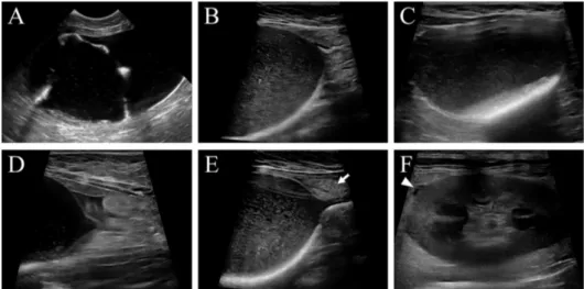
Ultrasonographic examination was performed using a com-mercially available ultrasound machine (RS80A, Samsung Medicine, Seoul, Korea) with blended frequencies of 4-9 MHz convex transducer and 3-16 MHz linear transducer. Moreover, ultrasound images were retrieved from the Picture Archiving and Communication System. Ultrasound images were evaluated blinded to the detailed history other than the urethral obstruction and laboratory findings. Ultrasonographic images of the urinary system were reviewed except for the urethra due to catheterization at the time of examination. Ultrasonographic findings are shown in Fig 1.
The presence of cystolithiasis (yes/no); urine echogenicity
classified as normal, mild, moderate, and severe echogenics; and the presence of urine sediment (yes/no), suspended lin-ear strand (yes/no), pericystic effusion (yes/no), and hypere-choic pericystic fat (yes/no) were the recorded urinary bladder findings. The bladder wall thickness was not measured because of overdistension with urine at the time of examina-tion. Additional abnormalities that were seen but not listed above were also recorded when identified. Moreover, uret-eral dilation, pyelectasia, and perirenal effusion (yes/no) were also recorded. Ureteral dilation is defined as the diameter of the ureter exceeding 4 mm and pyelectasia is defined as the dilated renal pelvis over 3.4 mm (2,10).
Urinalysis and serum chemistry profile
Urinalysis included hematuria, urine specific gravity, pH, sediment, and proteinuria. Urine was collected by urethral catheterization, and urinalysis was conducted within 10 min after collection. Consequently, urinalysis was performed using a urine-stick to check for hematuria, pH, and proteinuria. Urine-specific gravity was performed using a urinometer. Based on previous studies, a pH of 6.0-6.4 and urine-spe-cific gravity of > 1.035 was used as reference ranges (8,27). The sediment was microscopically examined after urine cen-trifugation at 1,000 rpm for 2 min.
Serum chemistry profile included blood urea nitrogen (BUN), creatinine, and potassium concentration using DRI-CHEM NX500 (Fujifilm, Tokyo, Japan). The normal range of BUN, creatinine and potassium are 15-32 mg/dL, 0.6-2.0 mg/dL and 3.6-4.6 mEq/L, respectively (14,19,21). This study defined azotemia and severe azotemia as the serum creati-nine concentrations of 2.1 and 5.0 mg/dL with increased BUN, respectively. Moreover, hyperkalemia and severe hyper-kalemia were defined as the serum potassium concentrations of 4.7 and 8 mEq/L, respectively (14,22,26). However, some examinations could not be performed in emergency cases.
Statistical analysis
Statistical analyses were commissioned by experts and
per-Fig 1. Ultrasonographic findings in cats with FLUTD. Suspended linear strand lined along bladder wall (A), severe urine echogenicity and sediment (B), hyperechoic cystolith with acoustic shadowing (C), and pericystic effusion (D) are noted. Hyperechoic pericystic fat (E, arrow) and a small amount of perirenal effusion (F, arrowhead) are also found.
formed using commercial software (SPSS 25.0, IBM SPSS statistics, USA). Multiple logistic regression analysis was performed to identify associations of reobstruction with ultra-sonographic findings, urinalysis, and serum chemistry pro-file in recurrence and nonrecurrence patients. A P-value of < 0.05 was considered statistically significant.
Results
The present study included 141 cats diagnosed with FLUTD. The predominant breeds were Domestic short hair (77/141, 54.6%), Russian blue (17/141, 12.1%), Persian (13/141, 9.2%) and Scottish fold (11/141, 7.8%). Some breeds also repre-sented were Siamese (3/141), Abyssinian (2/141), British short hair (2/141), Scottish straight (2/141), Turkish angora (4/141), American short hair (2/141), American curl (1/141), Bengal (1/141), British long hair (1/141), Kinkalow (1/141), Norwegian forest (1/141), Ragdoll (1/141), and mixed (2/ 141). The mean age of the 136 cats was 4.32 years (range, 0.67-13 years). Five stray cats had inaccurate age data. Of the cats, 139 were male (6 intact and 133 castrated) and two were spayed females. The mean body weight was 5.76 kg
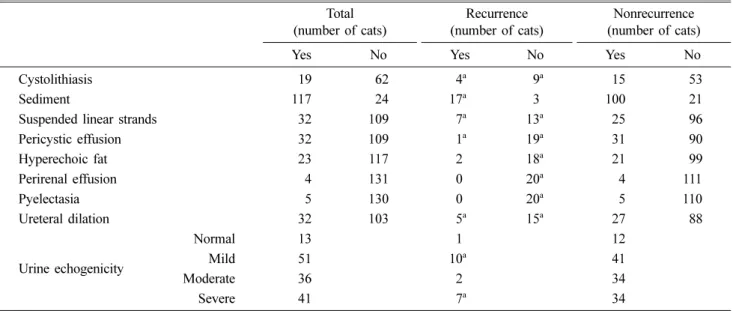
Table 1. Ultrasonographic findings of 141 cats, including 20 recurrent cats Total
(number of cats) (number of cats)Recurrence (number of cats)Nonrecurrence
Yes No Yes No Yes No
Cystolithiasis 019 062 04a 09a 015 053
Sediment 117 024 17a 03 100 021
Suspended linear strands 032 109 07a 13a 025 096
Pericystic effusion 032 109 01a 19a 031 090 Hyperechoic fat 023 117 02 18a 021 099 Perirenal effusion 004 131 00 20a 004 111 Pyelectasia 005 130 00 20a 005 110 Ureteral dilation 032 103 05a 15a 027 088 Urine echogenicity Normal 013 01 012 Mild 051 10a 041 Moderate 036 02 034 Severe 041 07a 034
aIncluded data on three cats having twice recurrence Table 2. Results of urine and blood analysis in cats
Recurrence Nonrecurrence
Total Mean ± SD (range) Total Mean ± SD (range)
USG 14 1.040 ± 0.013 (1.015-1.070) 060 1.032 ± 0.011 (1.009-1.053) pH 13 6.62 ± 0.52 (6-8) 062 6.89 ± 0.59 (5-8) Proteinuria 07 039 Hematuria 13 064 Urine sediment 10 017 BUN (mg/dL) 1861.27 ± 46.8a (18-140a) 11875.31 ± 49.30a (11-140a) Creatinine (mg/dL) 18 4.42 ± 3.96 (0.8-15.5) 117 6.83 ± 6.44a (0.8-24a) Potassium (mEq/L) 184.32 ± 1.09 (2.707.0) 109 5.00 ± 1.82 (3.0-10.0)
SD, standard deviation; USG, urine-specific gravity
aDiagnostic equipment cannot read more than 140 of BUN and 24 of creatinine
(range, 2.67-10.35 kg). All patients had dysuria. Other symp-toms included vomiting (17/141), diarrhea (1/141), anorexia (13/141), salivation (1/141), tachypnea (2/141), and over-grooming of the penis (2/141).
Only part of the ultrasonographic records in some cats could be reviewed because of the retrospective nature of the study. The abnormal ultrasonographic findings included cysto-lithiasis (19/81, 23.5%), echogenic urine (128/141, 90.1%), sediment (117/141, 83.0%), suspended linear strand (32/141, 22.7%), pericystic effusion (32/141, 22.7%), perirenal effu-sion (4/135, 3.0%), pyelectasia (5/135, 3.7%), ureteral dila-tion (32/135, 23.7%), and hyperechoic pericystic fat (23/140, 16.4%). The abnormal ultrasonographic findings in recur-rence and nonrecurrecur-rence patients are summarized in Table 1. The results of urinalysis and serum chemistry profile in recurrence and nonrecurrence patients are summarized in Table 2. The total number of cats performed with each test varied. Hematuria was observed in 77 of 97 cats (79.4%). The mean urine-specific gravity of 74 cats was 1.033 (range, 1.009-1.070) and mean pH of 75 cats was 6.84 (range, 5.0-8.0). Moreover, 46 of 52 cats (88.5%) were observed with proteinuria. Urine sediments were observed in 27 of 29 cats
(93.1%) and most sediments were identified as struvite on urine sediment examination. Of the 135 cats, 89 (65.9%) and 59 (43.7%) were azotemic and severely azotemic, respec-tively. In addition, 48 (37.8%) and 13 (10.2%) were hyperka-lemic and severely hyperkahyperka-lemic, respectively.
Reobstruction was observed in 20 cats. Recurrence was observed twice in three cats. No statistically significant asso-ciation was noted between reobstruction and ultrasonographic findings, serum chemistry profile, or urinalysis results at first hospitalization. The first reobstruction occurred most fre-quently after 1 (6/20, 30%) and 5 months (5/20, 25%). Three of the 20 cats experienced recurrence within 1 week (3/20, 15%). Consequently, another three of 20 cats experienced recurrence within 1 month (3/20, 15%). Each of the 20 cats experienced recurrence after 8, 12, and 16 months, respec-tively. In three cats who experienced reobstruction twice, each cat had recurrence at 1 and 2 months, 2 weeks and 1 month, and 5 and 7 months after the first time of hospitalization.
Small cystoliths were removed in 19 cats through catheter by flushing or managing by dietary prescription rather than cystectomy. There were 2 cats with clinical presentation con-sistent with Pandora syndrome. One cat was diagnosed as Pandora syndrome due to clinical presentation including loss of appetite, refractory cystitis, calicivirus infection, anemia, and cholecystitis. After treatment for 1 month, this cat im-proved but had mild clinical symptoms and did not develop reobstruction for 1 year. The other cat had increased hepatic enzyme concentration and calicivirus infection, and died of Pandora syndrome. These 2 cats with Pandora syndrome extended their hospitalization for 2 weeks and received symptomatic treatment including fluid therapy, antibiotics, adjuvants, urethrostomy or urethral catheter placement. Two cats with Pandora syndrome extended their hospitalization for 2 weeks. One cat died of Pandora syndrome and the other had improved clinical symptoms after 1 month. The mortal-ity due to FLUTD among 140 cats was 0.71% (1/140) except for the patient that succumbed due to a lung tumor. The mean time of hospitalization was 4.6 days (range, 0-18 days). Moreover, diets were mostly composed of wet food, and all patients were similarly treated with fluid therapy, and admin-istrations of antibiotics, analgesics, and adjuvants during hos-pitalization.
Discussion
Ultrasonography is a valuable technique for diagnosing urinary bladder calculi and/or plugs and distinguishing the different causes of FLUTD (30). The common ultrasono-graphic findings in cats with feline urethral obstruction are urine sediments, bladder wall thickening, hyperechoic peri-cystic fat, and periperi-cystic effusion (26). In addition, common ultrasonographic findings of nonobstructive FLUTD are urine sediments and bladder wall thickening (30). Moreover, the most common ultrasonographic findings in cats with FLUTD in the present study were echogenic urine and sediments. Because idiopathic cystitis is the main cause of FLUTD (12), and ultrasonographic findings which are common in cystitis may be also common in FLUTD patients. Most patients in the present study showed severe bladder dilation due to
ure-thral obstruction on ultrasonography. Thus, bladder wall thick-ness could not be evaluated. Perirenal effusion and pyelectasia were observed at a lower frequency and were not identified in recurrence patients. The dilation of the ureter and pelvis was associated with the duration of obstruction in humans (9). Therefore, if the duration and severity of obstruction are not severe in cats, renal findings (e.g., perirenal effusion and pyelectasia) may not be seen. In addition, identifying associ-ations may not be enough because of the small number of recurrent patients and only nine cats with abnormal renal findings.
Previous studies reported that lower urine-specific gravity and higher urine protein were detected in FLUTD patients (1,29). The results of the present study are similar to previ-ous studies, except for the absence of difference in urine-spe-cific gravity and frequency of proteinuria between recurrence and nonrecurrence patients. Moreover, urine sediments were microscopically found in 27 cats. Consequently, most sedi-ments, especially crystals, were identified as struvite by micro-scopic examination. Struvite is associated with urine pH and formed well at high pH. The acidic condition with low pH decreases precursors available to form uroliths and increases struvite solubility in urine (13). However, struvite and urine pH was not significantly associated with reobstruction in the present study. Struvite in urine can be normally detected in cats (25). Furthermore, the differences in handling and stor-ing urine samples may influence urinalysis interpretation (24). The urine collected by the catheter may vary in urine con-tents according to the time of collection. In addition, urine coming out immediately and at a later time after opening may have a lot of plugs and a lot of sediments, respectively. Although this study showed no reobstruction correlation with the results of urinalysis, prospective studies minimizing the dilution or contamination of urine are warranted excluding factors affecting urinalysis (3,18).
The mortality in this study was lower than 5.0-12.5% in previous studies (11,20). This may be because euthanasia was not performed in this study. The cat which succumbed to Pandora syndrome had chronic constipation and had many complications including viral infection and gastrointestinal disorders. These factors make treatment difficult. Cats with Pandora syndrome have a more sensitive and overactive sympathetic nervous system, which is thought to be associ-ated with abnormalities in intestinal, behavioral, dermato-logic, epithelial, neurodermato-logic, endocrine, or immune systems (5). These comorbidities can occur in any combination and some of them may precede the development of lower uri-nary tract signs. Thus, it is important to check not only the urinary system but also the other systems (5,6).
The previous as well as the present study did not show an association between reobstruction and ultrasonographic find-ings or urinalysis. Reobstruction was affected by many fac-tors (e.g., food and environment) and occurred with different causes for each occurrence (23). Each patient may be influ-enced by multiple different factors and these factors could result in different ultrasonographic findings and urinalysis results. Therefore, the evaluation of factors influencing reob-struction during the nonrecurrent period could be performed in further studies by using a questionnaire for food and the
environment. The recurrent interval in a previous study was 3-6 months (19). Although the number of recurrence patients was small, reobstruction occurred most frequently after 1 and 5 months. This result supported the importance of perform-ing a follow-up examination for at least 1 year.
The limitations of this study were missed examinations in several cats and a few recurrent patients. Patients who did not have all the information due to the nature of the retro-spective study were also included. In addition, the possibility of reobstruction can be influenced by perineal urethrostomy.
Conclusion
The ultrasonographic examination was useful for diagnos-ing and identifdiagnos-ing the causes of FLUTD. The results of this study may support the use of ultrasonographic examination for treatment planning in cats with FLUTD, but not as a method for predicting the risk of reobstruction. However, fur-ther prospective follow up study including FLUTD cats hav-ing similar environment is needed to identify correlation urethral reobstruction with ultrasonographic findings just prior to reobstruction.
Conflict of Interest
No conflicts of interest have been declared.
References
1. Bailiff NL, Westropp JL, Nelson RW, Sykes JE, Owens SD, Kass PH. Evaluation of urine specific gravity and urine sediment as risk factors for urinary tract infections in cats. Vet Clin Pathol 2008; 37: 317-322.
2. Berent AC. Ureteral obstructions in dogs and cats: a review of traditional and new interventional diagnostic and therapeutic options. J Vet Emerg Crit Care (San Antonio) 2011; 21: 86-103.
3. Bubenik LJ, Hosgood GL, Waldron DR, Snow LA. Frequency of urinary tract infection in catheterized dogs and comparison of bacterial culture and susceptibility testing results for catheterized and noncatheterized dogs with urinary tract infections. J Am Vet Med Assoc 2007; 231: 893-899.
4. Buffington CA. Idiopathic cystitis in domestic cats-beyond the lower urinary tract. J Vet Intern Med 2011; 25: 784-796. 5. Buffington CA. Pandora syndrome: rethinking our approach
to idiopathic cystitis in cats. Vet Med 2011; 106: 515-523. 6. Buffington CA, Westropp JL, Chew DJ. From FUS to Pandora
syndrome: where are we, how did we get here, and where to now? J Feline Med Surg 2014; 16: 385-394.
7. Cooper ES. Controversies in the management of feline urethral obstruction. J Vet Emerg Crit Care (San Antonio) 2015; 25: 130-137.
8. Cottam YH, Caley P, Wamberg S, Hendriks WH. Feline reference values for urine composition. J Nutr 2002; 132: 1754S-1756S.
9. Damen-Elias HA, Stigter RH, De Jong TP, Visser GH. Variability in dilatation of the fetal renal pelvis during a bladder filling cycle. Ultrasound Obstet Gynecol. 2004; 24: 750-755.
10. D'Anjou MA, Bédard A, Dunn ME. Clinical significance of renal pelvic dilatation on ultrasound in dogs and cats. Vet
Radiol Ultrasound 2011; 52: 88-94.
11. Defauw PA, Van de Maele I, Duchateau L, Polis IE, Saunders JH, Daminet S. Risk factors and clinical presentation of cats with feline idiopathic cystitis. J Feline Med Surg 2011; 13: 967-975.
12. Dorsch R, Remer C, Sauter-Louis C, Hartmann K. Feline lower urinary tract disease in a German cat population. A retrospective analysis of demographic data, causes and clinical signs. Tierarztl Prax Ausg K Kleintiere Heimtiere 2014; 42: 231-239.
13. Dru Forrester S, Roudebush P. Evidence-based management of feline lower urinary tract disease. Vet Clin North Am Small Anim Pract 2007; 37: 533-558.
14. Eisenberg BW, Waldrop JE, Allen SE, Brisson JO, Aloisio KM, Horton NJ. Evaluation of risk factors associated with recurrent obstruction in cats treated medically for urethral obstruction. J Am Vet Med Assoc 2013; 243: 1140-1146. 15. Gerber B, Boretti FS, Kley S, Laluha P, Müller C, Sieber N,
Unterer S, Wenger M, Flückiger M, Glaus T, Reusch CE. Evaluation of clinical signs and causes of lower urinary tract disease in European cats. J Small Anim Pract 2005; 46: 571-577.
16. Gunn-Moore D. Feline lower urinary tract disease. J Feline Med Surg 2003; 5: 133-138.
17. Hetrick PF, Davidow EB. Initial treatment factors associated with feline urethral obstruction recurrence rate: 192 cases (2004-2010). J Am Vet Med Assoc 2013; 243: 512-519. 18. Hugonnard M, Chalvet-Monfray K, Dernis J, Pouzot-Nevoret
C, Barthélémy A, Vialard J, Goy-Thollot I. Occurrence of bacteriuria in 18 catheterised cats with obstructive lower urinary tract disease: a pilot study. J Feline Med Surg 2013; 15: 843-848.
19. Jepson RE, Brodbelt D, Vallance C, Syme HM, Elliott J. Evaluation of predictors of the development of azotemia in cats. J Vet Intern Med 2009; 23: 806-813.
20. Kaul E, Hartmann K, Reese S, Dorsch R. Recurrence rate and long-term course of cats with feline lower urinary tract disease. J Feline Med Surg 2020; 22: 544-556.
21. Lee JA, Drobatz KJ. Characterization of the clinical char-acteristics, electrolytes, acid-base, and renal parameters in male cats with urethral obstruction. J Vet Emerg Crit Care (San Antonio) 2003; 13: 227-233.
22. Lee JA, Drobatz KJ. Historical and physical parameters as predictors of severe hyperkalemia in male cats with urethral obstruction. J Vet Emerg Crit Care (San Antonio) 2006; 16: 104-111.
23. Lund HS, Eggertsdottir AV. Recurrent episodes of feline lower urinary tract disease with different causes: possible clinical implications. J Feline Med Surg 2019; 21: 590-594.
24. Lund HS, Krontveit RI, Halvorsen I, Eggertsdóttir AV. Evaluation of urinalyses from untreated adult cats with lower urinary tract disease and healthy control cats: predictive abilities and clinical relevance. J Feline Med Surg 2013; 15: 1086-1097.
25. Malmasi A, Nazar T, Mojtahedzadeh M, Bokaei S, Mokhtari R, Babazadeh S, Tavallaie S. Therapeutic effects of parenteral vitamin C (ascorbic acid) on struvite crystalluria in domestic male cats. Iran J Vet Med 2019; 13: 233-242.
26. Nevins JR, Mai W, Thomas E. Associations between ultrasound and clinical findings in 87 cats with urethral obstruction. Vet Radiol Ultrasound. 2015; 56: 439-447.
27. Rishniw M, Bicalho R. Factors affecting urine specific gravity in apparently healthy cats presenting to first opinion practice for routine evaluation. J Feline Med Surg 2015; 17: 329-337.
28. Sævik BK, Trangerud C, Ottesen N, Sørum H, Eggertsdóttir AV. Causes of lower urinary tract disease in Norwegian cats. J Feline Med Surg 2011; 13: 410-417.
29. Treutlein G, Deeg CA, Hauck SM, Amann B, Hartmann K, Dorsch R. Follow-up protein profiles in urine samples during the course of obstructive feline idiopathic cystitis. Vet J 2013; 198: 625-630.
30. Vörös K, Wladár S, Marsi A, Vrabély T, Fenyves B, Németh
T. Ultrasonographic study of feline lower urinary tract diseases: 32 cases. Acta Vet Hung 1997; 45: 387-395.
31. Weichselbaum RC, Feeney DA, Jessen CR, Osborne CA, Dreytser V, Holte J. Urocystolith detection: comparison or survey, contrast radiographic and ultrasonographic techniques in an in vitro bladder phantom. Vet Radiol Ultrasound 1999; 40: 386-400.