.jejunu
.ac
.k
r/medsci/
The Journal of M
edicine and Lif
e S
cienc
e
1. 제주도 CADASIL 연구의 역사
CADASIL(cerebral autosomal dominant arteriopathy with
subcortical infarcts and leukoencephalopathy)은 상염색체 우
성으로 유전되는 대표적인 유전성 뇌혈관질환으로 염색체 19 번에 위치하는 NOTCH3 유전자의 돌연변이에 의해서 발생한
제주도에서 CADASIL 연구의 중요성: 역학, 진단 및 임상양상에
대한 고찰
최재철
1,2, 이정석
1, 김기태
3, 1제주대학교 의과대학 신경과학 교실, 2제주대학교 의과학연구소, 3분당서울대학교병원 신경과Importance of CADASIL research in Jeju: a review and update on epidemiology, diagnosis, and
clinical spectrum by Jay Chol Choi1,2, Jung Seok Lee1, Kitae Kim3 (1Department of Neurology, Jeju National
University Hospital, Jeju, Republic of Korea; 2Jeju National University Institue of Medical Science, Jeju, Republic of
Korea; 3Department of Neurology, Seoul National University Bundang Hospital, Gyeonggi, Republic of Korea)
Abstract Cerebral autosomal dominant arteriopathy with subcortical infarcts and leukoencephalopathy
(CADASIL) is a single-gene disease of the cerebral small blood vessels caused by mutations in the NOTCH3 gene on chromosome 19. Although CADASIL was known as a rare disease, recent research has suggested that the NOTCH variants could be found frequently even in the general population. The main clinical features included recurrent stroke, migraine, psychiatric symptoms, and progressive cognitive decline. On brain magnetic resonance imaging, patients with CADASIL showed multifocal white matter hyperintensity lesions, lacunar infarcts, microbleeds, and brain atrophy. Among them, lacunar infarcts and brain atrophy are important in predicting the clinical outcomes of patients with CADASIL. In the Jeju National University Hospital, we have diagnosed 213 CADASIL patients from
2004 to 2020. Most NOTCH3 mutations were located in exon 11(94.4%), and p.Arg544Cys was the most common
mutation. The mean age at diagnosis was 61.0±12.8 years. The most common presenting symptoms were ischemic
stroke(24.4%), followed by cognitive impairment(15.0%), headache(8.9%), and dizziness(8.0%). Although the exact
prevalence of CADASIL in Jeju is still unknown, the disease prevalence could be as high as 1% of the population considering the prevalence reported in Taiwan. Therefore, it is necessary to discover efficient biomarkers and genetic tests that can accurately screen and diagnose patients suspected of having CADASIL in this region. Ultimately, it is urgent to explore the exact pathogenesis of the disease to identify leading substances of treatment potential, and for this, multi-disciplinary research through active support from the Jeju provincial government as well as the national government is essential.
Key words: CADASIL, NOTCH3, Epidemiology, Ischemic stroke, Vascular dementia, Magnetic resonance imaging,
Biomarker
Received: November 22, 2020; Revised: December 16, 2020; Accepted: December 17, 2020 Correspondence to : Kitae Kim
Korea University, Center for Good Doctor, Seoul, Republic of Korea Tel: 82-31-787-7469, FAX: 82-31-787-4056
E-mail: kkt88@snu.ac.kr
.ac
.k
r/medsci/
The Journal of M
edicine and Lif
e S cienc e .ac .k r/medsci/ The Journal of M
edicine and Lif
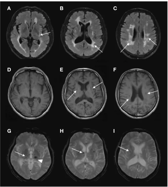
e S cienc e 다.1,2) 편두통, 반복적인 뇌졸중, 인지기능의 감퇴를 특징으로 하는 본 질환에 대한 국내 첫 보고는 2002년에 노낙경 등이 편 마비와 기억력장애로 방문한 52세의 환자에서 피부조직 전자 현미경검사에서 과립성 오스미움친화성물질(granular omio-philic material)을 발견하며 시작되었고 제주지역에서는 2004 년 갑자기 시작된 말더듬증을 주소로 제주대학교병원을 방문 한 44세의 남자에서 유전자검사를 통해서 p.Arg544Cys 돌연 변이가 발견되며 처음으로 진단되었다.3) 환자는 고혈압, 당뇨 등의 특별한 병력이 없었으나 이미 1년 전에 왼팔다리저림으 로 타병원을 방문하여 열공성뇌경색(lacunar infarction)이 확 인된 상태였고 환자의 어머니가 70대에 뇌졸중을 앓았다고 하 였다. 환자는 2년 후에 시행한 뇌자기공명영상에서 다발성의 열공성뇌경색, 백질고신호강도병변, 다수의 미세뇌출혈 외에 도 왼쪽 시상에서 뇌출혈이 관찰되었다(Fig. 1). 이를 시작으 로 제주지역에서 많은 환자들이 진단되기 시작하였고 대부분 이 p.Arg544Cys 돌연변이를 보유하였다. CADASIL 환자에서
Figure 1. Brain MRI of the first CADASIL patient diagnosed in Jeju. FLAIR(fluid attenuated inversion recovery) images(A,B, and C) show
multifocal white matter hyperintensity lesions on external capsule, periventricular and subcortical region. Multiple lacunar infarctions are seen on T1-weighted images at bilateral basal ganglia and corona radiata(D, E, and F). Gradient echo(GRE) images(G, H, and I) show multiple mi-crobleeds at bilateral thalamus and basal ganglia. An old thalamic hemorrhage is noted on the left thalamus(arrowhead).
A B C
D E F
.jejunu
.ac
.k
r/medsci/
The Journal of M
edicine and Lif
e S cienc e 이미 잘 알려진 허혈성 뇌졸중이 아닌 뇌출혈의 발생은 예상치 못한 임상증상으로 판단하여 최재철 등은 2006년에 그동안 진 단된 17가계의 20명의 환자들의 임상증상 및 뇌자기공명영상 을 조사하여 환자들의 25%에서 증상성 뇌출혈이 발생한 것을 확인하여 Neurology에 보고하였다.4) 이를 시작으로 하여 제주 지역의 CADASIL 환자들의 임상적, 영상학적, 유전학적 특징 에 대한 연구논문이 현재까지 꾸준히 발표되었다.2,5-10) 2004년 4월부터 2020년 10월까지 제주대병원에서 진단 된 CADASIL 환자는 모두 213명이었다. 환자들의 평균 연령 은 61.0±12.8세이고, 남자가 97명(45.5%)이었다. 엑손11번 (94.4%)에서 가장 흔하게 돌연변이가 발견되었고, 대부분 이 형접합체(heterozygote)였다(96.7%). 그중 p.Arg544Cys가 189개(88.7%)로 가장 흔하게 관찰되었다. 환자들이 진단에 이르게 된 주된 증상은 허혈성 뇌졸중(24.4%), 인지기능장애 (15.0%), 두통(8.9%), 어지럼증(8.0%), 보행장애의 순이었고 23.9%의 환자는 증상이 없는 상태에서 진단되었다(Table 1).
2. CADASIL의 역학
CADASIL은 가장 흔한 유전성 뇌혈관질환으로 아직까지 정 확한 유병률은 알려지지 않았다. 최근 특징적인 임상증상 및 뇌영상학적 소견이 점차 알려지고 유전학적 검사가 널리 사 용되면서 진단되는 환자의 수가 증가하고 있다. 2005년에 발 표된 스코틀랜드의 연구에 의하면 10만 명당 1.98명으로 추 정되었으나 당시에는 정확한 진단이 힘들었고 단지 4개의 엑 손만 분석이 되었다.11) 최근 대규모 유전자데이터베이스에 관 한 연구가 활발하게 진행되며 CADASIL의 유병률에 대한 새 로운 보고가 많이 발표되었다. 2016년 Rutten 등은 세계 각국 에서 수집된 60,706명의 엑손데이터를 분석하여 NOTCH3 돌 연변이를 무려 1000명 중에 3.4명에서 발견하였고 일반인에 서 발견된 돌연변이는 이미 보고된 CADASIL 환자들과 달리주로 NOTCH3 유전자의 epidermal growth factor-like repeat
(EGFr) 1-6에 위치하는 것을 밝혀냈다.12) 또한 같은 연구진 은 최근 영국의 유전자은행 자료의 분석을 통해서 cysteine-altering NOTCH3 변이가 1000명당 2.2명에서 발견하였고 CADASIL 환자들과 비교하여 대뇌백질 고신호강도의 병변은 경한 것을 확인하였다. 다른 연구에서 이와 같은 높은 빈도의 NOTCH3 변이는 다른 지역과 비교하여 동아시아 및 남아시아 에서 뚜렷하였다. 제주지역의 일반인을 대상으로 한 NOTCH3 유전자분석연구는 현재까지 없으며 다만 2011년 제주도의 허 혈성 뇌졸중 환자 151명을 대상으로 한 연구에서 NOTCH3 유 전자 변이는 4.0%였고 진행된 소동맥질환을 보인 환자만을 대 상으로 하는 경우 36%로 추정되었다.5) 최근 대만의 유전자은 행 자료를 이용한 약 8,000명의 대규모 연구에서 뇌졸중 및 치 매가 없는 대조군에서 NOTCH3 변이가 0.9% , 뇌졸중 환자군 에서는 2.1%, 소동맥유형의 뇌졸중 환자군에서는 6.5% 로 확 인되었다.13) 따라서, 최근의 여러 연구들을 종합하면 제주지역 의 NOTCH3 유전자 변이는 약 1% 정도로 추정되며 이를 확인 하기 위해서 건강한 일반인구를 대상으로 하는 유전자분석 연 구가 필요하다.
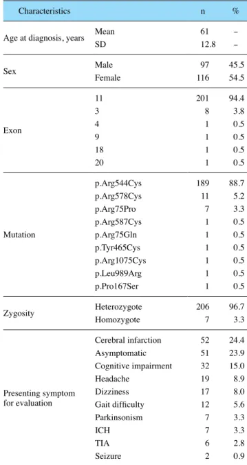
Table 1. Demographic, genetic, and presenting symptoms of 213
CADASIL patients in Jeju
Characteristics n %
Age at diagnosis, years MeanSD 6112.8 -
-Sex MaleFemale 11697 45.554.5
Exon 11 201 94.4 3 8 3.8 4 1 0.5 9 1 0.5 18 1 0.5 20 1 0.5 Mutation p.Arg544Cys 189 88.7 p.Arg578Cys 11 5.2 p.Arg75Pro 7 3.3 p.Arg587Cys 1 0.5 p.Arg75Gln 1 0.5 p.Tyr465Cys 1 0.5 p.Arg1075Cys 1 0.5 p.Leu989Arg 1 0.5 p.Pro167Ser 1 0.5
Zygosity HeterozygoteHomozygote 2067 96.73.3
Presenting symptom for evaluation Cerebral infarction 52 24.4 Asymptomatic 51 23.9 Cognitive impairment 32 15.0 Headache 19 8.9 Dizziness 17 8.0 Gait difficulty 12 5.6 Parkinsonism 7 3.3 ICH 7 3.3 TIA 6 2.8 Seizure 2 0.9
.ac
.k
r/medsci/
The Journal of M
edicine and Lif
e S cienc e .ac .k r/medsci/ The Journal of M
edicine and Lif
e S cienc e
3. 제주지역 CADASIL의 유전학적 특징
현재 약 200개 이상의 서로 다른 NOTCH3 돌연변이가 CADASIL을 일으키는 것으로 알려졌으며 그중 대부분은 점돌연변이(point mutation)였다.14) 대부분의 돌연변이는 NOTCH3
수용체의 세포외 영역을 코딩하는 엑손2-24에서 발견되며
EGFr의 시스테인 숫자(cysteine residue)에 영향을 미쳐, 정상
과 비교하여 CADASIL 환자는 홀수의 시스테인을 유도한다.15) 독일, 프랑스, 영국을 포함하는 유럽의 CADASIL 환자들 은 엑손3-4에서 가장 흔하게 NOTCH3 변이가 관찰되었다. 16-18) 그에 비해, 제주도와 대만의 CADASIL 환자군에서는 엑손 11에 위치하는 p.Arg544Cys 변이가 매우 높은 비율을 보였 다. p.Arg544Cys 변이는 최초 네덜란드에서 1999년에 보고되 었지만 여러 대규모 CADASIL 연구들(독일 411명, 영국 200 명, 핀란드 60명, 이탈리아 229명, 일본 70명)에서는 관찰되 지 않았다. 창시자효과는 유전적 부동(genetic drift)의 하나 로, 다양한 요인에 의해 소수의 개체가 새로운 개체군을 형성 할 때 창시자군이 갖고 있는 유전자에 의해 새로운 개체군이 형성되어 유전자 빈도의 차이를 설명하는 것이다. CADASIL 은 섬 지역인 제주와 대만 그리고 내륙 지역인 핀란드나 중부 이탈리아에서 창시자효과로 인하여 다른 지역보다 높은 유병 률을 보일 수 있고 또한 우세한 돌연변이가 존재할 수 있다. CADASIL은 상염색체 우성으로 유전되는 질환이기는 하나 일부 연구에서 동형접합체(homozygote)를 가진 환자에서 더 심한 질환을 나타낸다고 보고하였으나 또 다른 연구에서는 차이가 없는 것으로 나왔다.19-24) 적은 수의 환자만을 대상으 로 한 연구에서 이형접합체와 동형접합체의 임상양상의 차이 를 규명하기에는 한계가 있을 것으로 보인다. 지리적, 그리고 역사학적으로 뚜렷한 인구이동이 없는 제주 지역과 대만에서 동일한 돌연변이가 우세하게 발견된 이유는 현재로서는 정확하게 알 수 없다. 향후 일배체분석(haplotype analysis) 등을 통해서 유전적 근원이 일치하는지 확인이 가능 하며 이는 CADASIL의 유전적인 면에서 뿐만이 아니라 제주 와 대만의 문화인류학적인 면에서도 흥미로운 연구가 될 것 이다.
4. 제주지역 CADASIL 환자의 임상적 특징
NOTCH3 유전자의 돌연변이는 민무늬근육세포(smoothmuscle cell)와 혈관주위세포(pericyte)에 있는 NOTCH3 단
백질 수용체의 구조적 결함을 유도해서 비아밀로이드성
(non-amyloid), 비동맥경화성(non-atherosclerotic) 혈관병을 일
으킨다1). 유럽의 환자들에서 보고된 CADASIL의 5가지 주
요 임상증상은 조짐 편두통(migraine with aura), 피질하 허
혈성 뇌병변(subcortical ischemic events), 정동 장애(mood
disturbance), 무관심(apathy), 그리고 인지기능저하(cognitive
impairment)이다.15) (Fig. 2) 제주지역 CADASIL 환자의 임
.jejunu
.ac
.k
r/medsci/
The Journal of M
edicine and Lif
e S cienc e 상증상은 반복적인 허혈성 뇌졸중, 뇌출혈, 인지기능저하, 만 성 두통, 우울증 등 정동 장애, 발작 등이다.6,7,25) 제주도에서 는 p.Arg544Cys 돌연변이가 대부분을 차지하는데 EGFr 7-34 에 해당된다. 최근 엑솜(exome) 분석을 사용한 대규모 연구
에서 EGFr 7-34에 해당되는 CADASIL 환자가 EGFr 1-6 도
메인에 돌연변이가 위치하는 CADASIL 환자에 비하여 임상 증상의 발생이 12년 이상 늦고 생존 기간이 긴 것이 밝혀졌 다.26) 따라서 돌연변이가 주로 EGFr 1-6에 위치하는 유럽지역 의 CADASIL 환자들과 제주지역의 환자들은 임상적인 특징 및 예후에 많은 차이를 보인다. 제주지역의 CADASIL 환자들은 유럽 CADASIL 환자와 다 른 3가지 임상적인 특징이 있다. 우선 앞서 서술한 바와 같이 CADASIL 증상 발현이 훨씬 늦고, 유럽 CADASIL 환자에서 는 아주 드물게 보고되는 뇌출혈 발생률이 매우 높으며, 조짐
편두통(migraine with aura)의 발생률이 극히 낮다.4,7,25) 조짐
편두통은 유럽지역의 CADASIL 환자에서는 10대에 주로 시 작하여 환자들의 약 20~40%에서 발생한다.15) 그러나 제주 지역의 CADASIL 환자에서는 조짐 편두통이 극히 드물고 만 성 두통 중에서 긴장두통이 80% 이상으로 대부분을 차지하였 다.7) 또한 제주지역 CADASIL 환자의 약 25%에서 뇌출혈이 발 견되었고 뇌출혈 환자들은 뇌출혈이 없는 환자들과 비교하 여 뇌자기공명영상에서 발견된 대뇌미세출혈(cerebral micro-bleed)의 수가 많았으며 장애 정도가 심각하였다.4) 뇌출혈 발 생에 관한 후속 연구에서 뇌출혈 호발 부위는 기저핵, 시상, 그리고 대뇌엽(lobar area) 부위였으며 뇌출혈 호발 부위와 뇌 미세출혈 호발 부위는 일치하지 않았다.10)
인지장애(cognitive impairment)는 유럽지역의 CADASIL
환자에서 2번째로 흔한 증상이며 처음에는 전두엽기능저하 로 시작된다. 인지장애는 시간이 지남에 따라 서서히 악화되 어 전반적인 인지기능의 저하가 발생하여 60대가 되면 상당 수가 치매 상태로 진행한다고 알려져 있다. 또한 60대 중반부 터 자립 보행이 힘들어져서 침상 생활을 하고 남자는 60대 중 반, 여자는 70대 초반에 사망한다고 알려져 있다.15) 제주지역 의 CADASIL 환자에서도 인지장애는 뇌졸중 다음으로 흔한 임상증상이었다. 그러나 유럽 CADASIL 환자와는 달리 인지 장애의 발병 연령이 60대 중반으로 늦었고 70대 후반까지도 무증상 환자가 있는 등 유럽 CADASIL 환자에 비하여 전반 적으로 좋은 예후를 보였다.25) 일과성 허혈성 발작(transient
ischemic attack)을 포함하는 허혈성 뇌병변은 CADASIL에
서 가장 흔한 임상증상이며 60~85%의 환자에서 발생하고 평균 발생 연령은 49세였다.15) p.Arg544Cys 돌연변이 환자군 을 대상으로 한 제주도 연구에서는 허혈성 뇌졸중은 55%의 환자에서 발생하였고 평균 발병 연령이 50대 후반으로 유럽 CADASIL 환자보다 약 10년 정도 늦었다.25) 흥미롭게도 최근 연구에 의하면 제주지역의 CADASIL 환자들의 경우 아포지 단백E ε4 유전자형을 보이는 경우 치매의 발병 위험도가 10배 이상 증가된 것이 확인되었다.27) 따라서 혈관성치매의 대표적 인 질환으로 알려진 CADASIL에서 대뇌를 침범하는 대표적 인 퇴행성질환인 알츠하이머병리가 어떤 역할을 할지에 대해 서는 추후 대규모의 연구가 필요하다(Table 2).
5. 뇌영상을 이용한 CADASIL의 예후 예측
CADASIL과 같은 대뇌소혈관질환의 영상학적 표지인자(imaging biomarker)로는 급성 피질하 작은 뇌경색(recent
small subcortical infarcts), 열공성 경색(lacunes), 뇌미세출혈
(cerebral microbleeds), 백질 고신호강도(white matter
hyper-intensities), 증가된 혈관주위공간(enlarged perivascular
space), 그리고 뇌위축(brain atrophy)로 나눌 수 있다.28)
CADASIL에서 치매 또는 인지기능저하과 연관성이 가장 높은 영상학적 표지인자는 열공성 경색이다.29-31) 제주지역 CADASIL 환자를 대상으로 시행된 연구에서도 열공성 경색 은 인지기능저하와 연관성을 보였다.32) (Fig. 3) 뇌위축도 CADASIL을 비롯한 대뇌 미세혈관질환에서 인 지 및 장애 정도와 강한 연관성을 띤다고 알려져 있는데 이 는 뇌위축이 대뇌 미세혈관질환의 병태생리학적 최종 공통
경로(final common pathway)이기 때문이다. CADASIL 환자
에서 뇌위축 정도를 나타내는 뇌실질분율(brain parenchymal
Table 2. Genetic, clinical, and neuroimaging characteristics of
CA-DASIL patients in Jeju Genetic
Predominant p.Arg544Cys mutation on exon 116) Clinical
Later disease onset25)
Presence of intracerebral hemorrhage4,6) Rare migraine without aura7)
Less severe cognitive impairment25) Radiologic
Rare anterior temporal white matter hyperintensity4) Frequent cerebral microbleeds10,33)
Prognostic
Association of high blood pressure and incident stroke27) Association of APOE ε4 genotype and incident dementia27)
.ac
.k
r/medsci/
The Journal of M
edicine and Lif
e S cienc e .ac .k r/medsci/ The Journal of M
edicine and Lif
e S cienc e fraction, 두개내공간에서 뇌실질의 비율)은 인지기능의 저 하뿐만 아니라 장애 정도를 나타내는 수정 Rankin 척도 와 연관성이 높았다.31) 또한, CADASIL 환자에서 뇌실용적 (ventricular volume)의 증가와 뇌미세출혈도 단일 코호트 연 구에서 인지기능저하와 연관성을 보였다.29) 뇌미세출혈을 이용한 CADASIL의 예후 예측은 제주지역
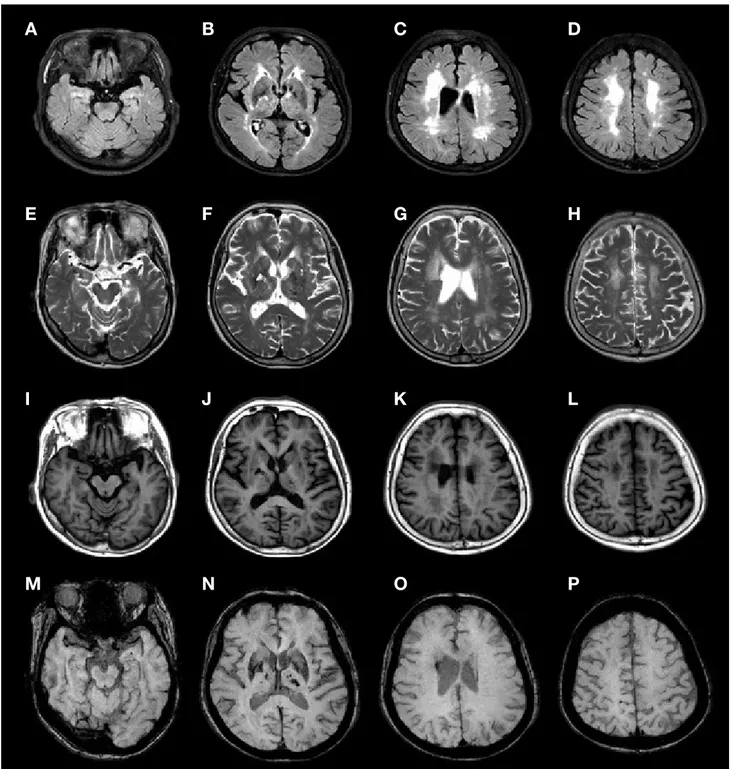
Figure 3. Representative brain MRI findings in patients with CADASIL. Fluid-attenuated inversion recovery images(A~D) show
periventricu-lar and deep white matter hyperintensities including anterior temporal area and external capsule. T2-weighted images(E~H). T1-weighte images (I~L) show subcortical lacune in the left basal ganglia. Susceptibility weighted images(M-P) show multiple cerebral microbleeds in various ar-eas and intracerebral hemorrhage in the right basal ganglia and left thalamus.
A B C D
E F G H
I J K L
.jejunu
.ac
.k
r/medsci/
The Journal of M
edicine and Lif
e S cienc e CADASIL 환자에서 활발하게 연구되었는데 이는 유럽지역 CADASIL 환자와 달리 제주지역 환자에서 뇌출혈의 빈도가 높았기 때문이다. 제주지역의 CADASIL 환자들의 경우 기저 핵 부위(basal ganglia) 뇌미세출혈은 유의하게 뇌졸중 발생과 연관성이 높았다.33) 또한, 뇌미세출혈이 9개 이상인 경우 뇌출 혈 발생과 의미있게 높은 연관성을 보였다.10)
6. CADASIL의 혈액바이오마커
열공경색과 뇌실질분율 등 영상분석을 이용한 CADASIL 의 예후 예측은 활발하게 연구가 진행되었다. 하지만 뇌자기 공명영상은 고가의 검사법이고 심각한 질환으로 인한 움직 임, 폐쇄공포증, 심박동기 등으로 인해서 검사에 제한이 있 다. 뇌영상검사에 비해 비용이 덜 들고, 손쉽게 검사가 가능 한 혈액, 뇌척수액 등을 이용한 CADASIL 관련 생물표지자 (biomarker) 관련 연구는 상대적으로 미진한 상태이나 최근 동물모델과 CADASIL 환자들을 대상으로 하여 혈액을 이용 한 다양한 생물표지자에 대한 연구가 발표되었다.34-36)Neurofilament light chain(Nfl)은 신경세포의 세포골격
(cytoskeleton)을 이루는 필수 골격단백질(scaffolding
pro-tein)로 축상 손상(axonal damage)이 있을 때 세포외영역
(extracellular space)로 분비되어, 뇌척수액과 혈액 등에 분포 하며 다양한 질환에서 신경세포 손상의 표지자로 사용되고 있다.37) 41명의 네델란드 CADASIL 환자들을 대상으로 한 전 향적인 연구에서 혈액 Nfl 농도는 환자들의 뇌자기공명영상 에서 관찰되는 병변의 심각도와 유의하게 관련성을 보였고 7 년간의 경과 관찰에서 장애발생 및 인지기능저하를 예측하는 데 유용하였다.36) 독일의 CADASIL 환자 53명을 대상으로 한 연구에서도 혈액 Nfl 농도는 환자들의 인지기능, 장애 정도 및 신경학적 증상과의 유의한 연관성을 보였다.35) CADASIL 동물모델에서는 손상된 혈관벽에 침착하는 것으 로 알려진 endostatin, CADASIL 모델의 대동맥에서 2배 이상
유전자발현이 증가된 high-temperature requirement A serine
protease(HTRA1), 그리고 NOTCH3 extracellular domain
(NOTCH3 ECD)이 유의하게 차이를 보였다.34)
NOTCH 수용체는 세포막에 존재하며 리간드(ligand)와 결
합에 의해서 세포 외 도메인(extracellular domain, ECD)과
세포 내 도메인(intracellular domain, ICD)으로 분리된다.38)
분리된 NOTCH ECD는 리간드와 결합된 채로 신호를 보내
는 세포막 내로 이송되며 이와 같은 세포내이입(endocytosis)
은 NOTCH 신호전달을 활성화하는 역할을 하는 것으로 알려
져 있다. NOTCH3 변이를 보인 생쥐의 혈액 내 NOTCH3
ECD 농도는 대조군과 비교하여 유의하게 감소되었기 때문에 NOTCH3 ECD는 향후 CADASIL 환자의 진단과 예후예측에
중요한 역할을 할 것으로 기대된다.
7. 제주지역에서 CADASIL 연구의 중요성
고찰을 통해서 살펴본 바와 같이 제주도는 대한민국에서 가장 많은 CADASIL 환자가 진단된 지역이고 뇌자기공명영 상과 유전자진단이 널리 사용되며 향후 진단되는 환자의 수 는 점차 증가할 것으로 예상된다. 동일한 우세 유전자형을 보 이는 대만의 예를 참고하면 전체 제주도 인구의 약 1%에서 CADASIL과 연관된 NOTCH3 유전자변이가 존재할 것으로 추정되며 이는 제주도민의 뇌건강에 중요한 영향을 미칠 수 있다. 따라서, 지역 내에서 CADASIL이 의심되는 환자들을 정확하게 감별 진단할 수 있는 바이오마커 및 유전자검사법 의 확립이 필요하고 진단된 환자들의 예후에 영향을 미치는 요인을 파악하여 질환의 악화를 막는 것이 중요하다. 궁극적 으로는 질환의 병태생리를 탐구하여 근본적인 치료 후보 물 질의 탐색이 시급하며 이를 위해서는 중앙정부 및 제주도 차 원의 적극적인 연구사업지원을 통한 다학제 연구가 필수적이 다.REFERENCES
1. Joutel A, Corpechot C, Ducros A, Vahedi K, Chabriat H, Mouton P, et al. Notch3 mutations in CADASIL, a hereditary adult-onset condition causing stroke and dementia. Nature 1996;383:707-10. 2. Choi JC. Cerebral autosomal dominant arteriopathy with
subcorti-cal infarcts and leukoencephalopathy: a genetic cause of cerebral small vessel disease. J Clin Neurol 2010;6:1-9.
3. Rho NK, Choi SJ, Lee ES. A Case of CADASIL(Cerebral Au-tosomal Dominant Arteriopathy with Subcortical Infarcts and Leukoencephalopathy) Diagnosed by Skin Biopsy. Korean J Der-matol 2002;40:1136-8.
4. Choi JC, Kang SY, Kang JH, Park JK. Intracerebral hemorrhages in CADASIL. Neurology 2006;67:2042-4.
5. Choi JC, Lee KH, Song SK, Lee JS, Kang SY, Kang JH. Screen-ing for NOTCH3 gene mutations among 151 consecutive Korean patients with acute ischemic stroke. J Stroke Cerebrovasc Dis 2013;22:608-14.
6. Choi JC, Song SK, Lee JS, Kang SY, Kang JH. Diversity of stroke presentation in CADASIL: study from patients harboring
.ac
.k
r/medsci/
The Journal of M
edicine and Lif
e S cienc e .ac .k r/medsci/ The Journal of M
edicine and Lif
e S
cienc
e
the predominant NOTCH3 mutation R544C. J Stroke Cerebro-vasc Dis 2013;22:126-31.
7. Choi JC, Song SK, Lee JS, Kang SY, Kang JH. Headache among CADASIL patients with R544C mutation: prevalence, character-istics, and associations. Cephalalgia 2014;34:22-8.
8. Kang SY, Oh JH, Kang JH, Choi JC, Lee JS. Nerve conduction studies in cerebral autosomal dominant arteriopathy with subcor-tical infarcts and leukoencephalopathy. J Neurol 2009;256:1724-7.
9. Lee JS, Choi JC, Kang SY, Kang JH, Lee SH, Kim JH, et al. Olfactory identification deficits in cerebral autosomal dominant arteriopathy with subcortical infarcts and leukoencephalopathy. Eur Neurol 2010;64:280-5.
10. Lee JS, Ko K, Oh JH, Park JH, Lee HK, Floriolli D, et al. Cere-bral microbleeds, hypertension, and intracereCere-bral hemorrhage in cerebral autosomal-dominant arteriopathy with subcortical in-farcts and leukoencephalopathy. Front Neurol 2017;8:203. 11. Razvi SS, Davidson R, Bone I, Muir KW. The prevalence of
ce-rebral autosomal dominant arteriopathy with subcortical infarcts and leucoencephalopathy(CADASIL) in the west of Scotland. J Neurol Neurosurg Psychiatry 2005;76:739-41.
12. Rutten JW, Dauwerse HG, Gravesteijn G, van Belzen MJ, van der Grond J, Polke JM, et al. Archetypal NOTCH3 mutations frequent in public exome: implications for CADASIL. Ann Clin Transl Neurol 2016;3:844-53.
13. Lee YC, Chung CP, Chang MH, Wang SJ, Liao YC. NOTCH3 cysteine-altering variant is an important risk factor for stroke in the Taiwanese population. Neurology 2020;94:e87-e96.
14. Mizuno T, Mizuta I, Watanabe-Hosomi A, Mukai M, Koizumi T. Clinical and Genetic Aspects of CADASIL. Front Aging Neurosci 2020;12:91.
15. Chabriat H, Joutel A, Dichgans M, Tournier-Lasserve E, Bousser MG. Cadasil. Lancet Neurol 2009;8:643-53.
16. Opherk C, Peters N, Herzog J, Luedtke R, Dichgans M. Long-term prognosis and causes of death in CADASIL: a retrospective study in 411 patients. Brain 2004;127:2533-9.
17. Adib-Samii P, Brice G, Martin RJ, Markus HS. Clinical spectrum of CADASIL and the effect of cardiovascular risk factors on phe-notype: study in 200 consecutively recruited individuals. Stroke 2010;41:630-4.
18. Tikka S, Mykkanen K, Ruchoux MM, Bergholm R, Junna M, Poyhonen M, et al. Congruence between NOTCH3 mutations and GOM in 131 CADASIL patients. Brain 2009;132:933-9.
19. Vinciguerra C, Rufa A, Bianchi S, Sperduto A, De Santis M, Malandrini A, et al. Homozygosity and severity of phenotypic pre-sentation in a CADASIL family. Neurol Sci 2014;35:91-3. 20. Tuominen S, Juvonen V, Amberla K, Jolma T, Rinne JO, Tuisku S,
et al. Phenotype of a homozygous cadasil patient in comparison to 9 age-matched heterozygous patients with the same R133C
Notch3 mutation. Stroke 2001;32:1767-74.
21. Soong B-W, Liao Y-C, Tu P-H, Tsai P-C, Lee IH, Chung C-P, et al. A homozygous NOTCH3 mutation p.R544C and a heterozy-gous TREX1 variant p.C99MfsX3 in a family with hereditary small vessel disease of the brain. J Chin Med Assoc 2013;76:319-24.
22. Ragno M, Pianese L, Morroni M, Cacchiò G, Manca A, Di Mar-zio F, et al. “CADASIL coma” in an Italian homozygous CADA-SIL patient: comparison with clinical and MRI findings in age-matched heterozygous patients with the same G528C NOTCH3 mutation. Neurol Sci 2013;34:1947-53.
23. Liem MK, Lesnik Oberstein SAJ, Vollebregt MJ, Middelkoop HAM, van der Grond J, Helderman-van den Enden ATJM. Ho-mozygosity for a NOTCH3 mutation in a 65-year-old CADASIL patient with mild symptoms. J Neurol 2008;255:1978-80. 24. Anamnart C, Songsaeng D, Chanprasert S. A large number of
cerebral microbleeds in CADASIL patients presenting with recur-rent seizures: a case report. BMC Neurology 2019;19:106. 25. Lee JS, Ko K, Oh JH, Park JH, Lee HK. Phenotypic features of
cerebral autosomal-dominant arteriopathy with subcortical in-farcts and leukoencephalopathy subjects with R544C mutation. Dement Neurocogn Disord 2016;15:15-9.
26. Rutten JW, Van Eijsden BJ, Duering M, Jouvent E, Opherk C, Pantoni L, et al. The effect of NOTCH3 pathogenic variant position on CADASIL disease severity: NOTCH3 EGFr 1-6 pathogenic variant are associated with a more severe phenotype and lower survival compared with EGFr 7-34 pathogenic variant. Genet Med 2019;21:676-82.
27. Lee JS, Ko KH, Oh J-H, Kim J-G, Kang C-H, Song S-K, et al. Apolipoprotein E ε4 Is Associated with the development of inci-dent dementia in cerebral autosomal dominant arteriopathy with subcortical infarcts and leukoencephalopathy patients with p.Arg-544Cys mutation. Front Aging Neurosci. 2020;12.
28. Pantoni L. Cerebral small vessel disease: from pathogenesis and clinical characteristics to therapeutic challenges. Lancet Neurol 2010;9:689-701.
29. Liem MK, Lesnik Oberstein SA, Haan J, van der Neut IL, Ferrari MD, van Buchem MA, et al. MRI correlates of cognitive decline in CADASIL: a 7-year follow-up study. Neurology 2009;72:143-8.
30. Liem MK, van der Grond J, Haan J, van den Boom R, Ferrari MD, Knaap YM, et al. Lacunar infarcts are the main correlate with cognitive dysfunction in CADASIL. Stroke 2007;38:923-8. 31. Viswanathan A, Gschwendtner A, Guichard JP, Buffon F,
Cumur-ciuc R, O’Sullivan M, et al. Lacunar lesions are independently as-sociated with disability and cognitive impairment in CADASIL. Neurology 2007;69:172-9.
32. Lee JS, Choi JC, Kang SY, Kang JH, Na HR, Park JK. Effects of lacunar infarctions on cognitive impairment in patients with
ce-.jejunu
.ac
.k
r/medsci/
The Journal of M
edicine and Lif
e S
cienc
e
rebral autosomal-dominant arteriopathy with subcortical infarcts and leukoencephalopathy. J Clin Neurol 2011;7:210-4.
33. Lee JS, Kang CH, Park SQ, Choi HA, Sim KB. Clinical signifi-cance of cerebral microbleeds locations in CADASIL with R544C NOTCH3 mutation. PLoS One 2015;10:e0118163.
34. Primo V, Graham M, Bigger-Allen AA, Chick JM, Ospina C, Quiroz YT, et al. Blood biomarkers in a mouse model of CADA-SIL. Brain Res 2016;1644:118-26.
35. Duering M, Konieczny MJ, Tiedt S, Baykara E, Tuladhar AM, Leijsen EV, et al. Serum neurofilament light chain levels are relat-ed to small vessel disease burden. J Stroke 2018;20:228-38.
36. Gravesteijn G, Rutten JW, Verberk IMW, Bohringer S, Liem MK, van der Grond J, et al. Serum Neurofilament light correlates with CADASIL disease severity and survival. Ann Clin Transl Neurol 2019;6:46-56.
37. Teunissen CE, Khalil M. Neurofilaments as biomarkers in multi-ple sclerosis. Mult Scler 2012;18:552-6.
38. Boucher J, Gridley T, Liaw L. Molecular pathways of notch sig-naling in vascular smooth muscle cells. Front Physiol 2012;3:81. 39. Parks AL, Klueg KM, Stout JR, Muskavitch MA. Ligand
endo-cytosis drives receptor dissociation and activation in the Notch pathway. Development 2000;127:1373-85.