ABSTRACT
Purpose: The aim of this study was to compare microRNA (miRNA) gene expression in saliva using miRNA polymerase chain reaction (PCR) arrays in healthy and aggressive periodontitis (AP) patients.
Methods: PCR arrays of 84 miRNAs related to the human inflammatory response and autoimmunity from the saliva samples of 4 patients with AP and 4 healthy controls were performed. The functions and diseases related to the miRNAs were obtained using TAM 2.0. Experimentally validated targets of differentially expressed miRNAs were obtained from mirTarBase. Gene ontology terms and pathways were analyzed using ConsensusPathDB. Results: Four downregulated miRNAs (hsa-let-7a-5p, hsa-let-7f-5p, hsa-miR-181b-5p, and hsa-miR-23b-3p) were identified in patients with AP. These miRNAs are associated with cell death and innate immunity, and they target genes associated with osteoclast development and function.
Conclusions: This study is the first analysis of miRNAs in the saliva of patients with AP. Identifying discriminatory human salivary miRNA biomarkers reflective of periodontal disease in a non-invasive screening assay is crucial for the development of salivary diagnostics. These data provide a first step towards the discovery of key salivary miRNA biomarkers for AP.
Keywords: Aggressive periodontitis; Biomarkers; MicroRNAs; Saliva
INTRODUCTION
MicroRNAs (miRNAs) are noncoding RNAs that are approximately 22 nucleotides long and can act as key regulators of diverse biological processes by mediating translational repression or messenger RNA (mRNA) degradation of their target genes [1-4]. The seed sequence of miRNAs specifically binds to the 3′ untranslated region of target mRNAs to exert its action [5,6]. Many studies have documented that miRNAs play a key role in certain infectious diseases [7], autoimmune diseases [8], chronic inflammatory diseases [9], cardiovascular diseases [10], cancers [11], and other diseases [12]. Furthermore, recent studies have reported that miRNAs have great potential as biomarkers for these diseases [13-15]. However,
Research Article
Received: Dec 30, 2019 Revised: May 10, 2020 Accepted: Jul 6, 2020 *Correspondence: Su-Hwan KimDepartment of Periodontics, Asan Medical Center, 88 Olympic-ro 43-gil, Songpa-gu, Seoul 05505, Korea.
E-mail: shkimperio@amc.seoul.kr Tel: +82-2-3010-5797
Fax: +82-2-3010-6967
Copyright © 2020. Korean Academy of Periodontology
This is an Open Access article distributed under the terms of the Creative Commons Attribution Non-Commercial License (https:// creativecommons.org/licenses/by-nc/4.0/). ORCID iDs Nam-Hun Lee https://orcid.org/0000-0002-5871-1927 Eunhye Lee https://orcid.org/0000-0001-9530-1508 Young-Sung Kim https://orcid.org/0000-0003-2674-3649 Won-Kyung Kim https://orcid.org/0000-0003-2557-1646 Young-Kyoo Lee https://orcid.org/0000-0001-7065-282X Su-Hwan Kim https://orcid.org/0000-0002-3431-453X Funding
This research was supported by Basic Science Research Program through the National Research Foundation of Korea (NRF) funded by the Ministry of Science, ICT and Future Planning (NRF-2013R1A1A2011974).
Nam-Hun Lee 1, Eunhye Lee 2, Young-Sung Kim 1,3, Won-Kyung Kim 1,
Young-Kyoo Lee 1, Su-Hwan Kim 1,3,*
1Department of Periodontics, Asan Medical Center, Seoul, Korea
2Department of Conservative Dentistry, School of Dentistry, Seoul National University, Seoul, Korea 3Department of Dentistry, University of Ulsan College of Medicine, Seoul, Korea
Differential expression of microRNAs in
the saliva of patients with aggressive
periodontitis: a pilot study of potential
biomarkers for aggressive periodontitis
Periodontal Science
Author Contributions
Conceptualization: Won-Kyung Kim, Young-Kyoo Lee, Su-Hwan Kim; Formal analysis: Young-Sung Kim, Nam-Hun Lee; Investigation: Nam-Hun Lee, Eunhye Lee, Su-Hwan Kim; Methodology: Nam-Hun Lee, Eunhye Lee; Project administration: Eunhye Lee, Su-Hwan Kim; Writing - original draft: Nam-Hun Lee, Su-Hwan Kim; Writing - review & editing: Young-Sung Kim, Su-Hwan Kim.
Conflict of Interest
No potential conflict of interest relevant to this article was reported.
most of those studies analyzed miRNA levels in subjects' serum or plasma, which requires invasive collection methods. To overcome this drawback, some recent studies have attempted to analyze miRNAs collected noninvasively from saliva, reporting promise for detecting pancreatic cancer [16], oral cancer [17], Sjögren syndrome [18], and esophageal cancer [19]. Periodontal remodeling and inflammation are regulated by miRNAs. Several studies have demonstrated that miRNAs play a role in osteogenesis by regulating bone morphogenic protein 7 (BMP-7); runt-related transcription factor 2 (RUNX2); osterix; and osteocalcin; in osteoclastogenesis by regulating protein inhibitor of activated STAT3 (PIAS3); nuclear factor of activated T-cells, cytoplasmic, calcineurin-dependent 1 (NFATc1); nuclear factor kappa B (NF-κB); and receptor activator of NF-κB ligand (RANKL); and in the inflammatory process by regulating inflammatory mediators such as interleukin-1β (IL-1β); IL-6; IL-8; tumor necrosis factor-α (TNF-α); and prostaglandin E2 (PGE2) [5].
Other studies have found that miRNAs were differentially expressed in healthy and diseased human gingival tissues [20-25]. Xie et al. [20], Stoecklin-Wasmer et al. [21], and Ogata et al. [22] found differentially expressed miRNAs in chronic periodontitis (CP) tissue using microarrays. Lee et al. [23], Perri et al. [24], and Na et al. [25] identified differentially expressed miRNAs in CP tissue using quantitative polymerase chain reaction (PCR) arrays. Microarray techniques have been used widely to profile miRNAs. The expression levels of thousands of miRNAs can be determined simultaneously in a single microarray experiment. However, the unique characteristics of miRNAs, such as their short length, differing melting temperatures, and the highly similar sequences between miRNA family members, make the design of microarray probes difficult [26]. Furthermore, a large amount of miRNAs are needed for microarray techniques [27], while PCR-based techniques can detect nanograms of miRNA with high sensitivity and specificity. Furthermore, more recently developed quantitative PCR-based arrays combine the high sensitivity of PCR-PCR-based techniques and the ability of microarray techniques to profile a large number of miRNA samples in a single experiment [27,28].
The studies discussed above used gingival biopsies, which are very invasive. To the best of our knowledge, only 1 study has evaluated the feasibility of using salivary miRNAs as a non-invasive diagnostic tool for CP [29].
Aggressive periodontitis (AP) is the most destructive type of periodontal disease [30]. While there are more genetic risk factors for AP than for CP, few studies have evaluated miRNA expression profiles in AP. Amaral et al. compared the expression patterns of miRNAs between CP and AP using gingival biopsies and PCR arrays [31], and Ghotloo et al. [32] compared expression levels of hsa-miR-146a between AP patients and healthy controls using gingival biopsies and real-time PCR. To our best knowledge, no study has utilized salivary miRNAs to identify AP. The aim of this study was to compare the miRNA gene expression profile in saliva using miRNA PCR arrays in healthy and AP patients.
MATERIALS AND METHODS
Study design
Eight subjects who visited the Department of Periodontics at Asan Medical Center between March and August 2012 were enrolled in this study. The test group for this study consisted of
4 patients who were diagnosed with AP, and the control group consisted of 4 patients with healthy periodontia. The inclusion criteria for the test group were as follows: 1) subjects who were younger than 40 years old and 2) subjects who were diagnosed with AP before 35 years of age in accordance with the classification system defined by the American Academy of Periodontology [33]. The inclusion criteria for the control group were as follows: 1) subjects who were younger than 40-years old and 2) subjects who had a healthy periodontium and who were not diagnosed with AP (had more than 20 permanent teeth, all probing pocket depths were 4 mm or less, and all clinical attachment levels were 2 mm or less). The exclusion criteria for both the control and test groups were as follows: 1) subjects who received periodontal treatment or antibiotic treatment within 3 months prior to participating in this study, 2) subjects who regularly received medication known to affect periodontal status, 3) subjects who required prophylactic antibiotics, 4) subjects who were pregnant or breastfeeding, 5) subjects who were alcoholics or who had a history of drug abuse, and 6) subjects with systemic diseases. This study was approved by the Institutional Review Board of Asan Medical Center (IRB No. 2012-0120). Informed consent was obtained from all subjects before they enrolled in the study.
Saliva collection
Unstimulated saliva samples were collected using a previously defined protocol [34]. Briefly, subjects were asked not to consume any food or water for at least 1 hour prior to saliva collection, and they were asked to rinse their mouth with sterile water for a minute. After 5 minutes, the subjects were asked to sit upright, and 2–3 mL of unstimulated whole saliva was collected in plastic tubes from each subject in the control and test groups. The whole saliva samples were stored at −70°C until use.
MicroRNA PCR array experiment
The miRNAs were isolated from 400-µL saliva samples using an Ambion mirVana miRNA isolation kit (Life Technologies, Carlsbad, CA, USA) according to the manufacturer's instruction. Complementary DNA (cDNA) was synthesized with a miScript II RT kit (Qiagen, Hilden, Germany) according to the manufacturer's instructions. Finally, PCR array experiments were performed using a miScript miRNA PCR Array for Human Inflammatory Response and Autoimmunity and miScript SYBR Green PCR kit (Qiagen). The miScript miRNA PCR Array for Human Inflammatory Response and Autoimmunity identifies 84 miRNAs related to inflammatory response and autoimmunity. A total volume of 1,100 µL of PCR reaction mix was prepared (550 µL of 2× QuantiTect SYBR Green PCR master mix, 110 µL of 10× miScript universal primer, 340 µL of RNase-free water and 100 µL of template cDNA) before addition of 10 µL of reaction mix to each well of the miScript miRNA PCR Array. Optical adhesive film was then sealed onto the array. The array was centrifuged for 1 minute at 1,000 ×g at room temperature to remove bubbles. The PCR real-time cycler was programmed as follows: the initial activation step was carried out for 15 minutes at 95°C, followed by 40 cycles of denaturation (15 seconds, 94°C), annealing (30 seconds, 55°C), and extension (30 seconds, 70°C).
Data analysis
Normalized gene expression values and fold changes between the test and control groups for each miRNA were calculated using the ΔΔCt method with the data from the PCR array. The Student t-test was used to evaluate the significance of differences in normalized gene expression levels between the test and control groups. Differences were considered significant when P<0.05. The enriched miRNA clusters, families, miRNA-associated diseases,
and functions of the differentially expressed miRNAs were determined using TAM 2.0 [35]. Experimentally validated target mRNAs were obtained from miRTarBase [36]. Finally, gene ontology (GO) terms and pathways were analyzed with ConsensusPathDB [37] using the selected target genes obtained from miRTarBase.
RESULTS
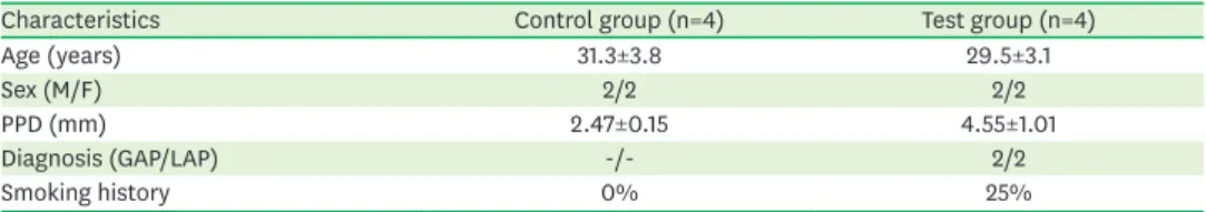
The demographic data for the control and test groups are summarized in Table 1.
We identified 4 significantly downregulated miRNAs in subjects with AP compared to healthy subjects (Fig. 1). The 4 miRNAs were let-7a-5p, let-7f-5p, miR-181b-5p, and hsa-miR-23b-3p. The fold change of those miRNAs was −3.81, −3.40, −5.91, and −3.66, respectively. The enriched miRNA clusters, families, miRNA-associated diseases, and functions for these 4 miRNAs were determined by TAM 2.0. Table 2 shows a total of 26 miRNA categories enriched by the 4 differentially expressed miRNAs (both adjusted P values less than 0.05 after adjustment for multiple testing by the Bonferroni and false discovery rate methods). Interestingly, 8 family members (7a-1, 7a-2, 7a-3, 7f-1, hsa-let-7f-2, hsa-mir-181b-1, hsa-mir-181b-2, and hsa-mir-23b) of all differentially expressed miRNAs (hsa-let-7a-5p, hsa-let-7f-5p, hsa-miR181b-5p, and hsa-miR-23b-3p) were associated with the cell death function, and 6 family members (let-7a-1, let-7a-2, let-7f-1, hsa-let-7f-2, hsa-mir-181b-1, and hsa-mir-181b-2) of 3 miRNAs (hsa-let-7a-5p, hsa-let-7f-5p, and hsa-miR181b-5p) were associated with innate immunity. There were no enriched disease categories known to be either directly or indirectly related to AP except diabetes mellitus. Table 1. Demographic data
Characteristics Control group (n=4) Test group (n=4)
Age (years) 31.3±3.8 29.5±3.1
Sex (M/F) 2/2 2/2
PPD (mm) 2.47±0.15 4.55±1.01
Diagnosis (GAP/LAP) -/- 2/2
Smoking history 0% 25%
PPD: probing pocket depth, GAP: generalized aggressive periodontitis, LAP: localized aggressive periodontitis.
Normaliz ed g ene e xpr ession 0 0.6 0.8 0.4 0.2 1.0
hsa-let-7a-5p hsa-let-7f-5p hsa-miR-181b-5p Test group Control group FC=−3.81 P=0.018 FC=−3.40 P=0.021 FC=−5.91 P=0.041 FC=−3.66 P=0.018 hsa-miR-23b-3p
Figure 1. Differentially expressed miRNAs. FC of statistically significantly downregulated miRNAs in subjects with aggressive periodontitis (P<0.05).
Experimentally validated target mRNAs were obtained from miRTarBase (Table 3). A total of 46 target genes that were targeted by more than 2 differentially expressed miRNAs and whose adjusted P values were lower than 0.05 were identified. Table 3 shows target genes that had adjusted P values lower than 0.02. CCNB2, SNRPC, TAB2, EIF3C, HNRNPUL1, and E2F1 were targeted by 3 differentially expressed miRNAs.
GO terms and pathways were identified with ConsensusPathDB using the 46 selected target genes from the above miRTarBase results (Tables 4 and 5).
Table 2. Identified enriched miRNA clusters, families, miRNA-associated diseases, and functions (both adjusted P<0.05)
Variables Factor # of miRNAs % P value Adj. P (Bonferroni) Adj. P (FDR)
Cluster hsa-let-7a-1 cluster 2 66.7 <0.001 0.016 0.005
Family let-7 family 5 41.7 <0.001 <0.001 <0.001
Function Cell death 8 10.3 <0.001 <0.001 <0.001
Innate immunity 6 14.3 <0.001 <0.001 <0.001
Adipocyte differentiation 5 12.2 <0.001 <0.001 <0.001
Neural stem cell differentiation 3 50.0 <0.001 <0.001 <0.001
Cell differentiation 5 8.9 0.014 0.002 0.001
Aging 5 7.9 0.003 0.003 0.002
Folliculogenesis 3 27.3 <0.001 0.005 0.002
Cell division 3 17.6 <0.001 0.020 0.006
Cardiac regeneration 3 13.6 <0.001 0.044 0.014
Disease Vascular hypertrophy 5 23.8 <0.001 <0.001 <0.001
Psoriasis 5 15.2 <0.001 <0.001 <0.001
Diabetes mellitus 6 8.1 <0.001 <0.001 <0.001
Asthma 5 12.2 <0.001 <0.001 <0.001
Hepatitis B virus infection 5 7.5 <0.001 0.005 0.002
ACTH-secreting pituitary adenoma 3 27.3 <0.001 0.005 0.002
Human papilloma virus infection 4 11.8 <0.001 0.006 0.003
Autoimmune diseases 4 11.1 <0.001 0.008 0.003
Spinal cord injuries 3 23.1 <0.001 0.008 0.003
Macular degeneration 3 21.4 <0.001 0.011 0.004
Dengue virus infection 3 20.0 <0.001 0.013 0.005
Diabetes mellitus, type 2 6 4.1 <0.001 0.016 0.005
Alzheimer's disease 5 5.0 <0.001 0.032 0.010
Hirschsprung's disease 3 13.6 <0.001 0.044 0.014
Liver cirrhosis 4 7.0 <0.001 0.048 0.014
miRNA: microRNA, FDR: false discovery rate.
Table 3. Experimentally validated target mRNAs using miRTarBase (adjusted P<0.02)
Gene symbol Gene name # of miRNAs P value Adj. P (FDR)
CCNB2 Cyclin B2 3 <0.001 <0.001
SNRPC Small nuclear ribonucleoprotein polypeptide C 3 <0.001 0.002
TAB2 TGF-beta activated kinase 1/MAP3K7 binding protein 2 3 <0.001 0.002
NUDT4 Nudix (nucleoside diphosphate linked moiety X)-type motif 4 2 <0.001 0.005
EIF3C Eukaryotic translation initiation factor 3, subunit C 3 <0.001 0.013
HNRNPUL1 Heterogeneous nuclear ribonucleoprotein U-like 1 3 <0.001 0.014
E2F1 E2F transcription factor 1 3 <0.001 0.014
ALDH7A1 Aldehyde dehydrogenase 7 family, member A1 2 <0.001 0.014
WWP2 WW domain containing E3 ubiquitin protein ligase 2 2 <0.001 0.014
GBF1 Golgi brefeldin A resistant guanine nucleotide exchange factor 1 2 <0.001 0.017
UBAP2 Ubiquitin associated protein 2 2 <0.001 0.017
CEP170B Centrosomal protein 170B 2 <0.001 0.017
DISCUSSION
In this study, 4 differentially expressed miRNAs were identified in saliva samples of patients with AP using PCR arrays. Common characteristics of the differentially expressed miRNAs, such as related functions, diseases, and their potential target genes, were analyzed using TAM 2.0 and mirTarBase. Based on the target genes, GO terms and pathways were further Table 4. GO terms with ConsensusPathDB (category BP level 4) (adjusted P<0.02)
GO term Set size Candidates contained P value Adj. P (FDR)
GO:0090304 Nucleic acid metabolic process 5,174 24 (0.5%) <0.001 0.005
GO:0034645 Cellular macromolecule biosynthetic process 4,867 23 (0.5%) <0.001 0.005
GO:0009889 Regulation of biosynthetic process 4,246 21 (0.5%) <0.001 0.005
GO:0009059 Macromolecule biosynthetic process 5,010 23 (0.5%) <0.001 0.005
GO:0044271 Cellular nitrogen compound biosynthetic process 4,906 22 (0.5%) <0.001 0.009
GO:0072359 Circulatory system development 1,033 9 (0.9%) <0.001 0.011
GO:0010467 Gene expression 5,440 23 (0.4%) <0.001 0.011
GO:0043902 Positive regulation of multi-organism process 161 4 (2.5%) <0.001 0.011
GO:0006338 Chromatin remodeling 167 4 (2.4%) <0.001 0.011
GO:0051851 Modification by host of symbiont morphology or physiology 75 3 (4.0%) <0.001 0.011
GO:0016569 Covalent chromatin modification 480 6 (1.2%) <0.001 0.011
GO:0044772 Mitotic cell cycle phase transition 495 6 (1.2%) <0.001 0.012
GO:0034654 Nucleobase-containing compound biosynthetic process 4,222 19 (0.5%) <0.001 0.013
GO:0016032 Viral process 721 7 (1.0%) <0.001 0.013
GO:0018130 Heterocycle biosynthetic process 4,285 19 (0.4%) 0.001 0.013
GO:0019438 Aromatic compound biosynthetic process 4,296 19 (0.4%) 0.001 0.013
GO:0043903 Regulation of symbiosis, encompassing mutualism through parasitism 206 4 (1.9%) 0.001 0.013
GO:0051276 Chromosome organization 1,211 9 (0.7%) 0.001 0.013
GO:0051171 Regulation of nitrogen compound metabolic process 5,904 23 (0.4%) 0.001 0.014
GO:1901362 Organic cyclic compound biosynthetic process 4,434 19 (0.4%) 0.002 0.016
GO:0044774 Mitotic DNA integrity checkpoint 106 3 (2.8%) 0.002 0.016
GO:0051817 Modification of morphology or physiology of other organism involved in
symbiotic interaction 111 3 (2.7%) 0.002 0.017
GO:0080090 Regulation of primary metabolic process 6,071 23 (0.4%) 0.002 0.018
GO:0044843 Cell cycle G1/S phase transition 252 4 (1.6%) 0.002 0.018
GO:0071826 Ribonucleoprotein complex subunit organization 253 4 (1.6%) 0.002 0.018
GO:0031323 Regulation of cellular metabolic process 6,130 23 (0.4%) 0.002 0.018
GO:0006974 Cellular response to DNA damage stimulus 850 7 (0.8%) 0.003 0.018
GO:0060255 Regulation of macromolecule metabolic process 6,156 23 (0.4%) 0.003 0.018
GO:0051101 Regulation of DNA binding 127 3 (2.4%) 0.003 0.019
GO: gene ontology, BP: blood pressure, FDR: false discovery rate. Table 5. Pathway analysis with ConsensusPathDB (adjusted P<0.02)
Pathway Source Percentage P value Adj. P (FDR)
Direct p53 effectors PID 6 (4.2%) <0.001 <0.001
E2F transcription factor network PID 4 (5.3%) <0.001 0.002
Regulation of TP53 expression and degradation Wikipathways 2 (50.0%) <0.001 0.002
FOXM1 transcription factor network PID 3 (7.1%) <0.001 0.006
HATs acetylate histones Reactome 4 (2.8%) <0.001 0.014
Integrated breast cancer pathway Wikipathways 3 (4.5%) <0.001 0.016
Polo-like kinase mediated events Reactome 2 (12.5%) <0.001 0.016
Herpes simplex infection - Homo sapiens (human) KEGG 4 (2.2%) 0.001 0.018
Cellular senescence Reactome 4 (2.1%) 0.001 0.018
C-MYB transcription factor network PID 3 (3.5%) 0.001 0.018
miRNA regulation of DNA damage response Wikipathways 3 (3.4%) 0.001 0.018
Cellular responses to stress Reactome 5 (1.5%) 0.002 0.018
B-WICH complex positively regulates rRNA expression Reactome 3 (3.2%) 0.002 0.018
Formation of the beta-catenin: TCF transactivating complex Reactome 3 (3.2%) 0.002 0.018 FDR: false discovery rate, PID: Pathway Interaction Database, KEGG: Kyoto Encyclopedia of Genes and Genomes.
analyzed using ConsensusPathDB. To the best of our knowledge, this is the first study to identify differentially expressed miRNAs in AP using saliva samples.
The 4 miRNAs (hsa-let-7a-5p, hsa-let-7f-5p, hsa-miR-181b-5p, and hsa-miR-23b-3p), which play a role in cell death and innate immunity by targeting CCNB2, SNRPC, TAB2, EIF3C, HNRNPUL1, and E2F1, were found to be significantly downregulated in AP in this study. Among these genes, the TAB2 gene encodes the activator of MAP3K7/TAK1, which is required for IL-1 induced activation of NF-κB and MAPK8/JNK. This protein participates in the signal transduction induced by RANKL through the activation of RANK, which regulates the development and function of osteoclasts [38]. Thus, the differentially expressed miRNAs in this study may play a role in the development of AP by regulating osteoclast development and function.
Some studies have identified differentially expressed miRNAs in healthy and diseased human gingival tissue. Xie et al. [20] found that 126, 20a, 142-3p, hsa-miR-19-a, hsa-let-7f, hsa-miR-203, hsa-miR-17, hsa-miR-223, hsa-miR-146b, and hsa-miR-146a were upregulated more than 2-fold, and hsa-miR-155 and hsa-miR-205 were downregulated more than 2-fold in CP tissue compared to healthy gingiva using a microarray. Stoecklin-Wasmer et al. [21] found that that hsa-miR-451, hsa-miR-223, hsa-miR-486-5p, and hsa-miR-3917 were upregulated, and hsa-miR-1246, hsa-miR-1260, hsa-miR-141, hsa-miR-1260b, hsa-miR-203, hsa-miR-210, and hsa-miR-205 were downregulated more than 2-fold in CP tissue using a microarray. Ogata et al. [22] reported that hsa-miR-150, hsa-miR-223, and hsa-miR-200b were upregulated, and that hsa-miR-379, hsa-miR-199a-5p, and hsa-miR-214 were downregulated in CP tissue using a microarray. Lee et al. [23] showed that hsa-let-7a, hsa-let-7c, hsa-miR-130a, hsa-miR-301a, hsa-miR-520d, and hsa-miR-548a were upregulated more than 8-fold in CP tissue compared to healthy gingiva using a PCR array. Perri et al. [24] showed hsa-miR-30e and hsa-miR-106b were upregulated in periodontitis tissue. Finally, Na et al. [25] showed that has-miR-128, has-miR-34a, and has-miR-381 were upregulated, and that has-miR-15b, has-miR-211, hsa-miR-372 and has-miR-656 were downregulated in periodontitis tissue.
Although genetics play a more important role in AP than CP [30], only a few studies have demonstrated associations between miRNA expression and AP, mostly using gingival biopsies. Amaral et al. [31] compared the expression patterns of 754 miRNAs between CP and AP, finding that hsa-miR-1274b, hsa-let-7b-5p, hsa-miR-24-3p, hsa-miR-19b-3p, hsa-miR-720, hsa-miR-126-3p, hsa-miR-17-hsa-miR-126-3p, and hsa-miR-21-3p were upregulated in both groups, suggesting that there were no significant differences in miRNA expression patterns between CP and AP. Ghotloo et al. [32] found an association between hsa-miR-146a and generalized aggressive periodontitis (GAP) and suggested that hsa-miR-146a may serve as an indicator of disease activity.
Recently, Fujimori et al. [29] performed the only previous study that utilized saliva samples as a non-invasive diagnostic tool for CP. They found that salivary hsa-miR-381-3p was upregulated in patients with severe periodontitis when compared to those with no or mild periodontitis and that it was positively correlated with the mean probing pocket depth. The studies mentioned above tried to distinguish miRNA expression profiles between patients with periodontitis and healthy patients. However, their results are not substantially consistent with each other or with our findings. Among these studies, hsa-miR-223 [20,21] and hsa-miR-381 [25,29] were found to be upregulated in periodontitis in only 2 studies, respectively, while hsa-miR-205 [20,21] was found to be downregulated in periodontitis in only 2 studies. These inconsistencies among studies may result from variation in the
inclusion criteria and differences in experimental designs. It is strongly recommended that genome-wide investigations using miRNAs in the future should be performed using standardized experimental designs and protocols.
The current pilot study has some limitations. The sample size of each group was small, so that the reliability of our study may be limited. The results of this study should be confirmed with a larger number of subjects in the future. The test group consisted of patients with GAP and localized aggressive periodontitis (LAP). In fact, 2 of the 4 test subjects were diagnosed with GAP, while the others were diagnosed with LAP. In patients with LAP, the miRNAs differentially expressed in AP in the whole saliva sample may be diluted by the miRNAs from healthy regions of the periodontium. The contents of the saliva samples might be affected by food or water intake, even though we instructed subjects not to consume any food or water for at least 1 hour prior to saliva collection. The effect of fasting time duration on salivary contents should be further investigated. Smoking history was not controlled, which might affect the results of this study. The correlation of smoking history and miRNA expression levels should be confirmed in the near future. Furthermore, few miRNA studies have been conducted in the field of dental research, so the databases utilized in this study (TAM 2.0, mirTarBase, and ConsensusPathDB) were biased towards medical terms, especially for pathological terms. In the future, dental research focusing on miRNAs should be performed, so that the databases can be gradually supplemented with dental terms. Finally, the results from saliva samples should be compared with those from other methods, such as gingival biopsies or gingival crevicular fluid, to show whether salivary diagnostics of AP could be a reliable method.
This study is the first analysis of miRNAs in the saliva of patients with AP. Identifying discriminatory human salivary miRNA biomarkers reflective of periodontal disease in a non-invasive screening assay is crucial for the development of salivary diagnostics. These data provide a first step towards the discovery of key salivary miRNA biomarkers for AP.
REFERENCES
1. Huntzinger E, Izaurralde E. Gene silencing by microRNAs: contributions of translational repression and mRNA decay. Nat Rev Genet 2011;12:99-110.
PUBMED | CROSSREF
2. Pasquinelli AE. MicroRNAs and their targets: recognition, regulation and an emerging reciprocal relationship. Nat Rev Genet 2012;13:271-82.
PUBMED | CROSSREF
3. Bartel DP. MicroRNAs: genomics, biogenesis, mechanism, and function. Cell 2004;116:281-97.
PUBMED | CROSSREF
4. Selbach M, Schwanhäusser B, Thierfelder N, Fang Z, Khanin R, Rajewsky N. Widespread changes in protein synthesis induced by microRNAs. Nature 2008;455:58-63.
PUBMED | CROSSREF
5. Irwandi RA, Vacharaksa A. The role of microRNA in periodontal tissue: a review of the literature. Arch Oral Biol 2016;72:66-74.
PUBMED | CROSSREF
6. Kim SH, Lee SY, Lee YM, Lee YK. MicroRNAs as biomarkers for dental diseases. Singapore Dent J 2015;36:18-22.
PUBMED | CROSSREF
7. Eulalio A, Schulte L, Vogel J. The mammalian microRNA response to bacterial infections. RNA Biol 2012;9:742-50.
8. Pauley KM, Cha S, Chan EK. MicroRNA in autoimmunity and autoimmune diseases. J Autoimmun 2009;32:189-94.
PUBMED | CROSSREF
9. Alexander M, O’Connell RM. Noncoding RNAs and chronic inflammation: micro-managing the fire within. BioEssays 2015;37:1005-15.
PUBMED | CROSSREF
10. Romaine SP, Tomaszewski M, Condorelli G, Samani NJ. MicroRNAs in cardiovascular disease: an introduction for clinicians. Heart 2015;101:921-8.
PUBMED | CROSSREF
11. Peng Y, Croce CM. The role of MicroRNAs in human cancer. Signal Transduct Target Ther 2016;1:15004.
PUBMED | CROSSREF
12. Luan X, Zhou X, Naqvi A, Francis M, Foyle D, Nares S, et al. MicroRNAs and immunity in periodontal health and disease. Int J Oral Sci 2018;10:24.
PUBMED | CROSSREF
13. Wang J, Chen J, Sen S. MicroRNA as biomarkers and diagnostics. J Cell Physiol 2016;231:25-30.
PUBMED | CROSSREF
14. Ai J, Zhang R, Li Y, Pu J, Lu Y, Jiao J, et al. Circulating microRNA-1 as a potential novel biomarker for acute myocardial infarction. Biochem Biophys Res Commun 2010;391:73-7.
PUBMED | CROSSREF
15. Mitchell PS, Parkin RK, Kroh EM, Fritz BR, Wyman SK, Pogosova-Agadjanyan EL, et al. Circulating microRNAs as stable blood-based markers for cancer detection. Proc Natl Acad Sci U S A 2008;105:10513-8.
PUBMED | CROSSREF
16. Xie Z, Yin X, Gong B, Nie W, Wu B, Zhang X, et al. Salivary microRNAs show potential as a noninvasive biomarker for detecting resectable pancreatic cancer. Cancer Prev Res (Phila) 2015;8:165-73.
PUBMED | CROSSREF
17. Park NJ, Zhou H, Elashoff D, Henson BS, Kastratovic DA, Abemayor E, et al. Salivary microRNA: discovery, characterization, and clinical utility for oral cancer detection. Clin Cancer Res 2009;15:5473-7.
PUBMED | CROSSREF
18. Alevizos I, Alexander S, Turner RJ, Illei GG. MicroRNA expression profiles as biomarkers of minor salivary gland inflammation and dysfunction in Sjögren's syndrome. Arthritis Rheum 2011;63:535-44.
PUBMED | CROSSREF
19. Xie ZJ, Chen G, Zhang XC, Li DF, Huang J, Li ZJ. Saliva supernatant miR-21: a novel potential biomarker for esophageal cancer detection. Asian Pac J Cancer Prev 2012;13:6145-9.
PUBMED | CROSSREF
20. Xie YF, Shu R, Jiang SY, Liu DL, Zhang XL. Comparison of microRNA profiles of human periodontal diseased and healthy gingival tissues. Int J Oral Sci 2011;3:125-34.
PUBMED | CROSSREF
21. Stoecklin-Wasmer C, Guarnieri P, Celenti R, Demmer RT, Kebschull M, Papapanou PN. MicroRNAs and their target genes in gingival tissues. J Dent Res 2012;91:934-40.
PUBMED | CROSSREF
22. Ogata Y, Matsui S, Kato A, Zhou L, Nakayama Y, Takai H. MicroRNA expression in inflamed and noninflamed gingival tissues from Japanese patients. J Oral Sci 2014;56:253-60.
PUBMED | CROSSREF
23. Lee YH, Na HS, Jeong SY, Jeong SH, Park HR, Chung J. Comparison of inflammatory microRNA expression in healthy and periodontitis tissues. Biocell 2011;35:43-9.
PUBMED
24. Perri R, Nares S, Zhang S, Barros SP, Offenbacher S. MicroRNA modulation in obesity and periodontitis. J Dent Res 2012;91:33-8.
PUBMED | CROSSREF
25. Na HS, Park MH, Song YR, Kim S, Kim HJ, Lee JY, et al. Elevated microRNA-128 in periodontitis mitigates tumor necrosis factor-α response via p38 signaling pathway in macrophages. J Periodontol 2016;87:e173-82.
PUBMED | CROSSREF
26. Barad O, Meiri E, Avniel A, Aharonov R, Barzilai A, Bentwich I, et al. MicroRNA expression detected by oligonucleotide microarrays: system establishment and expression profiling in human tissues. Genome Res 2004;14:2486-94.
PUBMED | CROSSREF
27. Chen Y, Gelfond JA, McManus LM, Shireman PK. Reproducibility of quantitative RT-PCR array in miRNA expression profiling and comparison with microarray analysis. BMC Genomics 2009;10:407.
28. Mestdagh P, Feys T, Bernard N, Guenther S, Chen C, Speleman F, et al. High-throughput stem-loop RT-qPCR miRNA expression profiling using minute amounts of input RNA. Nucleic Acids Res 2008;36:e143.
PUBMED | CROSSREF
29. Fujimori K, Yoneda T, Tomofuji T, Ekuni D, Azuma T, Maruyama T, et al. Detection of salivary miRNAs reflecting chronic periodontitis: a pilot study. Molecules 2019;24:1034.
PUBMED | CROSSREF
30. Van der Velden U. What exactly distinguishes aggressive from chronic periodontitis: is it mainly a difference in the degree of bacterial invasiveness? Periodontol 2000 2017;75:24-44.
PUBMED | CROSSREF
31. Amaral SA, Pereira TS, Brito JA, Cortelli SC, Cortelli JR, Gomez RS, et al. Comparison of miRNA expression profiles in individuals with chronic or aggressive periodontitis. Oral Dis 2019;25:561-8.
PUBMED | CROSSREF
32. Ghotloo S, Motedayyen H, Amani D, Saffari M, Sattari M. Assessment of microRNA-146a in generalized aggressive periodontitis and its association with disease severity. J Periodontal Res 2019;54:27-32.
PUBMED | CROSSREF
33. Lang N, Bartold PM, Cullinan M, Jeffcoat M, Mombelli A, Murakami S, et al. Consensus report: aggressive periodontitis. Ann Periodontol 1999;4:53.
CROSSREF
34. Yoshizawa JM, Wong DT. Salivary microRNAs and oral cancer detection. Methods Mol Biol 2013;936:313-24.
PUBMED | CROSSREF
35. Li J, Han X, Wan Y, Zhang S, Zhao Y, Fan R, et al. TAM 2.0: tool for MicroRNA set analysis. Nucleic Acids Res 2018;46:W180-5.
PUBMED | CROSSREF
36. Chou CH, Shrestha S, Yang CD, Chang NW, Lin YL, Liao KW, et al. miRTarBase update 2018: a resource for experimentally validated microRNA-target interactions. Nucleic Acids Res 2018;46:D296-302.
PUBMED | CROSSREF
37. Kamburov A, Stelzl U, Lehrach H, Herwig R. The ConsensusPathDB interaction database: 2013 update. Nucleic Acids Res 2013;41:D793-800.
PUBMED | CROSSREF
38. TAB2: TGF-beta activated kinase 1 (MAP3K7) binding protein 2 [Homo sapiens (human)] [Internet].
Bethesda (MD): National Center for Biotechnology Information; 2019 [cited 2019 Sep 1]. Available from: https://www.ncbi.nlm.nih.gov/gene/23118.