저작자표시-비영리-변경금지 2.0 대한민국 이용자는 아래의 조건을 따르는 경우에 한하여 자유롭게 l 이 저작물을 복제, 배포, 전송, 전시, 공연 및 방송할 수 있습니다. 다음과 같은 조건을 따라야 합니다: l 귀하는, 이 저작물의 재이용이나 배포의 경우, 이 저작물에 적용된 이용허락조건 을 명확하게 나타내어야 합니다. l 저작권자로부터 별도의 허가를 받으면 이러한 조건들은 적용되지 않습니다. 저작권법에 따른 이용자의 권리는 위의 내용에 의하여 영향을 받지 않습니다. 이것은 이용허락규약(Legal Code)을 이해하기 쉽게 요약한 것입니다. Disclaimer 저작자표시. 귀하는 원저작자를 표시하여야 합니다. 비영리. 귀하는 이 저작물을 영리 목적으로 이용할 수 없습니다. 변경금지. 귀하는 이 저작물을 개작, 변형 또는 가공할 수 없습니다.
A Thesis
For the Degree of Master of Veterinary Medicine
Phloroglucinol attenuates apoptosis
in γ-ray irradiated mice
GRADUATE SCHOOL
JEJU NATIONAL UNIVERSITY
Department of Veterinary Medicine
Suk Jae Park
Phloroglucinol attenuates apoptosis
in γ-ray irradiated mice
Suk Jae Park
(Supervised by professor Youngheun Jee)
A thesis submitted in partial fulfillment of the requirement for the degree of Master of Science
2010. 12
This thesis has been examined and approved by
Thesis director, Taekyun Shin, Prof. of Veterinary Medicine
Youngheun Jee, Prof. of Veterinary Medicine
Kyu-Kye Hwang, Prof. of Veterinary Medicine
Department of Veterinary Medicine
GRADUATE SCHOOL
JEJU NATIONAL UNIVERSITY
1
CONTENTS
LIST OF ABBREVIATIONS... 2
ABSTRACT
. ...3
INTRODUCTION... 4
MATERIALS AND METHODS... 6
RESULTS...12
DISCUSSION...17
REFERENCES...19
2
LIST OF ABBREVIATIONS
CFUs Colony-forming units
DAB 3,3'-diaminbenzidine
DAPI 4’6-diamidino-2-phenylindole dihydrochloride
DCF-DA 2'-7'-dichlorofluorescein diacetate
DNA deoxyribonucleic acid
DMEM Dulbecco's Modified Eagle's Medium
DMSO Dimethylformamide
DPBS Dulbecco’s phosphate-buffered saline
FBS Fetal bovine serum
FDA Fedral Drug Administration
GI syndrome Gastrointestinal syndrome
Gy Gray
H&E Hematoxylin and eosin
HRP Horseradish peroxidase
IHC Immunohistochemistry
IR Ionizing radiation
PBS Phosphate-buffered saline
PG Phloroglucinol
ROS Reactive oxygen species
SDS Sodium dodecyl sulfate
3
1. ABSTRACT
Phloroglucinol (PG), a polyphenol compound of Ecklonia cava known as brown algae abundant in Jeju island, has been proposed to exert the antioxidative and cytoprotective effects against oxidative stress. In this study, we confirmed that PG protected mice from damages caused by ionizing radiation and investigated its protection mechanism in detail. The results showed that PG significantly enhanced the survival and proliferation of splenocytes by decreasing the increment of sub-G1 DNA contents via the inhibition of reactive oxygen species production. In addition, PG significantly decreased DNA damage and the number of apoptotic fragments in lymphocytes against oxidative stress. Also, PG increased the counts of endogenous spleen CFUs and the regeneration of crypt cells known as hematopoietic cells in small intestine, compared with only ionizing radiation-irradiated controls. Moreover, PG down-regulated the expression of pro-apoptotic molecules such as p53, Bax, and Bak and up-regulated the expression of anti-apoptotic molecules such as Bcl-2 and Bcl-XS/L in small intestine. These results demonstrate the multi-faceted protection mechanisms of PG in mice against oxidative stress caused by ionizing radiation, providing the benefit of strengthening hematopoiesis and inhibiting apoptosis.
4
2. INTRODUCTION
The major detrimental effect of ionizing radiation (IR) on normal tissue cells is the induction of oxidative stress, the disturbance in the equilibrium status of pro-oxidant and antioxidant systems in favor of pro-oxidation in vivo [1]. In addition, oxidative stress induced by exposures to IR causes radiolysis of water in the cell, generating collectively reactive oxygen species (ROS) such as hydrogen peroxide, hydroxyl radial, and super oxide anion [1]. The produced ROS attacks diverse cellular macromolecules such as DNA, lipids, and proteins and induces cell death including apoptosis
in vitro or in vivo [1, 2, 3]. In particular, the exposure to IR leads to the destruction of the lymphoid
and hemopoietic systems by increasing apoptosis of not only proliferating stem cells such as jejuna crypt cells, but also radiosensitive lymphocytes causing ROS production in animals [4]. Due to these reasons, although IR is a useful tool in a wide variety of fields such as industry, agriculture, military operations, and medicinal researches, especially for cancer therapies, their practical applicability has been limited for many years [5, 6, 7, 8].
Efforts to develop effective agents to alleviate the harmful effects of IR have mounted correspondingly. This has been all the more prompted by the scarcity of products approved by Federal Drug Administration (FDA) [3]. The majority of radioprotectors under active investigation are designed to scavenge IR induced intracellular free radicals and inhibit apoptosis, averting initial cascades of radiochemical events in cells following IR exposure [3, 9, 10, 11]. In particular, various natural products with antioxidant and cyto-protective resources have begun to receive attention as possible radiation modifiers [10, 11, 12, 13]. Previous studies have reported that various natural products i.e. Elaeocarpus sylvestris, Peaonia japonica, Ecklonia cava and Panax ginseng induce the radioprotective effects as improving lymphoid or hemopoietic systems via decreasing apoptosis as well as ROS production [10, 11, 12, 13]. Considering the fact that most of the potent agents under study are experimental, the repertoire of putative natural product radioprotective agents needs further expansion, by incorporating more locally abundant materials.
5
Phlorolgucinol (PG) and its derivatives such as eckol and triphloroethol-A known as polyphenolic compounds are abundant in certain vegetables, fruits, seeds, and seaweeds and regarded as a class of semi-essential nutrients for humans [14]. Recent studies have indicated that Ecklonia
cava,brown algae abundant in Jeju island, contains PG and its derivatives which induce beneficial
effects by scavenging oxygen radicals, inhibiting apoptosis, and protecting cells against oxidative stress [15, 16, 17, 18, 19, 20]. Also, the biological mechanism of the radioprotective effects against damages from exposure to IR for some derivatives of PG has been reported [3, 10, 11]. However, the biological mechanism of PG itself has not been revealed yet, although a previous in vivo study has shown that PG protects intestinal stem cells in irradiated mice by inhibiting apoptosis. This study demonstrates that PG protects mice by inhibiting damages of radiosensitive cells such as splenocytes, peripheral blood lymphocytes, and intestinal stem cells by regulating apoptotic molecules in apoptosis caused by oxidative stress such as ionizing radiation.
6
3. MATERIALS AND METHODS
Preparation and treatment of PG compound
The PG used for this experiment was provided by Dr. Nam Ho Lee (Department of Chemistry, Jeju National University, Korea) and prepared as described in Moon et al. [5]. Purified PG that was extracted from brown alga Ecklonia cava was dissolved in phosphate buffer saline (PBS, pH 7.4). The PG was injected intraperitoneally (i.p.) twice into mice; 10 mg/kg b.w. dose first at 18 h and at 2 h before irradiation. In addition, as controls, sham irradiated and irradiated control groups of mice were injected i.p. with the same volume of PBS only as test mice.
Mice
C57BL/6 mice were purchased from Orientbio, Inc. (Sungnam, Korea). The mice were housed in conventional animal facilities with an NIH-07-approved diet and water ad libitum at a constant temperature(23 ± 1℃) according to the guidelines for the Care and Use of Laboratory Animals of the Institutional Ethical Committee of Jeju National University. Mice used for the experiments were 6-8 weeks of age and 18-25 g of weight. Mice were randomly separated into three groups (3 mice/group): a sham irradiated group, an irradiated control group, and a PG plus irradiation group
Irradiation with 60Co g-rays
A 60Co irradiator (Theratron-780 teletherapy unit, Applied Radiological Science Institute, Jeju National University) was used to irradiate splenocytes and mice. Splenocytes were exposed of 1.5 Gy/min, and each mouse was placed in a separate plastic container (3 × 3 × 11 cm) and exposed with 2 or 7 Gy whole body irradiation (WBI) of a dose rate of 1.5 Gy/min with the source-surface distance 150 cm as previously reported [3].
7
Preparation of splenocytes and peripheral blood lymphocytes
Single-cell suspensions were prepared by pressing the spleens through a cell strainer as described previously [3]. The purified splenocytes were suspended in RPMI-1640 medium (Gibco-BRL, Paisley, UK) that was supplemented with 10% fetal bovine serum (FBS) (Gibco-BRL) and 100 U/ml antibiotics (Gibco-BRL). And then, the purified cells were measured by Trypan blue dye exclusion (Sigma Aldrich, St. Louis, MI, USA), and the cells (viability > 90%) were used directly for additional experiments.Next, mice were killed by cervical dislocation and blood lymphocytes were isolated from the whole blood by using Ficoll-Hypaque (Sigma-Aldrich). The cells were used for DAPI staining and alkaline comet assay.
MTT assay
The effect of PG on the survival of splenocytes after exposure to gamma ray irradiation (2 Gy) was examined by MTT (3-(4,5-Dimethylthiazol-2-yl)-2,5-diphenyltetrazolium bromide) (Sigma-Aldrich) assay known as a colorimetric assay that is dependent on the conversion of yellow tetrazolium bromide to its purple formazan derivative by mitochondrial succinate dehydrogenase in viable cells. The non-irradiated or irradiated splenocytes (1 × 10⁵ cells/wells) were incubated with or without PG at various concentrations (from 25 to 100 μg/ml) for 24 h. Control cells were treated with only RPMI medium. Then, MTT stock solution (10 μl; 5 mg/ml) was applied to each of the wells for 4 h. The absorbance of formazan crystals dissolved in 100 μl of solublization buffer (pH 4.7) including 50% dimethylformamide (DMSO) (Sigma-Aldrich) and 10% sodium dodecyl sulfate (SDS) (Sigma-Aldrich) was measured at 540 nm using enzyme-linked immunosorbent assay (ELISA) plate reader. The optical density of the formazan generated in control cells was considered to represent 100% viability. The data are expressed as mean percentages of the viable cells versus the respective control.
8
3H-thymidine incorporation assay
3H-thymidine incorporation assay known as a standard assay based on the principle that the thymidine base of DNA sequences in the cells replaced with radioactive (3H)-thymidine (Amersham, Arlington Heights, IL, USA) was performed to determine whether PG stimulates the proliferation of splenocytes inhibited by gamma ray irradiation. For this assay, the 4 × 10⁵ cells were cultured with PG at final concentrations of 25, 50, or 100 μg/ml in 96-well round-bottom microtiter plates (Nunc, Copenhagen, Denmark). Concanavalin A (2 μg/ml) was used for positive control cells. After incubation for 72 h at 37℃, 95% humidity and 5% CO2, 1 Ci of 3H-thymidine (specific activity 42 Ci/mmol, Amersham, Arlington Heights IL, U.S.A.) was added to the cells, and the plates were incubated for an additional 18 h. The cells were then harvested on to glass fiber filters by an automatic cell harvester. The amount of radioactivity incorporated into DNA was determined in a liquid scintillation spectrometer (Wallac Micro Beta® TriLux, Perkin Elmer, Waltham, MA, U.S.A.).
DCF-DA (2'-7'-dichlorofluorescein diacetate) assay
To detect the production levels of intracellular reactive oxygen species (ROS), DCF-DA assay was performed. After irradiation of 2 Gy, splenocytes were directly seeded onto a 96 well culture plate at 1 × 10⁵ cells/well and incubated with PG at concentrations of 25, 50, and 100 μg/ml for 30 mins. After incubation, 25 μM of DCF-DA solution (Sigma-Aldrich) was added to each well for 10 min at 37°C. The intensity of 2, 7-dichlorofluorescein was measured at 585 nm and 620 nm using a Perkin Elmer LS-5B spectro-fluorometer (Becton Dickinson, Mountain View, CA, U.S.A.). The intracellular ROS scavenging activity (%) was calculated as 100 × [(optical density of irradiated group) - (optical density of irradiated group with SP treatment)] / (optical density of irradiated group).
9
4`6-diamidino-2-phenylindole, dihydrochloride (DAPI) staining
To confirm the cytoprotective effect of PG on the radiation-induced apoptosis of peripheral blood lymphocytes, DAPI staining was performed. The lymphocytes were suspended in RPMI-1640 medium (Gibco-BRL, Paisley, UK) supplemented with 10% fetal bovine serum (Gibco-BRL) and 1% antibiotics (100 U/ml penicillin–streptomycin, Gibco-BRL). The lymphocyte suspensions (containing 1~106 cells) were then washed with DPBS and fixed in Carnoy’s fixative solution. The cells were stained with 1 mg/ml of DAPI solution (Sigma-Aldrich) for 10 min in a dark room. Morphological changes of apoptotic cells, such as apoptotic body formation with fragmented nuclei and chromatin condensation, were viewed in a fluorescence microscope (Leica, Wetzler, Germany). The frequency of apoptotic cells was calculated as the ratio of apoptotic cells detected to total lymphocytes (400~500 cells in each mouse) inspected.
Alkaline comet assay
To determine the cytoprotective effects of PG on oxidative DNA damages induced in peripheral blood lymphocytes, alkaline comet assay was used. At 1 day after exposure to 2 Gy of ionizing radiation, peripheral blood lymphocytes isolated from mice of each group (3 mice/group) were used for this assay. The cells were lyzed in lysis buffer (2.5 M NaCl, 100 mM Na2-EDTA, 10 mM Tris, and 1% Triton X-100, pH 10) for 1 h at 4℃. After electrophoresis, the DNAs were observed under a fluorescence microscope and analyzed by using the Komet 5.5 program (Kinetic Imaging, Liverpool, UK). The percentage of fluorescence in the tail DNAs, tail lengths and olive tail movements of 100 cells per slide were recorded. Tail DNAs (%) was calculated as: 100-[the head optimal intensity/(the head optimal intensity + the tail optimal intensity) × 100]. Tail lengths (μm) are the distance of DNA migration from the body of the nuclear core and it is used to evaluate the extent of DNA damage. Vement (μm) is defined as the product of the tail length and the fraction of total DNA in the tail. Tail moment incorporates a measure of both the smallest detectable size of
10
migrating DNA and the number of relaxed / broken pieces. Olive tail movement (%) was calculated as: (the tail length mean – the head length mean) × Tail DNAs (%) / 100.
Endogenous hematopoietic colony forming units (CFUs) assay
To confirm the ability of PG to rescue and repopulate hemopoietic stem cells in irradiated mice, we examined the spleens to calculate endogenous colony forming units (CFUs). Spleens were removed from the mice, and their surfaces were examined with the naked eye to score for macroscopic colonies at 9 days after exposure to 7 Gy of irradiation [10].
Hematoxylin and eosin (H&E) staining in small intestine
To identify whether PG could rescue the intestinal stem cells from damages induced by gamma ray irradiation, H&E staining was performed. The small intestines were separated from mice at 9 days after 7 Gy WBI and fixed in 10% buffered formalin. After fixation, the tissues were vertically embedded in paraplast wax to prepare 5 μm sections for H&E staining.
Apoptotic fragmentation Assay
To identify the effect of PG on the g-ray irradiation-triggered apoptosis of jejunal crypt cells, small intestines were separated from mice at 24 h after 2 Gy WBI and fixed in 10% buffered formalin. The tissues were then embedded in paraplast wax to prepare 5 μm sections for H&E staining. Apoptosis was assessed on the morphological evidence of such characteristics as cell shrinkage, chromatin condensation and margination and cellular fragmentation as described by Park et al. [11].
Western blot analysis
To evaluate the molecular mechanism by which PG protects radiosensitive cells from irradiation-induced apoptosis, the expression patterns of various proteins associated with apoptosis
11
were studied in the small intestine of mice at 1 day after 7 Gy WBI. Tissue lysates from their small intestine were prepared, and cellular protein (60 µg/well) was loaded onto 10-15% SDS-PAGE gels and immunoblotted onto a nitrocellulose membrane (Bio-Rad, Hercules, CA, USA).The membranes were incubated with anti-p53 (1:500, Calbiochem, Darastadt, Germany), Bcl-2 (1:500, Santa Cruz Biotechnology, Santa Cruz, CA, USA), Bax (1:500, Cell Signaling Technology Inc. Beverly, MA, USA), and β-actin (1:500, Santa Cruz Biotechnology), followed by incubation with horseradish peroxidase (HRP)-conjugated anti-rabbit or anti-mouse IgG (Santa Cruz Biotechnology). The blots were developed by enhanced chemiluminescencere agents (iNtRON, Sungnam, Korea) according to the manufacturer’s instructions.
Immunohistochemistry (IHC)
For immunohistochemical localization of apoptosis-regulated protein, p53 protein, tissue sections were incubated with normal horse serum then reacted with antibody to p53 (1:200 dilutions, Calbiochem), Bax (1:500, Cell Signaling Technology), Bak (1:500, Cell Signaling Technology), Bcl-2 (1:500, Santa Cruz Biotechnology), and Bcl-XS/L (1:500, Santa Cruz Biotechnology) for 1 hour. Next came incubation of the sections with a second antibody, biotinylated anti-mouse IgG (Vector, Burlingame, CA, USA), and with horseradish peroxidase (HRP)-labeled VECTASTAIN ® ABC KIT (Vector). HRP-binding sites were detected with 3, 3'– diaminbenzidine (DAB; Vector) and a final counter stain with H&E.
Statistical analyses
The results are reported as means ± standard error (S.E.). All results represent three separate experiments. The results were analyzed using the Student’s t-test, and p < 0.05 was considered statistically significant.
12
4. RESULTS
PG increased the survival of splenocytes without cytotoxicity
The effect of PG on the survival of splenocytes after an exposure to gamma ray irradiation was examined with MTT assay. As shown in Figure 2A, treatment with PG induced a dose dependent increase in the cell survival rate; 125%, 150% and 165% at concentration 25, 50 and 100 μg/ml, respectively. Especially, the survival of cells markedly increased at 50 and 100 μg/ml (**; p < 0.01) of PG. These results indicate that PG has beneficial effects on the survival of splenocytes exposed to gamma ray irradiation without cytotoxicity.
PG enhanced the proliferation of splenocytes without cytotoxicity
Next, to determine whether PG stimulates splenocyte’s proliferation against gamma ray irradiation, the 3H-thymidine incorporation assay was performed. The proliferation of splenocytes was markedly reduced by an exposure to gamma ray irradiation when compared with non-irradiated cells (*; p < 0.05) (Figure 2B). Interestingly, the treatment of PG (25, 50 and 100 μg/ml) resulted in about two-, four-, and five-fold increases of splenocytes respectively in comparison with 2 Gy-irradiated non-treated cells (*; p < 0.05, **; p < 0.01). These results suggest that PG stimulated the splenocyte’s proliferation against gamma ray irradiation.
PG inhibited the production of intracellular ROS caused by gamma ray irradiation
We measured the radical scavenging effect of PG on ROS generated by gamma ray at 24 h after a radiation exposure of 2 Gy using DCF-DA assay in splenocytes. As illustrated in Figure 2C, ROS scavenging activity of PG was 19%, 31% and 35% at concentrations of 25, 50 and 100 μg/ml respectively (*; p < 0.05, **; p < 0.01). These results indicate PG might increase splenocyte’s survival and proliferation by inhibiting the production of intracellular ROS caused by gamma ray irradiation.
13
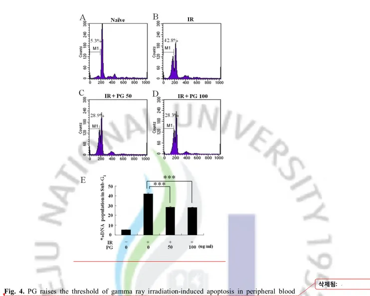
PG reduced the population of apoptotic DNA caused by gamma ray irradiation in splenocytes To identify whether PG decreases the proportion of apoptotic sub-G1 hypodiploid cells in 2 Gy-irradiated splenocytes, PI staining assay was used. As indicated in Figure 3A, B, and F, the formation of apoptotic DNA in sub-G1 peak was dramatically increased at 6 h after gamma ray irradiation up to 42.8%, compared to non-irradiated cells (5.3%) whereas PG treatment showed an interestingly lower percentage of cells in apoptotic peak at 50 and 100 μg/ml (28.9% and 28.3%, respectively) when compared with 2 Gy-irradiated untreated cells (42.8%) (Figure 3, ***; p < 0.001). These results indicate that PG enhanced splenocyte’s survival and proliferation by inhibiting the formation of apoptotic DNA caused by gamma ray irradiation.
PG inhibited apoptosis of peripheral blood lymphocyte
Since peripheral blood lymphocytes are one of the most sensitive cells to ionizing radiation (N.P. Harrington), we assessed the nuclear morphology of peripheral blood lymphocytes from irradiated mice with and without PG treatment to determine whether the inhibition of their DNA damage was attributable to apoptotic changes. As shown Figure 4A, DAPI staining revealed that nuclei of peripheral blood lymphocytes undergoing apoptosis had significant fragmentation and condensation of nuclei after an exposure to ionizing radiation. The recipients of PG manifested (Figure 4B) a dramatically decreased number of apoptotic nuclei compared to that of the sham irradiated group (3.85 ± 0.23 vs. 7.54 ± 0.28, *; p < 0.05). These results suggest that PG inhibited the damages of peripheral blood lymphocytes by reducing the formation of apoptosis caused by gamma ray irradiation.
PG reduced DNA damage in peripheral blood lymphocytes
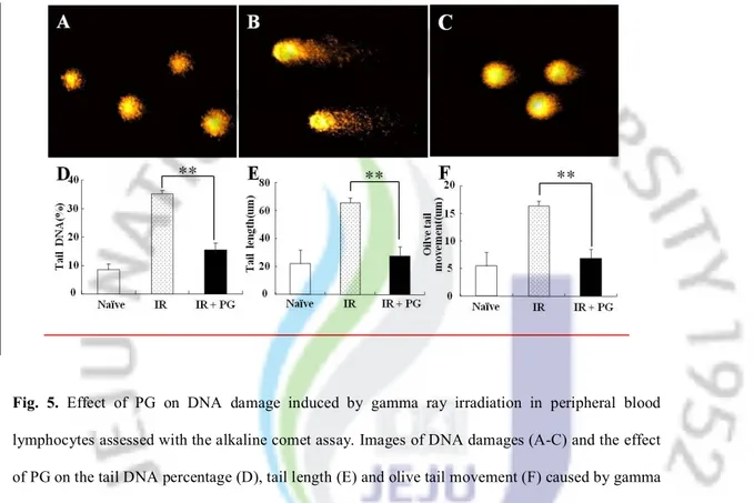
We studied the influence of PG on the DNA damage to peripheral blood lymphocytes caused by an ionizing radiation using alkaline comet assay. We measured the post-irradiation DNA damage in the tails of PG plus irradiated (Figure 5C) and non-irradiated (Figure 5A) mice, as
14
compared to the corresponding irradiated group (Figure 5B). Figure 5D, 5E and 5F show the changes in the levels of DNA damage (DNA in tail, tail length, and olive tail movement) in non-irradiated group, non-irradiated group, and PG plus non-irradiated group. Significant decrease in the levels of DNA damage was observed in PG treated group when compared with sham irradiated group. There was a significant decrease in the levels of DNA (%) in tail; 35.0 ± 1.30 vs. 15.5 ± 2.22 (**; p < 0.01), tail length (μm); 65.7 ± 3.57 vs. 27.3 ± 6.92 (**; p < 0.01) and olive tail moment (μm); 12.5 ± 1.58 vs. 4.9 ± 1.19 (**; p < 0.01). These results show that PG might reduce the DNA damages of the peripheral blood lymphocytes caused by ionizing radiation.
PG improved hemopoiesis in irradiated mice
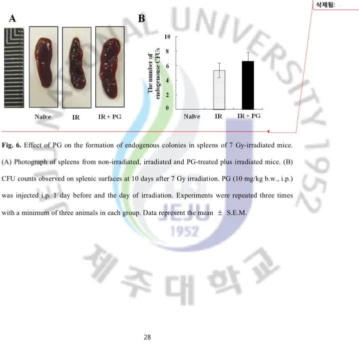
CFU assay was performed to assess whether PG can rescue and repopulate hemopoietic stem cells in gamma ray-irradiated mice. As Figures 6A and B depict, there are no colonies in spleens of mice of non-irradiated group. However, in spleens given with sub-lethal doses of gamma irradiation (7 Gy), few endogenous colonies were found (5.4 ± 1.00) and their sizes were markedly reduced, compared to non-irradiated mice. In contrast, the treatment of PG increased the size of spleen and the number of endogenous CFUs, when compared with the irradiated but non-treated mice (6.6 ± 1.20). These results indicate that PG led to the hemopoietic effects in gamma ray irradiated mice
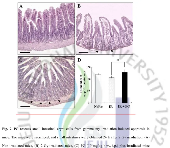
PG reduced the apoptosis of intestinal crypt cells
Since the gastrointestinal system is a major site of cell death induced by ionizing radiation, and intestinal crypt cells are one of the most sensitive cells to irradiation, we next investigated the potential of PG compound to rescue intestinal crypt cells from radiation-induced apoptosis. One day after WBI, the frequency of apoptotic fragments in crypt cells was reduced in the PG-treated group (Figure 7C) compared with sham irradiated group (Figure 7B). As shown in Figure 7D the number of intestinal crypt per circumference in the irradiated group was 107.6 ± 11.9 whereas a 16.19%
15
enlargement was observed in the PG plus irradiated group (128.4 ± 28.6, *; P < 0.05). These results show that PG enhanced the crypt survival of mice by reducing the damages of intestinal stem cells after an exposure to gamma ray irradiation.
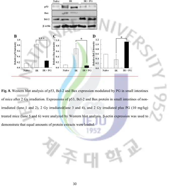
PG modulated the expression levels of apoptosis-related proteins in small intestine
As shown in Figure 8, exposure to gamma ray (2 Gy) irradiation considerably increased the expression of pro-apoptotic proteins, such as p53 and Bax whereas decreased the expression of anti-apoptotic Bcl-2 family proteins, compared with sham irradiated group. However, PG treatment apparently curtailed the increase of p53 and Bax expression levels and the decrease of Bcl-2 expression level in irradiated control group. β-actin was used to the equal amounts of protein extract were loaded. These results suggest that PG inhibited apoptosis of small intestine by modulating the expression levels of apoptosis-related with molecules in gamma ray-irradiated mice.
PG modulated the immunohistochemical localization and intensity of apoptosis-related proteins in intestine
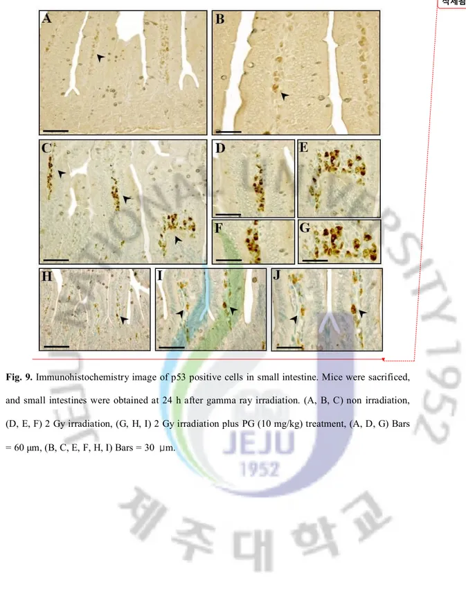
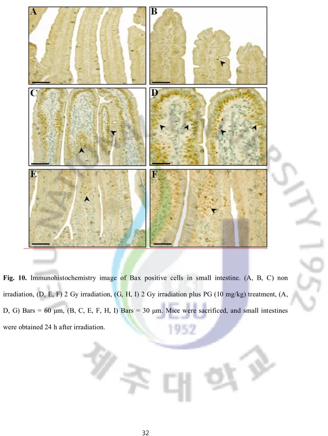
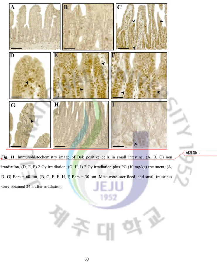
To pinpoint the mechanism of PG’s capacity to inhibit apoptosis after irradiation in vivo, we investigated the localization of regulatory proteins of apoptosis (p53, Bax, Bak, Bcl-Xs/L and Bcl-2) in intestines under PG-treated and untreated conditions using immunohistochemistry. As shown in Figures 9, 10, and 11, the immunoreactivities of p53, Bax, and Bak were markedly increased in small intestines of only-irradiated mice, but PG treatment decreased them. And, after an exposure to radiation, cells expressing p53 were highly expressed in lamina propria (**; p < 0.01) and crypt and Bax were highly overexpressed on apoptotic cells of intestinal epithelium (**; p < 0.01) and lamina propria (**; p < 0.01). In addition, the immunoreactivity of Bak was strongly expressed in intestinal epithelium (**; p < 0.01) and crypts (**; p < 0.01) as compared to untreated and unirradiated control mice. In contrast, PG treatment dramatically decreased the immunoreactivities of p53 (Figure 9H, I, and J), Bax (Figure 10E and F) and Bak (Figure 11G, H, and I), compared with sham irradiated mice.
16
Additionally, the staining intensities of anti-apoptotic Bcl-XS/L (Figure 12) and Bcl-2 (Figure 13) were weakly detected on laminar propria and intestinal epithelium in irradiated mice, compared with that of control mice. However, the application of PG significantly increased the localizations and intensities of Bcl-XS/L and Bcl-2 in intestinal epithelium (**; p < 0.01) and lamina propria (**; p < 0.01), respectively. Thus, the overexpression of Bcl-XS/L and Bcl-2 in fibrocytes and immune-related cells of intestinal epithelium and laminar propria after PG treatment was clearly involved in the repair of gastrointestinal cells. Also, the results were similar with those of Western blot assay. These results suggest that PG presumably inhibited the apoptosis of small intestine by regulating the expression of pro-apoptotic p53, Bax and Bak and anti-apoptotic Bcl-XS/L and Bcl-2 after a gamma ray irradiation.
17
5. DISCUSSION
Many researchers have reported that ionizing radiation commonly leads to oxidative stress, generating ROS in irradiated tissue and cells [21]. In addition, previous studies have demonstrated that oxidative stress induced by aqueous free radicals which are generated by the action of radiation on water in the cell causes the primary damage by reacting with cellular macromolecules such as DNA, protein and lipid membrane resulting in cell dysfunction and mortality [19, 22, 23]. In particular, apoptosis signaling, a form of programmed cell death, plays as an important role in the initiation of damages such as nuclear fragmentation and cytoplasmic shrinkage caused by oxidative stress [24, 25]. In this study, we showed that PG inhibited the increment of sub-G1 DNA contents and apoptotic fragments by reducing ROS generation from ionizing radiation without cytotoxicity in splenocytes and lymphocytes. Interestingly, previous studies have demonstrated that PG and its derivative, Eckol, led to free radical scavenging activities, which contributed to protect cells against damages caused by hydrogen peroxide in V79-4 lung fibroblast cells [10, 19, 26]. Also, polyphenols known as electron-rich compounds prone to enter into efficient electron-donation reactions with oxidizing agents to produce phenoxyl radical (PhO_) species which can undergo dimerization (‘‘phenol coupling’’) to produce new CC or CO linkage [19]. In view of these points, our results indicate that the intrinsic stability of phenolic structures in PG might induce its cytoprotective effects depending on its radical scavenging capacity from an exposure to oxidative stress.
In general, proliferating stem or progenitor cells are particularly vulnerable to radiation-induced cell damage, a prominent example being the stem cells in the small intestinal crypt [27]. Splenocytes and blood lymphocytes are known to be sensitive to oxidative stress from ionizing radiation [4]. Several studies have reported that the exposure of animals to radiation leads to the destruction of the lymphoid and hemopoietic systems by increasing apoptosis of not only proliferating stem cells such as jejuna crypt cells, but also lymphocytes [3, 4, 10, 11, 12]. Recent studies have also suggested that
18
two components of phlorotannins, PG and eckol, protect mice from damages by ionizing radiation by promoting the survival of jejunal crypt through the inhibition of apoptosis [5]. Moreover, our previous studies indicated that eckol and dieckol, two derivatives of PG, induced protective effects by inhibiting apoptosis and stimulating proliferation of hemopoietic cells such as intestinal stem cells, splenocytes and blood lymphocytes in mice [10, 11, 13]. This study demonstrated that PG enhanced the proliferation of splenocytes and the hematopoietic capacities of intestinal stem cells, splenocytes and blood lymphocytes, by decreasing the cellular damages through the inhibition of apoptosis.
In the apoptotic pathways, normally, the Bcl-2 family which includes both anti-apoptotic (Bcl-2 and Bcl-XS/L) and pro-apoptotic (Bax and Bak) members is an important factor in controlling the survival of cells under oxidative stress [28, 29, 30, 31]. Previous reports showed that dieckol induced the cytoprotective effect by decreasing the expression of pro-apoptotic p53 and Bax and correcting the radiation-induced decrease of anti-apoptotic Bcl-2 [3]. In our results, PG also regulated the expression of apoptotic molecules including both pro-apoptotic molecules such as Bax, Bak, p53 and anti-apoptotic molecules such as Bcl-2, Bcl-XS/L in intestinal stem cells. These results support our previous results that PG protected intestinal stem cells and rescued mice by inhibiting apoptosis against damages caused by ionizing radiation [5].
Previously, studies have suggested that PG stimulated lymphocytes by activating MAPK such as ERK, p38, and JNK and NFkB signaling and protected V79-4 cells by inhibiting apoptosis against oxidative damage and by enhancing the cellular catalase activity and modulating ERK signal pathway [19, 32]. These results suggest that the capacities of PG on the inhibition of apoptosis by regulating MAPK and NFkB signaling might affect to its protection against oxidative stress caused by ionizing radiation in mice. The possible involvement of ERK, p38, JNK, and/or NFkB offers an opportunity for future exploration.
In conclusion, these results demonstrate that PG protects mice against oxidative stress from ionizing radiation by enhancing hematopoiesis and promoting cell survival via inhibiting DNA damages through the regulation of apoptosis in peripheral blood lymphocytes, splenocytes and
19
intestinal stem cells. This study indicates that PG may serve as a beneficial protective agent for human recipients of ionizing radiation.
20
6. REFERENCES
1. Lee TK, O’Brien KF, Wang W, Sheng C, Wang T, Johnke RM, Allison RR. 2009. American ginseng modifies 137Cs-Induced DNA damage and oxidative stress in human lymphocytes. Open Nucl. Med. J. 1 (1): 1-8.
2. Kim MM, Kim SK. 2010. Effect of phloroglucinol on oxidative stress and inflammation. Food Chem. Toxicol. 48 (10): 2925-2933.
3. Park E, Ahn G, Yun JS, Kim MJ, Bing SJ, Kim DS, Lee J, Lee NH, Park JW, Jee Y. 2010. Dieckol rescues mice from lethal irradiation by accelerating hemopoiesis and curtailing immunosuppression. Int. J. Radiat. Biol. 86 (10): 848-859.
4. Andreson RW, Warner NL. 1976. Ionizing radiation and the immune response. Adv. Immunol. 24: 215-335.
5. Moon C, Kim SH, Kim JC, Hyun JW, Lee NH, Park JW, Shin T. 2008. Protective effect of phlorotannin components phloroglucinol and eckol on radiation-induced intestinal injury in mice. Phytother. Res. 22 (2): 238-242.
6. Moulder JE. 2002. Report on an interagency workshop on the radiobiology of nuclear terrorism. Molecular and cellular biology of moderate dose (1 – 10 Sv) radiation and potential mechanisms of radiation protection. Radiat. Res. 158 (1): 118-124.
7. Moulder JE. 2003. Pharmacological intervention to prevent or ameliorate chronic radiation injuries. Semin. Radiat. Oncol. 13 (1): 73-84.
8. Pellmar TC. 2005. Rockwell S. Radiological/Nuclear Threat Countermeasures Working Group. Priority list of research areas for radiological nuclear threat countermeasures. Radiat. Res. 163: 115-123.
9. Stone HB, Moulder JE, Coleman CN, Ang KK, Anscher MS, Barcellos-Hoff MH, Dynan WS, Fike JR, Grdina DJ, Greenberger JS, Hauer-Jensen M, Hill RP, Kolesnick RN, Macvittie TJ, Marks C, McBride WH, Metting N, Pellmar T, Purucker M, Robbins ME, Schiestl RH, Seed TM, Tomaszewski JE, Travis EL, Wallner PE, Wolpert M, Zaharevitz D. 2004. Models for
21
evaluating agents intended for the prophylaxis, mitigation and treatment of radiation injuries. Report of an NCI workshop, December 3-4, 2003. Radiat. Res. 162: 711-728.
10. Park E, Ahn GN, Lee NH, Kim JM, Yun JS, Hyun JW, Jeon YJ, Wie MB, Lee YJ, Park JW, Jee Y. 2008a. Radioprotective properties of eckol against ionizing radiation in mice. FEBS Lett. 582 (6): 925-930.
11. Park E, Lee NH, Joo HG, Jee Y. 2008b. Modulation of apoptosis of eckol against ionizing radiation in mice. Biochem. Biophys. Res. Commun. 372 (4): 792-797.
12. Park E, Hwang I, Song JY, Jee Y. 2009. Acidic polysaccharide of Panax ginseng as a defense against small intestinal damage by whole-body gamma irradiation of mice. Acta. Histochem. In press.
13. Park E, Lee NH, Ahn GN, Baik JS, Lee J, Hwang KK, Park JW, Jee Y. 2008c. Inhibition of Apoptosis by Elaeocarpus sylvestris in Mice Following Whole- body Exposure to Ionizing Radiation: Implications for Radioprotectors. Food Sci. Biotechnol. 17 (4): 718-722.
14. Nair MP, Mahajan S, Reynolds JL, Aalinkeel R, Nair H, Schwartz SA, Kandaswami C. 2006. The flavonoid quercetin inhibits proinflammatory cytokine (tumor necrosis factor alpha) gene expression in normal peripheral blood mononuclear cells via modulation of the NF-kappa beta system. Clin Vaccine Immunol. 13 (3): 319-328.
15. Kang HS, Chung HY, Kim JY, Son BW, Jung HA, Choi JS. 2004. Inhibitory phlorotannins from the edible brown alga Ecklonia stolonifera on total reactive oxygen species (ROS) generation. Arch. Pharm. Res. 27: 194-198.
16. Kang KA, Lee KH, Chae SW, Koh YS, Yoo BS, Kim JH, Ham YM, Baik JS, Lee NH, Hyun JW. 2005a. Triphlorethol-A from Ecklonia cava protects V79-4 lung fibroblasts against hydrogen peroxide induced cell damage. Free Radic. Res. 39: 883-892.
22
17. Kang KA, Lee KH, Chae SW, Zhang R, Jung MS, Lee YG, Kim SY, Kim HS. 2005b. Eckol isolated from Ecklonia cava attenuates oxidative stress induced cell damage in lung fibroblast cells. FEBS Lett. 579: 6295-6304.
18. Kang KA, Zhang R, Lee KH, Chae SW, Kin BJ, Kwak YS, Park JW, Lee NH, Hyun JW. 2006a. Protective effect of Triphoroethol-A from Ecklonia cava gainst ionidimg radiation. J. Radiat. Res. 47: 61-68.
19. Kang KA, Lee KH, Chae SW, Zhang R, Jung MS, Ham YM, Baik JS, Lee NH, Hyun JW. 2006b. Cytoprotective effect of phloroglucinol on oxidative stress induced cell damage via catalase activation, J. Cell. Biochem. 97 (3): 609-620.
20. Ahn GN, Kim KN, Cha SH, Song CB, Lee J, Heo MS, Yeo IK, Lee NH, Jee Y, Kim JS, Heu MS, Jeon YJ. 2007. Antioxidant activities of phlorotannins purified from Ecklonia cava on free radical scavenging using ESR and H2O2-mediated DNA damage. Eur. Food Res. Technol. 226: 71-79.
21. Ozben T. 2007. Oxidative stress and apoptosis: impact on cancer therapy. J Pharm Sci. 96: 2181-2196.
22. Tominaga H, Kodama S, Matsuda N, Suzuki K, Watanabe M. 2004. Involvement of reactive oxygen species (ROS) in the induction of genetic instability by radiation. J. Radiat. Res. 45: 181-188.
23. Mansour HH. 2006. Protective role of carnitine ester against radiation-induced oxidative stress in rats. Pharmacol. Res. 54: 165-171.
24. Meier P, Finch A, Evan G. 2000. Apoptosis in development. Nature 407: 796-801. 25. Vaux DL, Korsmeyer SJ. 1999. Cell death in development. Cell 96: 245-254.
26. Zhang R, Kang KA, Piao MJ, Ko DO, Wang ZH, Lee IK, Kim BJ, Jeong IY, Shin T, Park JW, Lee NH, Hyun JW. 2008. Eckol protects V79-4 lung fibroblast cells against gamma-ray radiation-induced apoptosis via the scavenging of reactive oxygen species and inhibiting of the
23
c-Jun NH(2)-terminal kinase pathway. Eur. J. Pharmacol. 591 (1-3): 114-123.
27. Potten CS, Grant HK. 1998. The relationship between ionizing radiation-induced apoptosis and stem cells in the small and large intestine. Br. J. Cancer. 78: 993-1003.
28. Jourdain A, Martinou JC. 2009. Mitochondrial outer-membrane permeabilization and remodelling in apoptosis. Int. J. Biochem. Cell Biol. 41: 1884-1889.
29. Haimovitz-Friedman A. 1998. Radiation-induced signal transduction and stress response. Radiat. Res. 150: S102-S108.
30. Rodel C, Haas J, Groth A, Grabenbauer GG, Sauer R, Rodel F. 2003. Spontaneous and radiation-induced apoptosis in colorectal carcinoma cells with different intrinsic radiosensitivities: survivin as a radioresistance factor. Int. J. Radiat. Oncol. Biol. Phys. 55: 1341-1347.
31. Kastan MB, Onyekwere O, Sidransky D, Vogelstein B, Craig RW. 1991. Participation of p53 protein in the cellular response to DNA damage. Cancer Res. 51: 6304-6311.
32. Ahn G, Park E, Park HJ, Jeon YJ, Lee J, Park JW, Jee Y. 2010. The classical NFkappaB pathway is required for phloroglucinol-induced activation of murine lymphocytes. Biochim. Biophys. Acta. 1800 (7): 639-645.
24
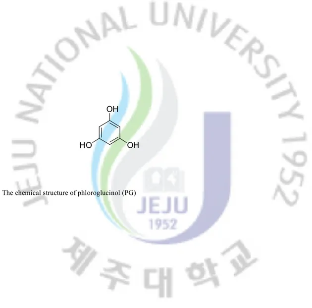
Fig. 1. The chemical structure of phloroglucinol (PG)
OH
OH
HO
25
Fig. 2. Effect of PG on the survival, proliferation and intracellular reactive oxygen species (ROS)
production in gamma ray-irradiated splenocytes. (A) One hundred thousand cells were exposed to 2 Gy irradiation and treated with PG (25, 50 and 100 μg/ml, respectively). After 24 h, the survival of cells was assessed by using MTT assay. (B) Four hundred thousand viable cells were exposed to 2 Gy irradiation and treated with PG (25, 50 and 100 μg/ml, respectively). After 72 h, the proliferation of splenocytes was measured by using the incorporation of 3H-thymidine. (C) The production levels of intracellular ROS were measured by using DCF-DA assay. Experiments were performed in triplicates, and data are expressed as average percent change from untreated controls ± S.E. *, p<0.05, **, p<0.01.
26
Fig. 4. PG raises the threshold of gamma ray irradiation-induced apoptosis in peripheral blood lymphocytes with 4'6-diamidino-2-phenylindole, dihydrochlodie (DAPI) staining. Mice received (A) 2 Gy irradiation or (B) 2 Gy irradiation plus PG (10 mg/kg) treatment. (C) Columns indicate the percentage of apoptotic fragments in 500 lymphocytes per mouse in each group. The cells were obtained at 24 h after irradiation and stained with DAPI. Arrows point to apoptotic cells with fragmented nuclei. Each data point represents the mean ± S.E. ***, p<0.005.
27
Fig. 5. Effect of PG on DNA damage induced by gamma ray irradiation in peripheral blood lymphocytes assessed with the alkaline comet assay. Images of DNA damages (A-C) and the effect of PG on the tail DNA percentage (D), tail length (E) and olive tail movement (F) caused by gamma ray irradiation in splenocytes. (A) Non-irradiated mice, (B) 2 Gy-irradiated mice, (C) PG plus irradiated mice (10 mg/kg b.w., i.p.) treated and irradiated mice. Experiments were performed in triplicates, and data are expressed as average percent change from untreated controls ± S.E. *,
28
Fig. 6. Effect of PG on the formation of endogenous colonies in spleens of 7 Gy-irradiated mice. (A) Photograph of spleens from non-irradiated, irradiated and PG-treated plus irradiated mice. (B) CFU counts observed on splenic surfaces at 10 days after 7 Gy irradiation. PG (10 mg/kg b.w., i.p.) was injected i.p. 1 day before and the day of irradiation. Experiments were repeated three times with a minimum of three animals in each group. Data represent the mean ± S.E.M.
29
Fig. 7. PG rescues small intestinal crypt cells from gamma ray irradiation-induced apoptosis in mice. The mice were sacrificed, and small intestines were obtained 24 h after 2 Gy irradiation. (A) Non-irradiated mice, (B) 2 Gy-irradiated mice, (C) PG (10 mg/kg b.w., i.p.) plus irradiated mice Bars = 30 μm. (C, F) Columns represent the number of apoptotic fragments per small intestinal crypt in each group. Values are means ± S.E. of 50 crypt sections per intestine and 5 small intestine sections from each mouse. (*, p < 0.05).
30
Fig. 8. Western blot analysis of p53, Bcl-2 and Bax expression modulated by PG in small intestines of mice after 2 Gy irradiation. Expressions of p53, Bcl-2 and Bax protein in small intestines of non-irradiated (lane 1 and 2), 2 Gy non-irradiated(lane 3 and 4), and 2 Gy non-irradiated plus PG (10 mg/kg) treated mice (lane 5 and 6) were analyzed by Western blot analysis. β-actin expression was used to demonstrate that equal amounts of protein extracts were loaded.
31
Fig. 9. Immunohistochemistry image of p53 positive cells in small intestine. Mice were sacrificed, and small intestines were obtained at 24 h after gamma ray irradiation. (A, B, C) non irradiation, (D, E, F) 2 Gy irradiation, (G, H, I) 2 Gy irradiation plus PG (10 mg/kg) treatment, (A, D, G) Bars = 60 μm, (B, C, E, F, H, I) Bars = 30 μm.
32
Fig. 10. Immunohistochemistry image of Bax positive cells in small intestine. (A, B, C) non irradiation, (D, E, F) 2 Gy irradiation, (G, H, I) 2 Gy irradiation plus PG (10 mg/kg) treatment, (A, D, G) Bars = 60 μm, (B, C, E, F, H, I) Bars = 30 μm. Mice were sacrificed, and small intestines were obtained 24 h after irradiation.
33
Fig. 11. Immunohistochemistry image of Bak positive cells in small intestine. (A, B, C) non irradiation, (D, E, F) 2 Gy irradiation, (G, H, I) 2 Gy irradiation plus PG (10 mg/kg) treatment, (A, D, G) Bars = 60 μm, (B, C, E, F, H, I) Bars = 30 μm. Mice were sacrificed, and small intestines were obtained 24 h after irradiation.
34
Fig. 12. Immunohistochemistry image of Bcl-XS/L positive cells in small intestine. (A, B, C) non irradiation, (D, E, F) 2 Gy irradiation, (G, H, I) 2 Gy irradiation plus PG (10 mg/kg) treatment, (A, D, G) Bars = 60 μm, (B, C, E, F, H, I) Bars = 30 μm. Mice were sacrificed, and small intestines were obtained 24 h after irradiation.
35
Fig. 13. Immunohistochemistry image of Bcl-2 positive cells in small intestine. (A, B, C) non irradiation, (D, E, F) 2 Gy irradiation, (G, H, I) 2 Gy irradiation plus PG (10 mg/kg) treatment, (A, D, G) Bars = 60 μm, (B, C, E, F, H, I) Bars = 30 μm. Mice were sacrificed, and small intestines were obtained 24 h after irradiation.
36
Fig. 14. Quantitative analysis result of immunohistochemistry for p53, Bax, Bak, Bcl-XS/L and Bcl-2 positive cells in intestine sections.
37
초록
Phloroglucinol 이 감마 방사선에 의해 유도된
세포자멸사의 억제에 미치는 영향
지도교수 : 지영흔 박석재 제주대학교 대학원 수의학과 제주지역에서 자생하고 있는 갈조류의 한 종류인 감태에서 추출된 Phloroglucinol (PG)은 산화적 스트레스에 대한 항산화 및 세포 보호효과 등의 생리활성을 나타내는 것으로 알려져 있다. 본 연구에서는 PG 가 방사선 조사와 같은 산화적 스트레스에 의해 야기되는 세포자멸사(apoptosis)를 감소시킴으로서 마우스를 보호한다는 것과 그 보호기전에 대하여 알아보았다. PG 는 방사선 조사에 의해 증가된 sub-G1 DNA 량을 감소시킴에 따라 마우스 비장세포의 생존과 증식을 유의적으로 증가시켰고, 뿐만 아니라 PG 는 마우스 림프구의 apoptotic 단편수를 감소시켰으며 DNA 회복을 촉진시켰다. 뿐만 아니라 PG 는 방사선 조사군에 비해, 조혈기능의 증가로 인한 비장 집락 형성의 수와 소장 내 줄기세포로 알려진 음와 세포의 재생을 증가시켰다. PG 는 소장에서 세포자멸화 유도단백질로 알려진 단백질 p53 과Bax 및 Bak 과 세포자멸화 억제와 관련된 단백질로 알려진 Bcl-2 and Bcl-XS/ L의 발현을 조절함으로 인해 방사선 조사가 야기한 손상들을 억제시켰다. 이상의 결과로부터 PG 가 방사선에 민감한 말초면역세포와 소장 줄기세포의 세포자멸화를 억제함과 더불어 조혈기능을 강화하여 방사선 조사에 의해 야기된 산화적 스트레스로부터 마우스를 보호할 수 있다는 사실을 확인하였다.