Insulin-Like Growth Factor-Binding Protein-3
Mediates High Glucose-Induced Apoptosis by
Increasing Oxidative Stress in Proximal Tubular
Epithelial Cells
Eun-Gyong Yoo, Woo Jung Lee, Jung Hyun Kim, Hyun-Wook Chae, Se Eun Hyun, Duk Hee Kim, Ho-Seong Kim, and Youngman Oh
Department of Pediatrics (E.-G.Y.), College of Medicine, CHA University, Sungnam 463-712, Korea; Department of Pediatrics (W.J.L., J.H.K., H.-W.C., S.E.H., D.H.K., H.-S.K.), Severance Children’s Hospital, Institute of Endocrinology, Yonsei University College of Medicine, Seoul 120-752, Korea; and
Department of Pathology (Y.O.), School of Medicine, Virginia Commonwealth University, Richmond, Virginia 23298
IGF-binding protein-3 (IGFBP-3) is the major circulating carrier protein for IGF, and also acts as a potent antiproliferative agent in various cell types. Recently, IGFBP-3 was reported to mediate high glucose-induced apoptosis in mesangial cells and podocytes. In this study, we investigated the role of IGFBP-3 in high glucose-induced apoptosis in proximal tubular epithelial cells (PTEC). Expression of IGFBP-3 protein and mRNA in a porcine PTEC line (LLC-PK1 cells) was measured after exposure to either standard (5.5 mM) or high-glucose (30 mM) medium. We quantified apoptosis after treat-ment with small interfering RNA against IGFBP-3 (siRNA:IGFBP-3) in high-glucose medium or in cells that overexpressed IGFBP-3. Oxidative stress was measured in high-glucose medium, in the pres-ence of siRNA:IGFBP-3, or in IGFBP-3-overexpressing cells. IGFBP-3 protein and mRNA expression in LLC-PK1 cells was higher in high-glucose medium than in standard-glucose medium. Exposure to high-glucose medium increased apoptosis, and high-glucose-induced apoptosis was abolished by siRNA:IGFBP-3. IGFBP-3 overexpression induced apoptosis in LLC-PK1 cells. Both high-glucose medium and IGFBP-3 overexpression increased reactive oxygen species, and siRNA:IGFBP-3 reduced this increase. Antioxidant treatment decreased IGFBP-3 expression and apoptosis, whereas oxidative stress from hydrogen peroxide increased IGFBP-3 expression, suggesting that oxidative stress increases IGFBP-3 expression. Our results suggest that increased IGFBP-3 expression by high glucose mediates high-glucose-induced apoptosis in PTEC. Increased oxi-dative stress from high glucose enhances IGFBP-3 expression, inducing apoptosis. Increased expression of IGFBP-3 by high glucose induces additional oxidative stress, which may result in amplification of hyperglycemic damage. (Endocrinology 152: 3135–3142, 2011)
D
iabetic nephropathy is the leading cause of end-stage renal disease (1). The pathophysiological mechanism of the development and progression of diabetic nephrop-athy has been studied extensively over the past few de-cades. The pathways that may be involved include in-creased oxidative stress, accumulation of advanced glycation end-product (AGE), and activation ofintracel-lular signaling molecules such as protein kinase C (2). Hy-perglycemia increases the expression of growth factors and cytokines, which may directly or indirectly contribute to the renal changes seen in diabetes (3, 4).
IGF-binding protein-3 (IGFBP-3) is a component of the IGF-IGFBP axis and major carrier protein for IGF. IGFBP-3 has intrinsic biological activity as well as the abil-ISSN Print 0013-7227 ISSN Online 1945-7170
Printed in U.S.A.
Copyright © 2011 by The Endocrine Society
doi: 10.1210/en.2010-1122 Received September 28, 2010. Accepted May 13, 2011. First Published Online June 7, 2011
Abbreviations: AGE, Advanced glycation end-product; BrdU, bromodeoxyuridine; Ct, cycle threshold; DCF, dichlorofluorescein; DCF-DA, dichlorodihydrofluorescein diacetate; DCFH, dichlorofluorescin; FITC, fluorescien isothiocyanate; GSH, glutathione; IGF-IR, IGF-I recep-tor; IGFBP-3, IGF-binding protein-3; NAC, N-acetyl-L-cysteine; PTEC, proximal tubular ep-ithelial cell; ROS, reactive oxygen species; siRNA:IGFBP-3, small interfering RNA against IGFBP-3.
ity to bind and sequester active IGF hormones, thereby modulating their bioactivity (5, 6). IGFBP-3 has a role as a potent IGF-IGF receptor-independent antiproliferative agent in various cell types, and acts by blocking the cell cycle and inducing apoptosis (7–9). In addition, IGFBP-3 may contribute to insulin resistance in adipocytes (10, 11) and inhibit adipocyte differentiation (12).
Apoptosis is rarely observed in the normal kidney but is reported in renal diseases including renal ischemia-rep-erfusion injury (13) and chronic renal failure (14). In di-abetic rats, apoptosis increases in renal tubular and inter-stitial cells, and this is reversed by insulin therapy (15). Apoptosis also occurs in the human diabetic kidney, es-pecially in the tubular epithelial cells (16). Although high ambient glucose induces mesangial IGF signaling and cel-lular hypertrophy, high glucose also induces programmed mesangial cell death in vitro by apoptosis (17).
IGFBP-3 may contribute to the pathogenesis of diabetic nephropathy by modulating the bioactivity of IGF via an IGF-dependent mechanism and by inducing apoptosis in kidney cells via both IGF-independent and IGF-dependent mechanisms (5). IGFBP-3 is reported to increase mesan-gial cell apoptosis under high ambient or standard levels of glucose, and an antisense IGFBP-3 oligodeoxynucleotide inhibits apoptosis induced by high glucose or by TNF-␣ (18). Recent human biopsy studies have proposed evi-dence that podocytes are functionally and structurally in-jured very early in the natural history of diabetic nephrop-athy (19). Exogenous IGFBP-3 induces IGF-independent podocyte apoptosis (5).
Both the glomerular and tubular compartments may be involved in the development of diabetic nephropathy. The proximal tubule plays a crucial role in the pathogenesis of diabetic kidney disease and is susceptible to a variety of metabolic and hemodynamic factors associated with dia-betes, especially hyperglycemia. Glucose entry into prox-imal tubular epithelial cells (PTEC) is insulin independent, which makes PTEC particularly sensitive to the deleteri-ous effects of chronic hyperglycemia, and the proximal tubules react with tubular hypertrophy, tubular atrophy, and reduced organic ion transport (20, 21). Tubulointer-stitial pathology, rather than glomerular pathology, cor-relates with renal dysfunction in diabetic nephropathy (20, 22).
Because the kidney has marked heterogeneity in cell populations, in vitro studies in individual renal cell types are essential. However, no study has focused on the role of IGFBP-3 in high-glucose-induced apoptosis in PTEC. In this study, we investigated IGFBP-3 expression and apo-ptosis in PTEC exposed to high-glucose medium, along with the specific role of IGFBP-3 in PTEC apoptosis. To investigate the mechanism of IGFBP-3-mediated PTEC
apoptosis in high-glucose medium, we also studied the relationship between oxidative stress and IGFBP-3 expression.
Materials and Methods Cell culture
The LLC-PK1 cells, a porcine PTEC line that does not contain any virus or oncogene for immortalization, was purchased from the American Type Culture Collection (Rockville, MD). LLC-PK1 cells were grown in 75-cm3tissue culture flasks in Life
Tech-nologies, Inc. Medium 199 (Invitrogen, Carlsbad, CA). Subcon-fluent cells were harvested and seeded into 100-mm dishes in 10 ml growth medium and allowed to adhere for 18 h in a humid-ified incubator at 37 C with 5% CO2in air.
Establishment of IGFBP-3-overexpressing PTEC line
An IGFBP-3-overexpressing LLC-PK1 cell line was estab-lished by transfection of IGFBP-3 cDNA containing 1.5-kb hu-man IGFBP-3 (equivalent to 133-1008 in the cDNA sequence), ligated into the pCMV6 vector. Transfection used Lipofectamine 2000 (Invitrogen). After 48 h, cells were split into selective me-dium containing G418 (800g/ml) for pCMV6/IGFBP-3, and foci were picked to generate a stable cell line.
Overexpression of GGG-IGFBP-3, a mutant IGFBP-3 that cannot bind to IGF, was also performed to verify the IGF-inde-pendent effect of IGFBP-3 on apoptosis in LLC-PK1 cells. The GGG-IGFBP-3 mutant cDNA was generated by site-directed mutagenesis at residues Ile 56, Leu 80, and Leu 81 to Gly 56, Gly 80, and Gly 81, as described previously (23). Binding studies, including BIAcore analysis, showed that the GGG-IGFBP-3 mu-tant protein, generated in Escherichia coli and baculovirus ex-pression systems, lacks affinity for IGF (23).
Small interfering RNA transfection
Small interfering RNA against IGFBP-3 (siRNA:IGFBP-3, siGENOME SMARTpool M-004777-01-0005), and mismatch control (ON-TARGETplus siCONTROL, D-001810-01-05) were from Dharmacon Research (Lafayette, CO), and 12.5 nM
siRNA:IGFBP-3 was transfected as above. Cells were incubated in growth medium with 10% fetal bovine serum for 24 h.
Reagents and antibodies
Antihuman IGFBP-3 antibody was from AbCam (Cam-bridge, MA). Horseradish peroxidase-labeled IgG (1:3000; Santa Cruz Biotechnology, Santa Cruz, CA) was used for sec-ondary antibody. Primer pairs for real-time PCR were from Ap-plied Biosystems (HS-00181211-m1 for IGFBP-3, Hs99999903 for-actin).
Annexin V-fluorescein isothiocyanate (FITC) apoptosis de-tection kits were from R&D Systems (Minneapolis, MN), and the cellular DNA fragmentation ELISA kit was from Roche Di-agnostics (Mannheim, Germany). The cell-permeable fluoro-genic probe dichlorodihydrofluorescein diacetate (DCF-DA), and the antioxidants, N-acetyl-L-cysteine (NAC) and
Western blot analysis
LLC-PK1 cells were analyzed after 72 h exposure to standard-glucose (5.5 mM) or high-glucose (30 mM) medium. For Western
blots, 20l medium from LLC-PK1 cells was electrophoresed on 11% acrylamide gels in the presence of sodium dodecyl sulfate and electroblotted onto polyvinylidene fluoride membranes. Af-ter blocking 1 h with 5% wt/vol skim milk, membranes were incubated overnight at 4 C with antihuman IGFBP-3 antibody. Secondary antibody was horseradish peroxidase-labeled IgG (1: 3000), and detection was by enhanced chemiluminescence (Am-ersham Biosciences, Little Chalfont, UK).
RT-PCR and real-time quantitative PCR
RT-PCR and real-time quantitative PCR were performed us-ing whole-cell lysates from LLC-PK1 cells after 48 h exposure to
standard- or high-glucose medium. In brief, total RNA was isolated using Trizol reagent (Invitrogen) and 2g total RNA subjected to reverse transcription using a Superscript III first-strand synthesis system (Invitrogen) with primers for RT-PCR[(IGFBP-3 (up): aaatggagga cacgctgaac, IGFBP-3 (down): tacttatcca cgcaccagca], according to the manufacturer’s instructions.
Amplification was in duplicate, using the ABI 7300 system (Applied Biosystems, Fos-ter City, CA) with the following profile: 94 C for 5 min, 35 cycles of 95 C for 30 sec, 50 C for 30 sec, and 72 C for 30 sec, followed by 72 C for 10 min.
Real-time quantitative PCR was with 20 l PCR amplification reaction mixture con-taining DNA, TaqMan primer pairs, and TaqMan universal PCR master mix (Ap-plied Biosystems). Amplification was in du-plicate as above with the following profile: 50 C for 2 min, 95 C for 10 min, and 40 cycles of 95 C for 15 sec with 60 C for 1 min. Gene expression for each sample was ex-pressed as cycle threshold (Ct) normalized to-actin (⌬Ct). ⌬Ct values were compared between samples from high-glucose-treated cells and control cells in serum-free me-dium, for⌬⌬Ct calculations. The final com-parison of transcript ratios between sam-ples is given as 2⫺⌬⌬Ct.
Quantification of apoptosis by annexin V-FITC staining
Subconfluent monolayers of LLC-PK1 cells were exposed to medium containing either standard or high glucose for 72 h. Detection of annexin V on the cell surface was done using a flow cytometer (Bekman Coulter Corp., Fullerton, CA). System II software was used for acquisition and anal-ysis. Annexin V-FITC conjugate (R&D Sys-tems) was used to stain cells. In brief, cells and supernatants were harvested, washed, and incubated for 15 min with annexin V-FITC and propidium iodide to differentiate between early apoptotic cells (annexin V-positive), late apoptotic and/or necrotic cells (annexin V and propidium iodide positive), and viable cells (unstained). The sum of early apoptotic and late apoptotic/necrotic cells were consid-ered as total apoptotic cells and was used for statistical analysis.
Quantification of apoptosis by DNA fragmentation
Cells were prelabeled by incubation with 10M
bromode-oxyuridine (BrdU) for 12 h at 37 C. Labeled cells were incubated in 5.5 or 30 mMglucose or 30 mMmannitol for an osmotic
control. After 48 h, cells were centrifuged, and the supernatant was analyzed for apoptosis. After adsorbing anti-DNA antibody onto the wells of a microplate, sample was added to the micro-plate, allowing BrdU-labeled DNA fragments to bind to the im-mobilized antibody. Immunocomplexed BrdU-labeled
DNA-FIG. 1. High glucose increased 3 expression and apoptosis in LLC-PK1 cells, and
IGFBP-3 mediated glucose-induced apoptosis. A, Western blot of IGFBP-IGFBP-3 in standard and high-glucose medium for 72 h with or without siRNA:IGFBP-3. Representative blot and means⫾SD of six independent experiments are shown. B and C, RT-PCR (B) and real-time PCR (C) showing increased IGFBP-3 mRNA expression after 48 h exposure to high-glucose medium (n⫽ 4). D, Representative data and means ⫾SDof annexin V-FITC staining showing increased apoptosis in LLC-PK1 cells after 72 h exposure to high-glucose medium. High-glucose-induced apoptosis was prevented by siRNA:IGFBP-3. The sum of early apoptotic (lower right) and late apoptotic and/or necrotic cells (upper right) are presented (n⫽ 4). E, High glucose increased DNA fragmentation compared with standard glucose and osmotic control. High-glucose-induced apoptosis was prevented by treatment with siRNA:IGFBP-3 (n⫽ 3). *, P ⬍ 0.05.
fragments were denatured by microwave irradiation, and anti-BrdU antibody peroxidase conjugate was added to form an immunocomplex. Bound anti-BrdU-peroxidase was quantified using an ELISA plate reader.
Oxidative stress measurement
The cell-permeable fluorogenic probe DCF-DA diffuses across cell membranes and is hydrolyzed by nonspecific cellular esterases to the nonfluorescent compound dichlorofluorescin (DCFH), which is predominantly trapped within the cell. In the presence of reactive oxygen species (ROS), DCFH rapidly un-dergoes one-electron oxidation to the highly fluorescent com-pound dichlorofluorescein (DCF). Our assay was a modification of the Bestwick and Milne (24) procedure. Glucose-exposed cell monolayers were incubated with 10MDCF-DA in PBS (pH 7.4)
for 15 min at 37C. Cells were washed once with PBS, and cell debris was cleared by centrifugation at 3000 rpm for 5 min. Cells were treated with trypsin and washed once with PBS, and cell debris were cleared by centrifugation at 1000 rpm for 5 min. Fluorescence was monitored using a flow cytometer as above, using 480-nm excitation and 530-nm emission wavelengths. Flu-orescent levels are expressed as the percent increase over the control.
Data expression and statistical analysis
Results of three or more independent ex-periments are expressed as mean⫾SD.
Sta-tistical comparison among groups were per-formed using one-way ANOVA followed by Tukey’s post hoc analysis. P⬍ 0.05 was considered significant.
Results
High glucose increases IGFBP-3 expression in PTEC
We examined whether high glucose increases IGFBP-3 expression in LLC-PK1 cells. Western blots revealed that IGFBP-3 protein is increased in LLC-PK1 cells exposed to high-glucose me-dium, compared with cells exposed to standard-glucose medium (Fig. 1A). IGFBP-3 expression was decreased in LLC-PK1 cells in high-glucose medium after cotreatment with siRNA:IGFBP-3 (Fig. 1A). IGFBP-3 mRNA expression was also increased in cells in high glu-cose, as measured by RT-PCR (Fig. 1B) or real-time quantitative PCR (Fig. 1C). To confirm the effect of high-glucose medium on IGFBP-3 expression, we ex-amined the expression of IGFBP-3 at different glucose concentrations and time courses. IGFBP-3 expressions in 30 and 45 mMglucose media were sig-nificantly increased compared with those in 5.5 mM glu-cose medium (Supplemental Fig. 1, A and B, published on The Endocrine Society’s Journals Online web site at http://endo.endojournals.org). IGFBP-3 protein expres-sion was significantly increased by 24 h exposure to high-glucose medium, which was inhibited by siRNA:IGFBP-3 (Supplemental Fig. 1, C and D). IGFBP-3 mRNA expres-sion was significantly increased after 12, 24, and 48 h exposure to high-glucose medium, which was also effec-tively inhibited by siRNA:IGFBP-3 (Supplemental Fig. 1, E and F).
IGFBP-3 mediates high-glucose-induced apoptosis in PTEC
We investigated whether high glucose increases apoptosis in LLC-PK1 cells. Annexin V-FITC staining revealed that the percentage of apoptotic cells in high-glucose medium was higher than in standard-glucose medium (18.3 ⫾ 1.2 vs. 5.8⫾ 1.7%, P ⬍ 0.05), indicating that high glucose increases
FIG. 2. IGFBP-3 overexpression increased apoptosis in LLC-PK1 cells. A, Western blot of
IGFBP-3 protein expression (n⫽ 6). B and C, RT-PCR (B) and real-time PCR (C) showing increased IGFBP-3 mRNA expression (n⫽ 6). D, Annexin V-FITC staining showing increased PTEC apoptosis by IGFBP-3 overexpression. GGG-IGFBP-3 overexpression also increased apoptosis. Representative data and means⫾SDof four independent experiments are shown. E, IGFBP-3 overexpression and exposure to high glucose increased apoptosis measured by DNA fragmentation (n⫽ 3). *, P ⬍ 0.05.
apoptosis in LLC-PK1 cells (Fig. 1D). The apoptosis-induc-ing effect of high glucose was abolished by cotreatment with siRNA:IGFBP-3, suggesting that IGFBP-3 mediates high-glucose-induced apoptosis (Fig. 1D). Moreover, measure-ment of apoptosis by DNA fragmeasure-mentation demonstrated the same pattern in LLC-PK1 cells in high glucose, with or with-out siRNA:IGFBP-3 (Fig. 1E). Treatment with 30 mM man-nitol did not increase apoptosis in LLC-PK1 cells (Fig. 1E), suggesting that the increased apoptosis in high glucose did not result from high osmolarity.
To confirm the effect of high-glucose medium on PTEC apoptosis, we performed annexin V-FITC staining under different glucose concentrations and exposure time. Ap-optosis in LLC-PK1 cells were significantly increased after exposure to 15, 30, and 45 mMglucose media compared with 5.5 mMglucose medium. Because apoptosis was most prominent in 30 mMglucose medium, we labeled 30 mM glucose as a high-glucose condition throughout the exper-iment (Supplemental Fig. 2A). Apoptosis was significantly increased in LLC-PK1 cells after 24, 48, and 72 h exposure to high-glucose medium, which was most prominent at 72 h (Supplemental Fig. 2B). Preincubation with siRNA:IGFBP-3 significantly inhibited apoptosis after 12, 24, and 48 h exposure to high-glucose medium (Supplemental Fig. 2C).
IGFBP-3 overexpression increases apoptosis in PTEC
To confirm the effect of increased IGFBP-3 expression on apoptosis in LLC-PK1 cells, we established an IGFBP-3-overexpressing PTEC line by transfection with IGFBP-3 cDNA. Figure 2, A–C, shows the increase in IGFBP-3 pro-tein and mRNA in the IGFBP-3-overexpressing PTEC line. Annexin V-FITC staining showed that IGFBP-3 overex-pression increased apoptosis (empty vector control, 8.1⫾ 0.7%, vs. IGFBP-3 overexpression, 21.8 ⫾ 4.2%; P ⬍ 0.01) (Fig. 2D). GGG-IGFBP-3 overexpression also in-creased apoptosis (empty vector control, 8.1⫾ 0.7%, vs. GGG-IGFBP-3 overexpression, 18.4⫾ 3.6%; P ⬍ 0.01), suggesting that IGFBP-3 promotes PTEC apoptosis in an IGF-independent manner (Fig. 2D). Measurement of ap-optosis by DNA fragmentation also demonstrated that GGG-IGFBP-3 overexpression, as well as IGFBP-3 over-expression, increased apoptosis (P⬍ 0.05 compared with cells in standard glucose, Fig. 2E).
IGFBP-3 is involved in oxidative stress induction by high glucose in PTEC
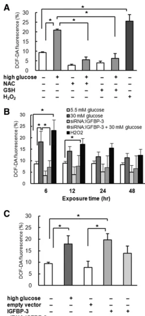
We investigated the effect of high glucose and IGFBP-3 expression on oxidative stress in LLC-PK1 cells. Exposure to high-glucose medium increased oxidative stress, as measured by DCF-DA staining (standard glucose, 9.7⫾ 0.4%, vs. high glucose, 20.8⫾ 0.7%, P ⬍ 0.01; Fig. 3A).
NAC and GSH, the antioxidants, prevented ROS increase in high-glucose medium (P ⬍ 0.01, Fig. 3A). ROS was significantly increased by high glucose at 6 h, and treat-ment with siRNA:IGFBP-3 reduced oxidative stress in LLC-PK1 cells exposed to high-glucose medium (siRNA: IGFBP-3 with high glucose, 7.2⫾ 2.9%, vs. high glucose only, 18.2 ⫾ 4.6%, P ⬍ 0.01; Fig. 3B). Moreover, IGFBP-3 overexpression increased oxidative stress (empty vector, 7.9⫾ 2.6%, vs. IGFBP-3 overexpression, 19.7 ⫾ 2.7%, P⬍ 0.01; Fig. 3C), suggesting that IGFBP-3 is in-volved in high-glucose-induced ROS generation in LLC-PK1 cells.
FIG. 3. A, Exposure to high-glucose medium for 6 h increased
oxidative stress measured by DCF-DA fluorescence. NAC and GSH, the antioxidants, prevented ROS increase by high-glucose medium (n⫽ 4). B, ROS was significantly increased by high glucose at 6 h, and treatment with siRNA:IGFBP-3 reduced oxidative stress in cells exposed to high-glucose medium (n⫽ 4). C, IGFBP-3 overexpression increased oxidative stress in LLC-PK1 cells (n⫽ 4). *, P ⬍ 0.05.
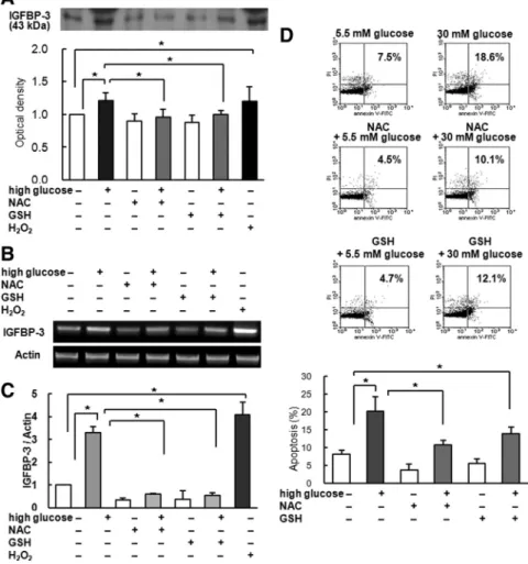
Oxidative stress increases IGFBP-3 expression and mediates PTEC apoptosis
We further investigated the effect of oxidative stress on IGFBP-3 expression and high-glucose-induced apoptosis in LLC-PK1 cells. Preincubation with antioxidants NAC and GSH abolished increase in IGFBP-3 protein expres-sion by high-glucose media, whereas elevating oxidative stress with hydrogen peroxide increased IGFBP-3 expres-sion (Fig. 4A). IGFBP-3 mRNA expresexpres-sion was also re-duced by NAC and GSH, whereas it was increased by hydrogen peroxide (Fig. 4, B and C). These findings sug-gest that increased oxidative stress by high glucose may stimulate IGFBP-3 expression.
We also investigated whether antioxidant prevented high-glucose-induced apoptosis in LLC-PK1 cells. An-nexin V-FITC staining showed that NAC and GSH de-creased apoptosis in high-glucose medium (NAC with high glucose, 10.9⫾ 1.2%, and GSH with high glucose,
13.9 ⫾ 1.9%, vs. high glucose only, 20.2 ⫾ 4.0%, P ⬍ 0.01; Fig. 4D). Taken together, these results imply that increased oxidative stress from high glucose increased IGFBP-3 expression, resulting in increased apoptosis.
Discussion
Chronic hyperglycemia is the central initiating factor in diabetic nephropa-thy. Although all diabetic cells are ex-posed to elevated levels of plasma glu-cose, most cells are able to reduce glucose transport into the cell during hyperglycemia, so that internal glucose concentrations remain constant. In contrast, in cells damaged by hypergly-cemia, the glucose transport rate does not decline rapidly during hyperglyce-mia (2). PTEC, like mesangial cells and podocytes, develop intracellular hyper-glycemia, which can result in hypergly-cemic damage (25). Samikkannu et al. (26) demonstrated that acute exposure of human PTEC to high glucose induces a time-dependent dual effect of early proliferation and late apoptosis.
Cellular responses to high glucose are numerous and varied but ultimately result in functional changes and often cell death. High glucose induces oxida-tive and nitrosaoxida-tive stress, causing ac-tivation of proteins involved in apopto-tic cell death, which is important to the development of diabetic complications (27). In this study, high-glucose medium increased IGFBP-3 protein and mRNA levels in PTEC. IGFBP-3 mRNA expression is reported to be in-creased in the proximal tubules of diabetic rat kidney (28), but few studies have reported on IGFBP-3 expression in high-glucose medium in a PTEC line.
Initially, IGFBP-3 was considered to modulate only the actions of circulating IGF, but it also exhibits clear, dis-tinct biological effects independent of the IGF/IGF-I re-ceptor (IGF-IR) axis. Apart from modulating IGF actions (IGF/IGF-IR-dependent actions), IGFBP-3 clearly exerts intrinsic bioactivity either in the absence of IGF or without triggering IGF-IR signaling (IGF/IGF-IR-independent ac-tions) (6). IGFBP-3 inhibits growth and induces apoptosis by both IGF-dependent and IGF-independent mecha-nisms. GH and tumor suppressors such as p53 induce
FIG. 4. Effect of antioxidants (NAC and GSH) on IGFBP-3 expression and apoptosis. A,
Preincubation with 10 mMNAC for 15 min or 4 mMGSH for 30 min inhibited IGFBP-3 expression, whereas exposure to 0.1% hydrogen peroxide (H2O2) for 6 h increased IGFBP-3 expression. (n⫽ 6). B and C, RT-PCR (B) and real-time PCR (C) showing decreased IGFBP-3 mRNA after preincubation with NAC or GSH. Exposure to H2O2increased IGFBP-3 mRNA (n⫽ 4). D, Preincubation with NAC or GSH reduced PTEC apoptosis induced by high-glucose conditions, as measured by annexin V-FITC staining (n⫽ 4). Representative blots and means⫾SDof four experiments are shown. *, P⬍ 0.05.
IGFBP-3 expression. Physiological stimuli, including DNA damage by irradiation and hypoxia, are reported to induce IGFBP-3, suggesting that IGFBP-3 is involved in physiological protection against aberrant cell growth (29). In this study, high glucose increased IGFBP-3 expres-sion and apoptosis in PTEC, and high-glucose-induced apoptosis was abolished by cotreatment with siRNA: IGFBP-3, suggesting that IGFBP-3 mediates high-glucose-induced apoptosis in PTEC. In addition, IGFBP-3 over-expression increased PTEC apoptosis. Moreover, overexpression of GGG-IGFBP-3, a mutant IGFBP-3 that cannot bind to IGF, also increased apoptosis. These find-ings suggest that increased IGFBP-3 by high glucose in-duces PTEC apoptosis, at least partly by an IGF-indepen-dent mechanism. Although similar results have been reported in mesangial cells (18) and podocytes (5), to our knowledge, this is the first report on the independent role of IGFBP-3 in high-glucose-induced PTEC apoptosis.
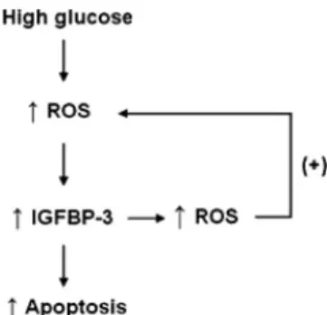
Changes in cellular function by hyperglycemia that re-sult in oxidative stress are crucial in the development and progression of diabetic nephropathy. A unifying hypoth-esis suggests that mitochondrial overproduction of ROS initiates hyperglycemia-induced damage in the diabetic kidney, leading to activation of four major biochemical pathways: AGE formation, protein kinase C isoforms, and flux through the polyol and hexosamine pathways. Each of these pathways can contribute to the perpetuation of, and in some cases initiate, cellular ROS generation (25). Increased levels of ROS in hyperglycemia induce re-nal cell apoptosis and diabetic nephropathy (30). Ver-zola et al. (31) reported that hyperglycemia induces ap-optotic changes in human tubular cells by increasing oxidative stress. In this study, high glucose or IGFBP-3 overexpression increased oxidative stress, and siRNA: IGFBP-3 abolished the increase in oxidative stress caused by high glucose or IGFBP-3 overexpression, sug-gesting that IGFBP-3 is involved in inducing oxidative stress by high glucose in PTEC. On the other hand, antioxidants decreased IGFBP-3 expression and high-glu-cose-induced apoptosis, and elevation of oxidative stress by hydrogen peroxide increased IGFBP-3 expression, sug-gesting that oxidative stress increases IGFBP-3 expression. In addition, ROS increase by high glucose or IGFBP-3 overexpression was significant at 6 h, whereas IGFBP-3 mRNA expression was significantly increased at 12– 48 h, and apoptosis was most prominent at 72 h exposure to high-glucose medium. Taken together, these results sug-gest a positive loop between ROS and IGFBP-3, amplify-ing ROS production and resultamplify-ing in amplification of hy-perglycemic damage (Fig. 5).
In the present study, treatment of siRNA:IGFBP-3 un-der standard-glucose conditions also resulted in decreased
apoptosis. PTEC itself normally secrete IGFBP-3 in some degree. It is possible that that inhibition of intrinsic IGFBP-3 secretion by siRNA:IGFBP-3 resulted in subnor-mal IGFBP-3 expression, resulting in decreased apoptosis. Although it is well documented that IGFBP-3 promotes apoptosis, the underlying mechanism of action remains uncertain (7). Kim et al. (8) reported that IGFBP-3 induces apoptosis through the activation of caspases, via a death receptor-mediated pathway in breast cancer cells. It was also demonstrated that IGFBP-3 significantly enhances apoptosis by inhibiting nuclear factor-B activation in co-lonic carcinoma-derived cell lines (32). However, the ac-tual mechanism of IGFBP-3 action on PTEC apoptosis is still unclear and needs to be elucidated.
In conclusion, this study found that high glucose in-creases IGFBP-3 expression and induces oxidative stress and apoptosis in PTEC. The results suggest that increased IGFBP-3 expression by high glucose mediates high-glu-cose-induced apoptosis in PTEC. We propose that ele-vated levels of oxidative stress from high glucose increase IGFBP-3 expression, thereby inducing apoptosis. In addi-tion, increased expression of IGFBP-3 by high glucose in-duces additional oxidative stress, which may result in am-plification of hyperglycemic damage in PTEC.
Acknowledgments
Address all correspondence and requests for reprints to: Ho-Seong Kim, M.D, Ph.D., Professor, Department of Pediatrics, College of Medicine Yonsei University, 250 Sungsan-ro, Seo-daemun-gu, Seoul 120-752, Korea. E-mail: [email protected].
This investigation was supported by LG Life Science Grant (to H.-S.K.) and Green Cross Grant (to H.-S.K.).
Disclosure Summary: E.-G.Y., W.J.L., J.H.K, H.-W.C., S.E.H., D.H.K., and Y.O. have nothing to declare. H.-S.K. re-ceived grants from LG Life Science and Green Cross.
FIG. 5. Schematic diagram showing ROS generation by high glucose
stimulates IGFBP-3 expression, resulting in apoptosis. Increased expression of IGFBP-3 induces ROS, which can result in amplified hyperglycemic damage.
References
1. Perneger TV, Brancati FL, Whelton PK, Klag MJ 1994 End-stage renal disease attributable to diabetes mellitus. Ann Intern Med 121: 912–918
2. Brownlee M 2005 The pathobiology of diabetic complications: a unifying mechanism. Diabetes 54:1615–1625
3. Schrijvers BF, De Vriese AS, Flyvbjerg A 2004 From hyperglycemia to diabetic kidney disease: the role of metabolic, hemodynamic, in-tracellular factors and growth factors/cytokines. Endocr Rev 25: 971–1010
4. Balakumar P, Arora MK, Reddy J, Anand-Srivastava MB 2009 Pathophysiology of diabetic nephropathy: involvement of multifac-eted signalling mechanism. J Cardiovasc Pharmacol 54:129 –138 5. Vasylyeva TL, Ferry Jr RJ 2007 Novel roles of the IGF-IGFBP axis
in etiopathophysiology of diabetic nephropathy. Diabetes Res Clin Pract 76:177–186
6. Firth SM, Baxter RC 2002 Cellular actions of the insulin-like growth factor binding proteins. Endocr Rev 23:824 – 854
7. Jogie-Brahim S, Feldman D, Oh Y 2009 Unraveling insulin-like growth factor binding protein-3 actions in human disease. Endocr Rev 30:417– 437
8. Kim HS, Ingermann AR, Tsubaki J, Twigg SM, Walker GE, Oh Y 2004 Insulin-like growth factor-binding protein 3 induces caspase-dependent apoptosis through a death receptor-mediated pathway in MCF-7 human breast cancer cells. Cancer Res 64:2229 –2237 9. Kim HS, Lee WJ, Lee SW, Chae HW, Kim DH, Oh Y 2010
Insulin-like growth factor binding protein-3 induces G1 cell cycle arrest with inhibition of cyclin-dependent kinase 2 and 4 in MCF-7 human breast cancer cells. Horm Metab Res 42:165–172
10. Chan SS, Twigg SM, Firth SM, Baxter RC 2005 Insulin-like growth factor binding protein-3 leads to insulin resistance in adipocytes. J Clin Endocrinol Metab 90:6588 – 6595
11. Kim HS, Ali O, Shim M, Lee KW, Vuguin P, Muzumdar R, Barzilai
N, Cohen P 2007 Insullike growth factor binding prote3
in-duces insulin resistance in adipocytes in vitro and in rats in vivo. Pediatr Res 61:159 –164
12. Chan SS, Schedlich LJ, Twigg SM, Baxter RC 2009 Inhibition of adipocyte differentiation by insulin-like growth factor-binding pro-tein-3. Am J Physiol Endocrinol Metab 296:E654 –E663
13. Daemen MA, van ’t Veer C, Denecker G, Heemskerk VH, Wolfs TG,
Clauss M, Vandenabeele P, Buurman WA 1999 Inhibition of
apo-ptosis induced by ischemia-reperfusion prevents inflammation. J Clin Invest 104:541–549
14. Schelling JR, Cleveland RP 1999 Involvement of Fas-dependent ap-optosis in renal tubular epithelial cell deletion in chronic renal fail-ure. Kidney Int 56:1313–1316
15. Kumar D, Zimpelmann J, Robertson S, Burns KD 2004 Tubular and interstitial cell apoptosis in the streptozotocin-diabetic rat kidney. Nephron Exp Nephrol 96:e77– e88
16. Kumar D, Robertson S, Burns KD 2004 Evidence of apoptosis in human diabetic kidney. Mol Cell Biochem 259:67–70
17. Kang BP, Frencher S, Reddy V, Kessler A, Malhotra A, Meggs LG 2003 High glucose promotes mesangial cell apoptosis by oxidant-dependent mechanism. Am J Physiol Renal Physiol 284:F455–F466 18. Vasylyeva TL, Chen X, Ferry Jr RJ 2005 Insulin-like growth factor binding protein-3 mediates cytokine-induced mesangial cell apo-ptosis. Growth Horm IGF Res 15:207–214
19. Wolf G, Chen S, Ziyadeh FN 2005 From the periphery of the glo-merular capillary wall toward the center of disease: podocyte injury comes of age in diabetic nephropathy. Diabetes 54:1626 –1634 20. Thomas MC, Burns WC, Cooper ME 2005 Tubular changes in early
diabetic nephropathy. Adv Chronic Kidney Dis 12:177–186 21. Magri CJ, Fava S 2009 The role of tubular injury in diabetic
ne-phropathy. Eur J Intern Med 20:551–555
22. Thomson SC, Vallon V, Blantz RC 2004 Kidney function in early diabetes: the tubular hypothesis of glomerular filtration. Am J Physiol Renal Physiol 286:F8 –F15
23. Buckway CK, Wilson EM, Ahlse´n M, Bang P, Oh Y, Rosenfeld RG 2001 Mutation of three critical amino acids of the N-terminal do-main of IGF-binding protein-3 essential for high affinity IGF bind-ing. J Clin Endocrinol Metab 86:4943– 4950
24. Bestwick CS, Milne L 2001 Quercetin modifies reactive oxygen levels but exerts only partial protection against oxidative stress within HL-60 cells. Biochim Biophys Acta 1528:49 –59
25. Forbes JM, Coughlan MT, Cooper ME 2008 Oxidative stress as a major culprit in kidney disease in diabetes. Diabetes 57:1446 –1454 26. Samikkannu T, Thomas JJ, Bhat GJ, Wittman V, Thekkumkara TJ 2006 Acute effect of high glucose on long-term cell growth: a role for transient glucose increase in proximal tubule cell injury. Am J Physiol Renal Physiol 291:F162–F175
27. Allen DA, Yaqoob MM, Harwood SM 2005 Mechanisms of high glucose-induced apoptosis and its relationship to diabetic compli-cations. J Nutr Biochem 16:705–713
28. Bach LA, Cox AJ, Mendelsohn FA, Herington AC, Werther GA,
Jerums G 1992 Focal induction of IGF binding proteins in proximal
tubules of diabetic rat kidney. Diabetes 41:499 –507
29. Grimberg A, Coleman CM, Burns TF, Himelstein BP, Koch CJ,
Cohen P, El-Deiry WS 2005 p53-Dependent and p53-independent
induction of insulin-like growth factor binding protein-3 by deoxy-ribonucleic acid damage and hypoxia. J Clin Endocrinol Metab 90: 3568 –3574
30. Wagener FA, Dekker D, Berden JH, Scharstuhl A, van der Vlag J 2009 The role of reactive oxygen species in apoptosis of the diabetic kidney. Apoptosis 14:1451–1458
31. Verzola D, Bertolotto MB, Villaggio B, Ottonello L, Dallegri F,
Salvatore F, Berruti V, Gandolfo MT, Garibotto G, Deferrari G
2004 Oxidative stress mediates apoptotic changes induced by hy-perglycemia in human tubular kidney cells. J Am Soc Nephrol 15(Suppl 1):S85–S87
32. Williams AC, Smartt H, H-Zadeh AM, Macfarlane M, Paraskeva C,
Collard TJ 2007 Insulin-like growth factor binding protein 3
(IGFBP-3) potentiates TRAIL-induced apoptosis of human colorec-tal carcinoma cells through inhibition of NF-B. Cell Death Differ 14:137–145